
Combined Valvular Heart Disease: A Comprehensive Clinical Review Of Etiology, Diagnosis, And Management
1. Shefali Roshan Meshram
2. Chandan Sadar
(1. Student, International Medical Faculty, Osh state university, Kyrgyzstan.
2. Student, International Medical Faculty, Osh State University, Kyrgyzstan)
Abstract
Background: Combined (mixed) heart disease is characterized by the presence of both stenotic and regurgitant lesions in one or more cardiac valves. It is primarily a complication of rheumatic heart disease, manifests with complicated hemodynamics that often mask the traditional clinical presentation.
Objective: To investigate the etiology, pathophysiology, clinical presentation, diagnostic approach and treatment of combined heart disease.
Methods: A structured literature-based review was performed using standard cardiology references including textbooks, clinical guidelines, and peer-reviewed journal articles. Relevant published data on combined valvular lesions were collected and examined.
Results: The most frequently observed combined lesions are mitral stenosis with mitral regurgitation, followed in frequency by lesions of the aortic valve and multivalvular disease. Rheumatic heart disease is the most common cause of combined heart disease worldwide. Patients typically present with dyspnea, fatigue, palpitations and signs/symptoms of heart failure. Echocardiography is the preferred modality to assess valve morphology and severity. The management of combined heart disease consists of medical therapy for symptom control and surgical or percutaneous intervention for definitive treatment.
Conclusion: Combined heart disease presents as a complex valvular disorder requiring careful clinical evaluation and an echocardiographic assessment for diagnosis and treatment of the disorder. Early diagnosis and timely intervention improve prognosis and decrease complications associated with combined heart disease.
Introduction
Mixed (combined) heart disease describes the existence of both stenotic and regurgitant lesions occurring in one (or more) of the body’s heart valves at the same time. These various types of heart valve problems can impact how your blood flows through your body, creating complications in the way that your heart works.
Valvular heart disease continues to be one of the highest causes of morbidity throughout the world, especially among people in developing nations. In those locations, rheumatic heart disease (RHD) is often the reason for developing valvular problems. People with RHD suffer from multiple episodes where one or more of their heart valves become inflamed. The result of this ongoing inflammation produces progressive thickening of the heart valve(s), as well as various degrees of scarring and calcification or hardening and binding of heart valves to adjacent structures (commissural fusion). The result can lead to patients developing complex types of valvular disease such as mitral stenosis and mitral regurgitation—both of which will be present in many individuals with combined heart disease.
In addition to RHD, combined heart disease may occur as a result of aging, calcific degeneration of heart valves, infective endocarditis (endocarditis that results in the destruction of a heart valve after the person has been infected), congenital heart valve abnormalities, and trauma from surgical procedures or other medical treatments designed to repair or replace heart valves.
Combined heart disease has a high degree of clinical relevance because of its complex diagnostic features and unpredictable combination of symptoms. The presence of one lesion can obscure or alter the classical findings of auscultation of any other lesions. One example of this is the presence of mitral regurgitation, which can reduce the intensity of the mitral stenotic murmurs by lowering the left atrial pressure gradients. Similarly, there are combinations of both aortic stenosis and aortic regurgitation that have features of both volume overload and pressure overload, and this can complicate the clinical assessment.
In terms of pathophysiology, combined lesions present an individual cardiac chamber with two burdens, both a pressure overload because of the stenosis and a volume overload because of the regurgitation. The result of these two burdens is the progressive dilation of the cardiac chambers, development of hypertrophy, atrial fibrillation, pulmonary hypertension, and if not treated will ultimately lead to congestive heart failure.
The most reliable means of making an accurate diagnosis of combined heart disease is through the use of two-dimensional echocardiography with Doppler studies, which allows for direct visualization of the morphology of the valvular structures, an assessment of the degree of disease severity, and an assessment of the hemodynamic impact of the combined lesions. Just a clinical examination will often provide insufficient information to make an accurate diagnosis due to overlapping physical exam findings. Managing combined heart disease is complex and requires individualized evaluation based on all aspects of the patient's condition, such as the dominant lesion, the severity of the lesions, patient symptoms, and ventricular function. Therapeutic options include pharmacological treatment, percutaneous intervention, or surgical valve repair or replacement.
Given the complexity of combined heart disease, the increasing prevalence of this condition in rheumatic populations, and its growing importance in the field of cardiovascular medicine, early recognition and comprehensive management are critical for optimizing patient outcomes.
Methods
Study Design
This article aims to provide an organized/classified narrative review of mixed heart disease including its causes, biology of the disease, symptoms and ways in which to diagnose and manage it. This review brings together existing literature found on the internet from widely read and used textbooks, clinical practice guidelines, and peer-reviewed journal articles.
Data Sources
Information needed to complete this article was obtained from the following trusted and accepted sources of information in cardiology/internal medicine:
● Harrison’s Principles of Internal Medicine
● Braunwald’s Heart Disease: A Textbook of Cardiovascular Medicine
● API Textbook of Medicine
● European Society of Cardiology (ESC) Guidelines on Valvular Heart Disease
● American Heart Association (AHA/ACC) Guidelines for Valvular Heart Disease
● Peer-reviewed articles from indexed journals (PubMed, Google Scholar)
Search strategy
The literature search was completed using electronic search engines: PubMed, Google Scholar and Cochrane Library, using search terms: "combined valvular heart disease", "mixed mitral valve disease", "multivalvular heart disease", "rheumatic valvular lesions". The use of Boolean operators (AND, OR ) helped limit the outcomes to relevant studies which addressed combined valvular pathology, and its associated clinical issues.
Inclusion criteria
Studies that described combined or mixed valvular heart disease, the clinical features of combined or mixed valvular heart disease, the methods used to diagnose mixed valvular heart disease, and the management of mixed valvular heart disease were included. Inclusion was limited to studies published in English and studies containing human subjects. Review articles, clinical guidelines, and original research studies that showed a strong link to combined valvular lesions had preference.
Exclusion criteria
All studies which focused only on isolated valvular lesions with no mixed involvement (including pure mitral disease) were excluded. All case studies that only involved isolated valvular disease were also excluded due to lack of broader clinical relevance; all non-English studies that could not be reliably translated were excluded. Finally, studies with inadequate information regarding combined valvular disease were also excluded from review.
Data extraction and synthesis
Take Information out from Literature, Group By Major Area: Etiology, Epidemiology, Pathophysiology, Clinic, Diagnosis, and Treatment, then Develop an Overall Picture of Combined Heart Disease with the Evidence that is Available to Support that Picture.
Outcome Parameters Reviewed
Distribution of Valve Combinations, Hemodynamic Effect of Mixed Lesions, Diagnostic Accuracy of Echocardiogram, Available Treatment Options and Results (Prognosis) in Patients with Multiple Valve Involvement.
Limitations
Limitations of This Review Include Reliance on Secondary Data Sources, Likelihood of Publication Bias and Differences in the Definition/Grading of Valvular Lesions Across Studies; Access to Non-English Articles was Also Limited.
Result
The findings from this systematic review are based on an integration of widely quoted cardiology literature including Harrison’s Principles of Internal Medicine, Braunwald’s Heart Disease, API Textbook of Medicine, and the ESC/AHA/ACC Valvular Heart Disease Guidelines.
The Cause of Combined Heart Disease:The reviewed literature demonstrates that rheumatic heart disease (RHD) is a major contributor to the presence of combined valvular lesions in developing countries. Harrison’s Principles of Internal Medicine indicates that rheumatic fever causes repeated episodes of inflammatory injury to the endocardium, resulting in progressive fibrosis, commissural fusion, shortening of the chordae tendineae, and calcification. These alterations in the structure of the valves are responsible for the frequent occurrence of both stenotic and regurgitant lesions in the playing structures of the same valve; the mitral valve in particular.
Braunwald’s Heart Disease goes on to describe that degenerative valve disease is the second most common cause of combined valvular lesions, particularly among individuals over 65 years of age. In these populations, calcific degeneration causes stiffness of the valve leaflets (stenosis) and the inability of the leaflets to come together (regurgitation). As such, Infective endocarditis is yet another significant etiology of combined valvular lesions according to the ESC guidelines; this occurs when the leaflets and chordae tendineae are acutely destroyed leading to sudden anomalies of both conditions.The less common forms of mixed lesions include those caused by congenital defects (e.g. bicuspid aortic valve with mixed lesions); connective tissue disorders; and post-operative valvular dysfunction following surgical repair or replacement.
Common Valvular Combinations:As outlined in the API Textbook of Medicine, the most frequently seen combinations of valves both in practice and literature are MS + MR (mitral stenosis + mitral regurgitation) due to the influence of rheumatic disease on both leaflet motion causing stenosis and chordae/papillary muscle injury causing regurgitation.. The second most frequently seen combination is AS + AR (aortic stenosis + aortic regurgitation), particularly in degenerative and rheumatic pathologies. Furthermore, the ESC guidelines also recognize the presence of multivalvular disease (mitral + aortic ± tricuspid) as a sign of advanced rheumatic disease. In some cases, triple valve disease (MS + MR + AR ± TR) can be present which typically occurs in patients who have experienced long-standing rheumatic fever and have significant pulmonary hypertension.
Pathophysiology: As stated in Braunwald’s Heart Disease, combined valvular lesions create an unusual and complex hemodynamic state as they create both pressure and volume overload on the heart. In the case of mitral valve disease, the presence of stenosis will increase left atrial pressure whereas the presence of regurgitation will create a volume overload in the left atrium thus leading to markedly dilated atria. The presence of these two pathologies concurrently will predispose the patient to atrial fibrillation as outlined in both Harrison's Internal Medicine and ESC guidelines. In regard to aortic valve disease, stenosis will create a pressure overload of the left ventricle and lead to left ventricular concentric hypertrophy while the presence of regurgitation will lead to a volume overload of the left ventricle and result in left ventricular eccentric hypertrophy. The presence of both will create a situation that will cause the left ventricle to hypertrophy in an accelerated manner.
As explained in "Braunwald's Heart Disease," patients with advanced mitral valve disease can develop pulmonary hypertension secondary to long-standing elevated left atrial pressures. As the left atrial pressures continue to elevate, it eventually results in right ventricular overload causing right ventricular failure and tricuspid regurgitation.
Clinical Presentation: The clinical presentation of Combined Valvular Heart Disease is variable and not always reliable. The type of symptoms will depend on the dominant lesion and the ability of the heart to compensate (Harrison's Principles of Medicine A21, pg 1297).
Common Symptoms Include:
●Dyspnea on exertion from pulmonary congestion.
● Fatigue and decreased exercise tolerance from low cardiac output.
● Palpitations (may be related to atrial fibrillation).
● Orthopnea and paroxysmal nocturnal dyspnea (advanced cases).
● Hemoptysis from pulmonary venous hypertension with severe mitral stenosis.
The physical exam often appears misleading because of overlapping lesions. In "Braunwald's Heart Disease" it is stated the intensity of the murmur may be softer or obscured. As an illustration, severe mitral regurgitation can decrease the intensity of the diastolic murmur of mitral stenosis, by decreasing the transmitral pressure gradient. Similarly, in the case of a patient with both aortic stenosis (AS) and aortic regurgitation (AR), a variety of unusual or atypical pattern of murmurs would be produced, complicating the clinician’s ability to make a clinical diagnosis and rely on imaging studies.
Diagnostic Results:According to the main reference documents, such as the European Society of Cardiology (ESC) and American Heart Association/American College of Cardiology (AHA/ACC) guidelines, two-dimensional echocardiography with Doppler is the ideal method to evaluate combined valvular disease.
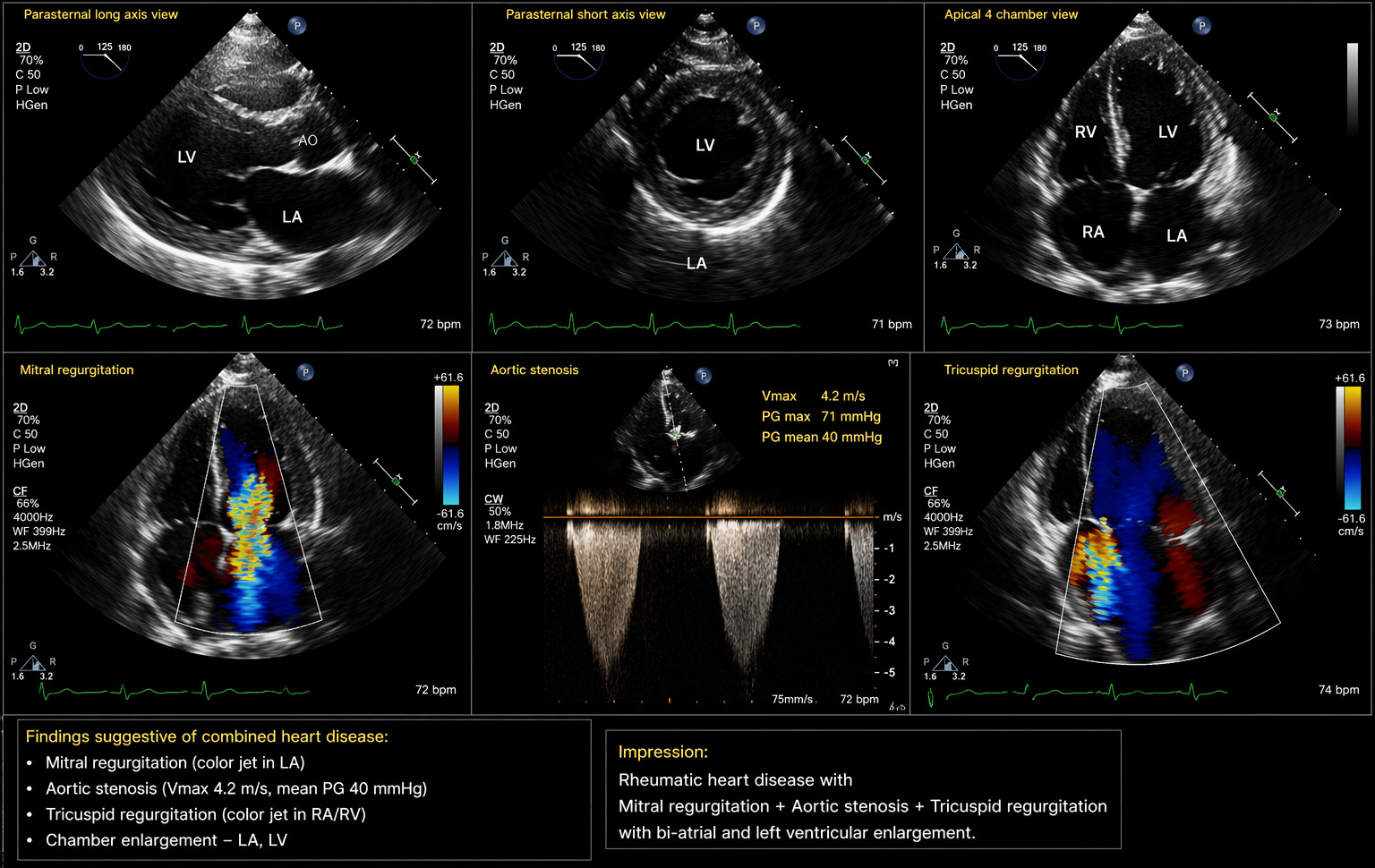
Echocardiography provides clinical information for the following
● Structural evaluation of valves - thickening/thickening of leaflets, calcification, and restriction of leaflets.
● Functional evaluation of valves - regurgitant jets, flow velocities and gradients.
● Quantification of severity of disease - calculated valve area, calculated valve gradients and calculated regurgitant fraction.
● Evaluation of cardiac chambers - evaluation of atrial and ventricular sizes and functions.
The management approach of valvular heart disease is determined by the recommendations/guidelines from the European Society of Cardiology (ESC) and the American Heart Association/American College of Cardiology (AHA/ACC) as well as by the severity of the patients’ symptoms, the dominant lesion, and the ventricular function.
There are two options for the medical management of valvular heart disease: diuretics for pulmonary congestion or systemic congestion; beta-blockers for rate control of atrial fibrillation and symptom relief; ACE inhibitors in select cases of ventricular dysfunction, and anticoagulation for patients who have atrial fibrillation or at risk of thrombosis in the left atrium.
Interventional methods of treating valvular heart disease include balloon mitral valvotomy in appropriate patients who have isolated or a dominant mitral stenosis, percutaneous valve interventions in select cases of degenerative changes.
Surgical repair is considered to be the definitive management of valvular heart disease and can be accomplished by performing valve repairs (when indicated); valve replacement (mechanical or bioprosthetic); double or triple valve replacement in patients with multiple valve disease.Surgical decision-making is complicated in patients who have combined lesions since the treatment of one lesion may unmask the severity of the second lesion, according to Braunwald's Heart Disease.
Prognostic Determinants: A patient's prognosis is determined by the extent of valvular involvement, degree of functional impairment and potential for developing complications. Harrison's Principles of Medicine lists multiple valves affected as having worse outcomes, also including those with A-fib and pulmonary hypertension. According to ESC guidelines, the longer a patient is treated nonoperatively the greater their risk of developing irreversible LV dysfunction and thus having a poorer prognosis. Early diagnosis and prompt treatment enhance the likelihood of surviving and having a better quality of life after surgery.
Discussion
Combined heart disease is a complicated range of valve problems with both stenosis and regurgitation occurring at one valve or multiple valves. This review article emphasizes that Rheumatic heart disease is by far the most common cause of combined heart disease globally, especially in underdeveloped nations as has been documented regularly in Harrison's Principles of Internal Medicine and Braunwald's Heart Disease. Chronic inflammation from rheumatic fever results in progressively increasing levels of fibrous tissue, fusion between commissures, and shortened chordae which result in mixed lesion (stenosis and regurgitation) formation at the mitral valve to a greater extent than in other valves.
A major diagnostic problem with combined heart disease is the modification and/or masking of classic clinical signs. Braunwald's Heart Disease stresses that the presence of regurgitant lesions alters the intensity of murmurs usually associated with a corresponding stenotic lesion by changing the transvalvular pressure gradient. For instance, in patients with severe MR; there will be diminished mutation of the mid-diastolic murmur of MS due to diminished left atrial pressure gradient. Likewise, combined AS and AR will yield overlapping auscultatory findings that may not correctly characterize the degree of either stenosis or regurgitation. This diagnostic complication makes a clinical exam alone an inadequate method to make an accurate clinical diagnosis.
In terms of pathophysiology, the effects of both pressures and volume on the heart are compounded by having multiple areas of disease. (Braunwald, 1977) Both of these types of load affect progressive changes in size and shape of chambers (e.g., enlargement of left atrium with blockage at mitral valve; left ventricular hypertrophy with obstruction at aortic valve). When there is long-term pressure overload to the heart due to hypertension, there will be concentric hypertrophy; however, without any form of pressure or preload increase, there will be eccentric hypertrophy and perhaps those changes won’t improve ventricular function unless proper medical treatment is given. Permanent elevation in left atrial pressure over time combined with pulmonary congestion from blood returning to the heart can lead to pulmonary venous hypertension, which then leads to or results in worsening pulmonary arterial hypertension and subsequent failure of the right ventricle/pulmonary arterial hypertension and development of functional tricuspid regurgitation based on right-sided heart failure.Echocardiography is still the primary way to diagnose CVD and echocardiography following the guidelines established by the ESC and the AHA/ACC. Echocardiography allows you to see and measure the stiffness and amount of backward flow of a valve, and measure the size and function of cardiomyocytes. In addition, Doppler echocardiography is useful for distinguishing the predominant lesions from the other types in cases of combined pathology. This is very important for making clinical decisions. Transesophageal echocardiography may also improve the accuracy of the diagnosis in cases of both complex and pre-operative pathology.
Manifestations of functional status are often nonspecific and therefore dependent on what the predominant lesion is and any compensatory mechanism one may have developed as a result of their disease. The most common complaint is shortness of breath caused by pulmonary congestion; however, many people will complain of palpitations when there is atrial fibrillation due to enlargement of the atrium caused by dilating around the atrium. Because of this variability and the inability to perform echocardiography due to limited resources (e.g., unable to obtain imaging), diagnosis may frequently be delayed in countries where there is limited access.
Outcomes of combined heart disease are usually not as good as those seen with an isolated valvular disease as noted by Harrison's Internal Medicine. How well a patient does with their combined heart disease will be dependent on the number of valves involved, the severity of their dysfunction, presence of atrial fibrillation, pulmonary hypertension, and their ventricular function. Combined multivalvular involvement especially when there are advanced heart failure or pulmonary hypertension is associated with a poor survival rate. Therefore, obtaining early diagnosis and performing timely surgery is the primary way to improve prognosis.
In conclusion, combined heart disease is a challenging diagnosis and has complex hemodynamic effects on patients. In order to improve the patient outcomes for combined heart disease, clinicians need to have a high index of suspicion, echocardiography evaluation and guideline based management.
Conclusion
Combined heart disease is a complex disease of the heart that contains both a narrowing (stenosis) and from the value not closing properly (regurgitation) in one or more of the heart's valves. Rheumatic heart disease is the most common cause of this heart disease; it causes damage to the heart's structure over time (chronic), which leads to the mixed function of the valves with different pressures (pressure overload; stenosis) and volume overload (volume overload; regurgitation); therefore, the combination of these two conditions or entities creates a unique (stress-induced) interaction on the body physiologically and often alters (or masks) the classical signs of heart disease and presents a challenge with the diagnosis of these patients at the medical level.
Echocardiography with Doppler evaluation (Doppler echocardiography) is the gold standard for diagnosing the condition of combined heart disease, the severity of the heart disease, and identifying the most significant lesion (valve), which is critical in making management decisions. Recognition of disease characteristics early, ongoing monitoring/follow-up, and prompt intervention (medical, percutaneous, or surgical) are vital in preventing irreversible changes in the heart from remodeling, atrial fibrillation, pulmonary hypertension, or congestive heart failure.
The prognosis of the patient in combined heart disease is based on the number of valves involved, severity of the dysfunction of each of the valves, and when (timely) you perform the intervention. Multidisciplinary and guideline-based treatment strategies will significantly improve the patients' clinical outcome (i.e., quality of life) with combined heart disease.
References
● Braunwald, E. (Ed.). (2022). Braunwald’s heart disease: A textbook of cardiovascular medicine (12th ed.). Elsevier.
● Jameson, J. L., Fauci, A. S., Kasper, D. L., Hauser, S. L., Longo, D. L., & Loscalzo, J. (Eds.). (2022). Harrison’s principles of internal medicine (21st ed.). McGraw Hill
● Kasper, D. L., Fauci, A. S., Hauser, S. L., Longo, D. L., & Jameson, J. L. (2021). Harrison’s principles of internal medicine (20th ed.). McGraw Hill.
● Dewan, B., & API Medicine Committee. (2023). API textbook of medicine. Association of Physicians of India.
● Otto, C. M., & Bonow, R. O. (2020). Valvular heart disease. In Braunwald’s heart disease: A textbook of cardiovascular medicine (11th ed., pp. 1345–1430). Elsevier.
● Vahanian, A., Beyersdorf, F., Praz, F., et al. (2021). 2021 ESC/EACTS guidelines for the management of valvular heart disease. European Heart Journal, 43(7), 561–632. https://doi.org/10.1093/eurheartj/ehab395
● Otto, C. M., Nishimura, R. A., Bonow, R. O., et al. (2021). 2020 ACC/AHA guideline for the management of patients with valvular heart disease. Journal of the American College of Cardiology, 77(4), e25–e197. https://doi.org/10.1016/j.jacc.2020.11.018
● World Health Organization. (2023). Rheumatic heart disease. https://www.who.int
● Unger, P., Galloo, X., & Pibarot, P. (2025). Mixed valvular heart disease: Diagnosis and management. European Heart Journal, 46(24), 2261–2274. https://doi.org/10.1093/eurheartj/ehaf116
● Duggal, B., Varada, V. K., Mukherjee, A., Chawla, R., & Saha, R. N. (2025). Combined rheumatic mitral and aortic stenosis: Hemodynamics and tailored management. JACC: Case Reports, 30(24), 104670. https://doi.org/10.1016/j.jaccas.2025.104670
● Mantovani, F., Fanti, D., Tafciu, E., et al. (2021). When aortic stenosis is not alone: Epidemiology, pathophysiology, diagnosis and management in mixed and combined valvular disease. Frontiers in Cardiovascular Medicine, 8,744497.https://doi.org/10.3389/fcvm.2021.744497
● Vahanian, A., Beyersdorf, F., Praz, F., et al. (2021). 2021 ESC/EACTS guidelines for the management of valvular heart disease. European Heart Journal, 43(7), 561–632.https://doi.org/10.1093/eurheartj/ehab395
● Otto, C. M., & Bonow, R. O. (2020). Valvular heart disease. In Braunwald’s heart disease: A textbook of cardiovascular medicine (11th ed.). Elsevier.https://www.elsevier.com/books/braunwalds-heart-disease
●Zipes, D. P., Libby, P., Bonow, R. O., Mann, D. L., & Tomaselli, G. F. (2022). Braunwald’s heart disease (12th ed.). Elsevier. https://www.elsevier.com/books/braunwalds-heart-disease