
Duodenal Ulcer: Pathophysiology, Diagnosis, and Contemporary Management
1. Kebekova Aizat Ashimzhanovna
2. Shiek Kadar Rizwana
3. Mariappan Muthumari
(1. Lecturer, International Medical Faculty, Osh State University, Osh, Kyrgyz Republic
2. Student, International Medical Faculty, Osh State University, Osh, Kyrgyz Republic
3. Student, International Medical Faculty, Osh State University, Osh, Kyrgyz Republic.)
Abstract
Duodenal ulcer, the most common anatomical form of peptic ulcer disease, remains a clinically significant condition despite dramatic advances in acid suppression and the near eradication of its principal infectious cause in many developed countries. This article presents a comprehensive, clinically oriented review of duodenal ulcer disease in the modern era. The Introduction traces the historical evolution of understanding from the acid centric dogma of Schwarz to the discovery of Helicobacter pylori by Marshall and Warren, and situates the current epidemiology in the context of widespread nonsteroidal anti-inflammatory drug use and rising antimicrobial resistance. The Methods section describes the narrative review methodology. The Results section systematically addresses etiology and pathophysiology, including H. pylori infection, NSAID induced mucosal injury, acid pepsin imbalance, and emerging entities such as H. pylori negative idiopathic ulcers; clinical presentation and complications encompassing hemorrhage, perforation, penetration, and gastric outlet obstruction; diagnostic strategies spanning endoscopy and noninvasive testing; and contemporary management including proton pump inhibitor therapy, potassium competitive acid blockers, the landmark 2024 American College of Gastroenterology H. pylori treatment guideline, endoscopic hemostasis, and surgical options for refractory disease. The Discussion critically examines treatment challenges, including antimicrobial resistance, the growing proportion of idiopathic ulcers, the long term safety debate surrounding acid suppression, and future directions. The review concludes that duodenal ulcer has been transformed from a chronic relapsing surgical disease into a largely curable medical condition, yet remains a source of substantial morbidity when diagnosis is delayed, eradication fails, or ulcerogenic exposures persist.
Keywords: duodenal ulcer; peptic ulcer disease; Helicobacter pylori; proton pump inhibitors; vonoprazan; gastrointestinal bleeding; bismuth quadruple therapy
Introduction
The story of the duodenal ulcer is one of the great narratives of twentieth century medicine, a condition that moved from the surgeon's domain to the physician's prescription pad and then, in its infectious majority, toward genuine cure. For the first half of the last century, the governing maxim was Schwarz's dictum of 1910: no acid, no ulcer. Duodenal ulcer was understood as a disorder of aggressive acid secretion, managed with bed rest, bland diets, antacids, and, when these failed, formidable operations including vagotomy, antrectomy, and partial gastrectomy, procedures that relieved ulcer pain at the cost of dumping syndromes, weight loss, and nutritional disturbance. The introduction of the H2 receptor antagonist cimetidine in 1976, followed by the proton pump inhibitor omeprazole in 1989, progressively retired the ulcer surgeon, but the deeper conceptual revolution came from Perth, Australia, where Robin Warren and Barry Marshall identified a curved bacterium colonizing the gastric mucosa and, in a legendary act of self-experimentation, Marshall drank a culture of the organism, developed gastritis, and thereby helped establish the infectious etiology of peptic ulceration, work recognized with the Nobel Prize in Physiology or Medicine in 2005 [1,2]. Eradication of H. pylori converted duodenal ulcer from a chronic relapsing illness with lifetime recurrence rates exceeding fifty percent into a condition cured by a two-week course of antibiotics.
The epidemiological picture has shifted accordingly, though the disease is far from vanquished. Peptic ulcer disease still affects millions worldwide, with lifetime prevalence estimates historically around five to ten percent in Western populations. Duodenal ulcers outnumber gastric ulcers in most series, particularly in younger patients. In high income countries, declining H. pylori prevalence, driven by improved sanitation and childhood acquisition patterns, has reduced ulcer incidence, while the aging population's consumption of nonsteroidal anti-inflammatory drugs, low dose aspirin, anticoagulants, and corticosteroids has sustained a large reservoir of drug induced ulceration. A notable and concerning trend is the rising proportion of ulcers attributable to neither H. pylori nor NSAIDs; multicenter observations have reported that idiopathic peptic ulcers now constitute roughly eighteen percent of cases in some Japanese cohorts, with similar trends reported elsewhere [3]. These idiopathic ulcers behave more aggressively, heal more slowly, bleed more often, and recur more frequently, and they have restored some humility to a field that briefly believed the problem solved. Meanwhile, the therapeutic landscape has evolved again with the arrival of potassium competitive acid blockers such as vonoprazan, now recommended as first line acid suppression for peptic ulcer disease in the 2026 Japanese evidence based guidelines [4], and with the 2024 American College of Gastroenterology guideline fundamentally restructuring H. pylori eradication strategy in response to clarithromycin resistance rates exceeding thirty percent [5].
This article aims to provide medical students, residents, and practicing clinicians with a coherent, current account of duodenal ulcer disease: its causes and mechanisms, its clinical faces and complications, the rational use of diagnostic tests, and the evidence-based management of both uncomplicated and complicated disease. The guiding conviction throughout is that this common condition rewards mechanistic understanding; the clinician who grasps why acid, bacteria, and drugs conspire to breach the duodenal mucosa is best placed to heal the ulcer, prevent its return, and recognize the patient in whom the story is more sinister than simple peptic disease.
Methods
This article is structured as a narrative review with systematic elements. The literature informing the review was assembled from searches of PubMed/MEDLINE, the Cochrane Library, and guideline repositories covering publications through November 2026. Search terms combined duodenal ulcer and peptic ulcer disease with pathophysiology, Helicobacter pylori, NSAID, epidemiology, diagnosis, urea breath test, proton pump inhibitor, vonoprazan, bismuth quadruple therapy, eradication, gastrointestinal bleeding, perforation, endoscopic hemostasis, and surgical treatment. Priority was given to randomized controlled trials, systematic reviews and meta-analyses, and current guidelines from the American College of Gastroenterology, the American College of Gastroenterology and Canadian Association of Gastroenterology joint guideline on upper gastrointestinal bleeding, the Maastricht VI consensus, the Japanese Gastroenterological Association, and the National Institute for Health and Care Excellence. Foundational historical literature was retained where it remains definitive, notably the work of Marshall and Warren and the Cochrane analyses establishing eradication therapy as curative. Regulatory documents informed discussion of newer agents including vonoprazan and rifabutin based therapy. Because the aim is clinical education rather than quantitative synthesis, no formal meta-analysis was undertaken; healing rates, eradication rates, and outcome figures are quoted from the source trials and pooled analyses most commonly cited in guideline literature. The Results section is organized by clinical theme, from etiology through diagnosis to treatment, with cross cutting controversies reserved for the Discussion.
Results
Etiology and Pathophysiology
The duodenal mucosa survives in an environment of extraordinary hostility, bathed intermittently in gastric effluent of pH near one together with activated pepsin. Ulceration results when aggressive forces overwhelm mucosal defense, and the modern understanding recognizes three dominant mechanisms: H. pylori infection, NSAID exposure, and, in a shrinking but consequential minority, hypersecretory states and idiopathic disease. H. pylori, a gram-negative spiral bacterium acquired almost universally in childhood and persisting for life unless treated, colonizes the gastric antrum of most infected individuals and provokes chronic active gastritis. The organism's urease generates ammonia that buffers its microenvironment; its flagella permit penetration of the mucus layer; and its virulence factors, notably the cytotoxin associated gene A product and vacuolating cytotoxin, injure epithelial cells and recruit inflammation. Critically for duodenal disease, antral predominant infection suppresses somatostatin producing D cells and thereby disinhibits gastrin release from G cells, producing hypergastrinemia, exaggerated parietal cell acid output, and increased duodenal acid load. The duodenal mucosa responds with gastric metaplasia, patches of duodenal epithelium adopting gastric phenotype, upon which H. pylori can then colonize and directly inflame, completing the pathway to ulceration [1,6]. This elegant sequence explains the long-observed association between duodenal ulcer and high acid output, and explains why eradication of the bacterium, by normalizing gastrin and acid physiology, cures the diathesis rather than merely healing the individual lesion.
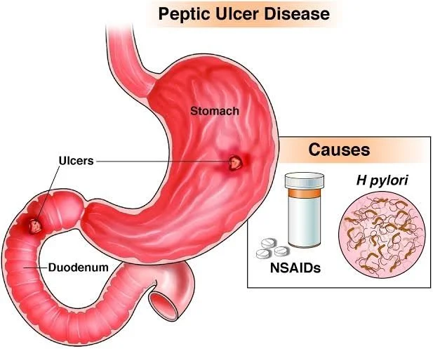
Fig.1 Peptic Ulcer Disease
NSAID induced ulceration proceeds by a different route. Nonselective cyclooxygenase inhibition depletes mucosal prostaglandins, the lipid mediators that maintain mucosal blood flow, mucus secretion, and bicarbonate output. The prostaglandin depleted mucosa loses its defensive barrier while acid secretion continues unabated, and topical irritation from the drugs themselves adds direct injury. Risk escalates with advanced age, prior ulcer history, high dose or multiple NSAIDs, and concurrent use of anticoagulants, antiplatelet agents, corticosteroids, or serotonin reuptake inhibitors; H. pylori and NSAIDs act at least additively and probably synergistically [7]. Selective COX-2 inhibitors reduce but do not abolish ulcer risk, and their cardiovascular liabilities constrain their use. Zollinger Ellison syndrome, gastrin driven hypersecretion from a gastrinoma, accounts for a small fraction of duodenal ulcers, classically multiple, postbulbar, refractory, or associated with diarrhea, and must be considered when ulcers recur despite eradication and NSAID avoidance. Smoking impairs mucosal blood flow and healing and remains an independent risk factor; the role of alcohol and dietary factors is more modest than popularly believed; and psychological stress, though not a primary cause of uncomplicated peptic ulcer, contributes to the severe stress ulceration of critical illness through mucosal ischemia in shock, sepsis, burns, and central nervous system injury.
The idiopathic ulcer category deserves emphasis because it is growing. Rigorous studies excluding surreptitious NSAID use, a common confounder, confirm a genuine subset of patients with H. pylori negative, drug negative ulcers whose natural history is markedly worse: higher recurrence, more frequent bleeding, and higher mortality than conventional ulcers [8]. Observational data also suggest these ulcers are relatively refractory to standard proton pump inhibitor therapy, with healing rates around eighty percent after six weeks of vonoprazan compared with over ninety percent for H. pylori associated ulcers [3]. Whether idiopathic ulcers reflect occult hypersecretion, impaired mucosal defense, vascular insufficiency, or unidentified infections remains unresolved.
Clinical Presentation and Complications
The classic presentation of duodenal ulcer is epigastric pain with a characteristic temporal signature: burning or gnawing discomfort arising two to three hours after meals and in the night, when the empty stomach delivers unbuffered acid to the duodenum, and relieved transiently by food or antacids. This food relief pattern contrasts with the food provoked pain more typical of gastric ulcer, though the distinction is too unreliable to guide management and both demand objective investigation. Nausea, bloating, and early satiety are common companions. Physical examination in uncomplicated disease is often unremarkable apart from epigastric tenderness, and herein lies a clinical trap: the severity of symptoms correlates poorly with the severity of disease. NSAID induced ulcers in particular may be clinically silent until they announce themselves with hemorrhage, and the first manifestation of ulcer disease in an elderly aspirin user may be melena or hematemesis without any antecedent pain.
The complications of duodenal ulcer define its continued clinical importance. Bleeding is the most common, occurring when the ulcer erodes into a vessel, classically the gastroduodenal artery posterior to the duodenal bulb, where erosion can produce catastrophic arterial hemorrhage. Upper gastrointestinal bleeding from peptic ulcer carries a mortality that has declined with modern endoscopic and pharmacological therapy but remains around two to ten percent depending on comorbidity, and mortality is driven less by exsanguination than by decompensation of cardiovascular and other comorbid disease. Perforation, occurring in roughly two to ten percent of ulcer patients over a lifetime, presents with sudden severe abdominal pain, board like rigidity, and free subdiaphragmatic air on upright radiography; the leaked gastroduodenal contents provoke chemical then bacterial peritonitis, and mortality rises steeply with delayed presentation, particularly in the elderly and in NSAID users whose drug may mask early pain. Penetration describes erosion into an adjacent structure, most often the pancreas from a posterior ulcer, producing pain that radiates to the back, becomes continuous, and loses its food relief pattern, with elevation of serum amylase. Gastric outlet obstruction, now rare, arises from edema or cicatrization of the duodenal bulb or pyloric channel and presents with early satiety, weight loss, and vomiting of stale, nonbilious food, sometimes with the succussion splash of a dilated stomach; it causes hypochloremic, hypokalemic metabolic alkalosis through loss of gastric acid. Unlike gastric ulcer, duodenal ulcer carries no malignant potential, a distinction of practical importance: gastric ulcers mandate biopsy and healing surveillance, whereas duodenal ulcers do not, and malignancy in the duodenal bulb is sufficiently rare that its presence should prompt reconsideration of the diagnosis.
Diagnosis
Upper gastrointestinal endoscopy is the definitive diagnostic investigation. It directly visualizes the ulcer, permits assessment of size, depth, and stigmata of recent hemorrhage, allows biopsy of gastric lesions and histological detection of H. pylori, and, crucially, is therapeutic in bleeding. Duodenal ulcers are overwhelmingly located in the first part of the duodenum, the bulb, most often on the anterior wall; postbulbar, giant, or multiple ulcers should raise suspicion of hypersecretory states. Because duodenal ulcers are almost never malignant, routine biopsy is not required, but tissue sampling for H. pylori via rapid urease testing or histology is standard when infection status is unknown. Endoscopy also stratifies bleeding risk through the Forrest classification, which ranges from spurting arterial hemorrhage through visible vessel and adherent clot to clean based ulcer, and thereby guides the intensity of endoscopic and pharmacological intervention.
Testing for H. pylori should accompany essentially every duodenal ulcer diagnosis, because eradication is curative and failure to test forfeits the single most important intervention. Noninvasive tests are preferred for initial diagnosis in uninvestigated dyspepsia and for confirming eradication after treatment. The urea breath test exploits bacterial urease: the patient ingests urea labeled with carbon 13 or 14, and hydrolysis by the organism yields labeled carbon dioxide detectable in expired air, with sensitivity and specificity both exceeding ninety five percent. The stool antigen test offers comparable performance at lower cost and is the most practical option in many settings. Serology detects antibodies and therefore cannot distinguish active from past infection, limiting its utility for test of cure, though it is not confounded by recent acid suppression or antibiotics and retains value when other tests risk false negatives.
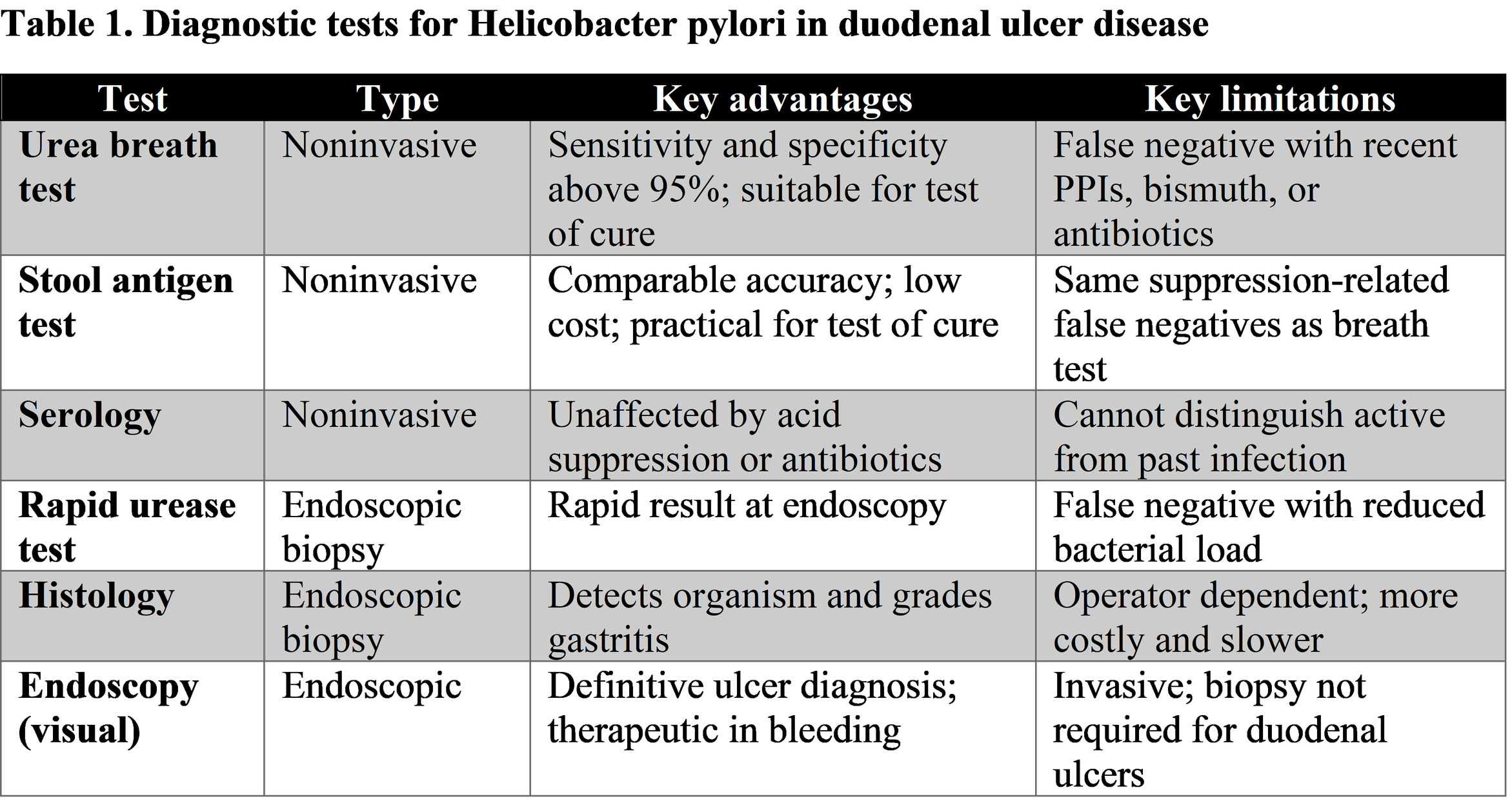
All urea based and antigen-based tests share an important vulnerability: proton pump inhibitors, bismuth, and antibiotics suppress bacterial load and produce false negative results, so testing should be deferred at least two weeks after stopping PPIs and four weeks after antibiotics and bismuth. Test of cure is now strongly emphasized in the 2024 ACG guideline, which recommends confirming eradication in every treated patient, typically by urea breath test or stool antigen at least four weeks after completing therapy [9]. Supporting laboratory evaluation includes a complete blood count for anemia, and serum gastrin measurement, performed after withholding acid suppression, when Zollinger Ellison syndrome is suspected.
Management of Uncomplicated Disease
The management of uncomplicated duodenal ulcer rests on three pillars: acid suppression, eradication of H. pylori when present, and removal of ulcerogenic exposures. Proton pump inhibitors, which irreversibly inhibit the gastric hydrogen potassium ATPase, remain the global standard of care. Standard doses, such as omeprazole 20 mg, esomeprazole 20 to 40 mg, or lansoprazole 30 mg once daily, heal the great majority of duodenal ulcers within four weeks, with meta-analytic healing rates around ninety percent at four weeks, superior to H2 receptor antagonists in both speed and completeness [10]. Treatment duration is typically four weeks for duodenal ulcer; continued therapy beyond healing is unnecessary once H. pylori has been eradicated and NSAIDs withdrawn, though patients requiring ongoing NSAID or aspirin therapy need maintenance gastroprotection. The potassium competitive acid blockers represent the most important pharmacological advance in acid suppression in three decades. Vonoprazan binds the proton pump reversibly and competitively at the potassium site, requires no acid activation, achieves full effect from the first dose, and produces more profound and more stable acid inhibition than PPIs, unaffected by CYP2C19 polymorphism. A network meta-analysis of twenty-five randomized trials confirmed that vonoprazan is at least equivalent to PPIs for ulcer healing, with lansoprazole 30 mg and pantoprazole 40 mg ranking highly among comparators [11], and the 2026 Japanese guidelines have moved vonoprazan to first line status for peptic ulcer and for NSAID and low dose aspirin induced ulceration [4]. In NSAID users who must continue therapy, vonoprazan reduced ulcer recurrence by roughly half compared with lansoprazole (risk ratio 0.55) and reduced gastrointestinal bleeding (risk ratio 0.40) in a 2025 meta-analysis of over 2500 patients, with a safety profile comparable to PPIs [12].
For H. pylori positive ulcers, and historically more than ninety percent of duodenal ulcers were H. pylori related, eradication therapy is the single most important treatment, converting a relapsing disease into a cured one; Cochrane evidence established that eradication therapy is superior to acid suppression alone for healing and prevents recurrence almost completely [13]. The choice of regimen has been transformed by antimicrobial resistance. Clarithromycin resistance now exceeds thirty percent in North America, rendering the traditional clarithromycin triple therapy unacceptably ineffective, and the 2024 ACG guideline accordingly recommends optimized bismuth quadruple therapy as the preferred first line regimen: a proton pump inhibitor twice daily, tetracycline 500 mg four times daily, metronidazole 500 mg three or four times daily, and bismuth four times daily, all for fourteen days, achieving eradication in eighty five to eighty seven percent of patients compared with seventy six to eighty percent for clarithromycin triple therapy [5]. The guideline emphasizes optimization details that matter: tetracycline must not be replaced by doxycycline, which reduces eradication by ten to seventeen percent; bismuth dosing must be adequate; and fourteen days is the recommended duration [14]. Alternative first line options include rifabutin triple therapy and vonoprazan based regimens, with vonoprazan amoxicillin dual therapy validated by the pivotal US and European randomized trial of Chey and colleagues [15]. Crucially, the guideline recommends against empiric clarithromycin or levofloxacin regimens unless susceptibility testing demonstrates a sensitive strain, a mandate reflecting the resistance era and differing somewhat from the Maastricht VI consensus, which is more permissive of levofloxacin second line use and more conservative about rifabutin [5]. For refractory infection, the guideline directs clinicians toward regimens the patient has not received, bismuth quadruple therapy where naive to it, rifabutin triple therapy after bismuth failure, and susceptibility guided selection wherever testing is available. For penicillin allergic patients, bismuth quadruple therapy conveniently avoids amoxicillin entirely. Adjunctive and historical therapies retain niche roles: sucralfate forms a protective barrier over the ulcer base, misoprostol replaces mucosal prostaglandins but is limited by diarrhea and cramping, and H2 receptor antagonists are acceptable when PPIs are unavailable or poorly tolerated, though they are inferior for healing and for NSAID ulcer prevention. Lifestyle measures are modest but real: smoking cessation accelerates healing and reduces recurrence, NSAID and alcohol exposure should be minimized, and patients should be counseled that eradication, not indefinite acid suppression, is the goal in H. pylori disease.
Management of Complicated Disease
Bleeding duodenal ulcer is a medical emergency governed by principles codified in international guidelines. Initial management is resuscitation: hemodynamic assessment, large bore intravenous access, blood product transfusion guided by a restrictive threshold of hemoglobin around 7 g/dL (higher in cardiovascular disease), reversal or mitigation of anticoagulation, and risk stratification with scores such as Glasgow Blatchford. Pre-endoscopic high dose proton pump inhibitor therapy is standard. Endoscopy should be performed within twenty four hours, and urgently in unstable patients, both to diagnose and to treat: stigmata of high risk hemorrhage (active spurting or oozing, nonbleeding visible vessel, adherent clot) mandate endoscopic hemostasis with epinephrine injection combined with a second modality such as thermal coagulation or clips, and the 2024 ACG guideline recommends combination therapy over injection alone [16]. Following successful hemostasis of high-risk lesions, continuous infusion or twice daily high dose PPI for seventy two hours, followed by standard oral therapy, reduces rebleeding and mortality, as established by the landmark intravenous esomeprazole trial of Sung and colleagues [17]. Recurrent bleeding warrants repeat endoscopy; failure of two endoscopic attempts or massive uncontrolled hemorrhage directs the patient to transarterial embolization or surgery, with embolization increasingly preferred as first salvage. Over-the-scope clips and hemostatic powders have expanded the endoscopic armamentarium for large or fibrotic ulcers. Every bleeding ulcer patient must subsequently be evaluated for H. pylori and for ongoing need of NSAIDs, aspirin, or anticoagulation, balancing ulcer recurrence risk against cardiovascular indications; resuming aspirin for secondary cardiovascular prevention with PPI cover is generally safer than withholding it.
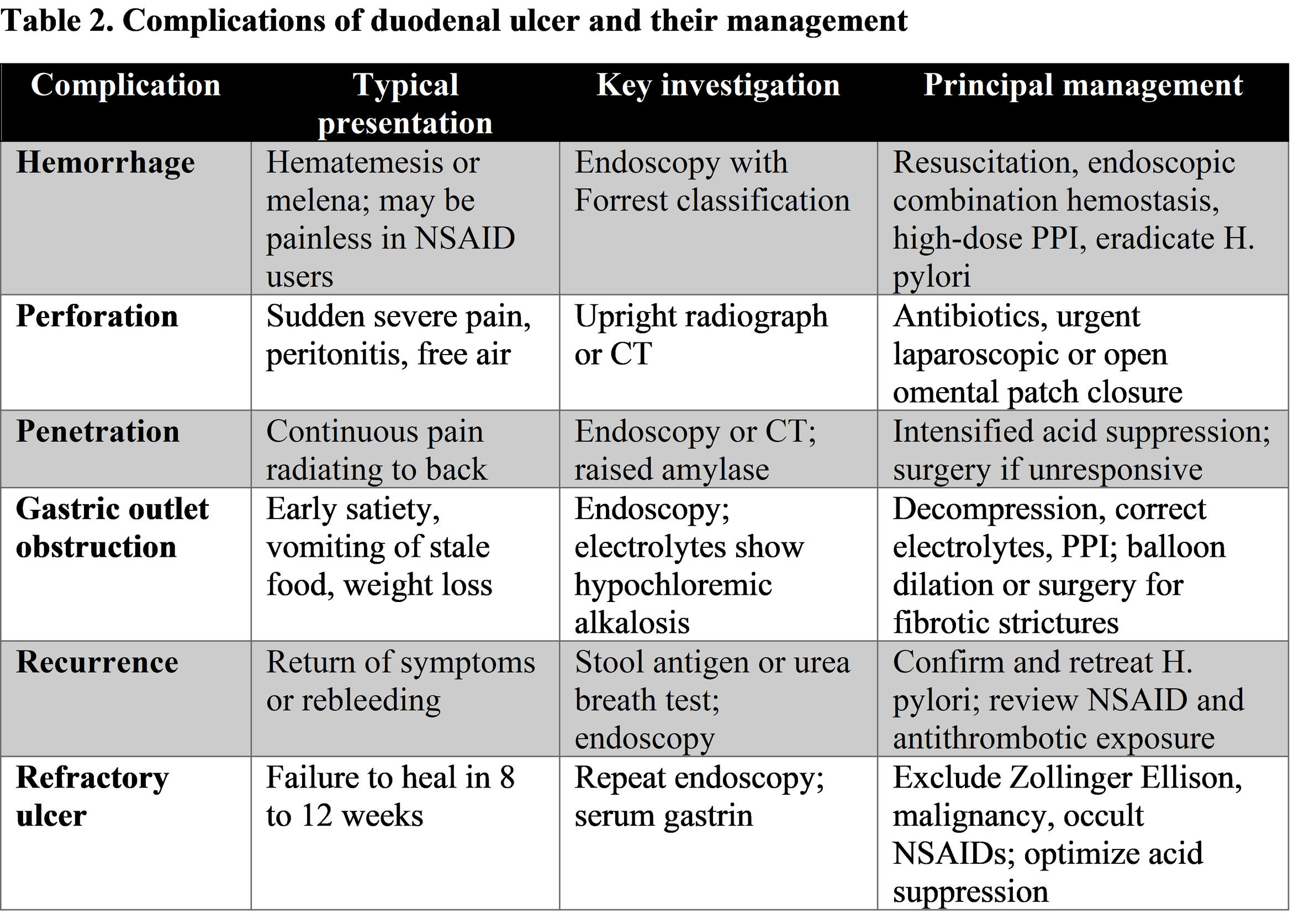
Perforated duodenal ulcer requires urgent surgery in most cases. After resuscitation, broad spectrum antibiotics, and nasogastric decompression, the standard operation for a perforated duodenal ulcer is omental (Graham) patch closure, increasingly performed laparoscopically with outcomes comparable to open surgery and reduced wound morbidity; simple closure rather than definitive acid reducing surgery is appropriate in the modern era because postoperative H. pylori eradication and PPI therapy address the underlying diathesis. Selected stable patients with contained perforations, no peritonitis, and contrast studies showing sealing may be managed nonoperatively with close observation, though failure demands prompt surgery. Gastric outlet obstruction is managed initially with decompression, correction of the characteristic hypochloremic hypokalemic alkalosis, nutritional support, and acid suppression; edema related obstruction may resolve, while fibrotic strictures require endoscopic balloon dilation or surgical drainage procedures. In all complicated disease, the surgical maxim of the past century has inverted: the operation now treats the complication, and the medicine treats the disease.
Discussion
Several themes deserve critical reflection. First, the success story of eradication therapy is under genuine threat from antimicrobial resistance, and the 2024 ACG guideline's pivot away from clarithromycin is less a refinement than an acknowledgment that the most widely prescribed regimen in history no longer works in much of the world. The practical consequence is a more complicated therapeutic calculus: clinicians must now know local resistance patterns, take antibiotic histories, consider susceptibility testing as molecular assays become available, and verify eradication in every patient, a test of cure discipline that surveys suggest remains incompletely implemented. The arrival of vonoprazan based regimens offers more potent acid suppression as a platform for eradication, with the pharmacological logic that higher intragastric pH renders H. pylori more metabolically active and thus more antibiotic susceptible, and meta-analyses support superiority of vonoprazan based over PPI based eradication, particularly for clarithromycin resistant strains [15]. Second, the rise of the idiopathic ulcer is a genuine epidemiological puzzle with clinical teeth. As H. pylori prevalence falls and NSAID use is better managed, the residual ulcer population is enriched for patients whose ulcers heal slowly, bleed more, recur more, and die more often [8], and some evidence suggests even potent acid suppression heals fewer than ninety percent of these lesions in standard courses [3]. These patients may need prolonged therapy, vigilant follow up, and scrupulous exclusion of surreptitious NSAID use, Crohn disease, and hypersecretory states. Third, the long-term safety of acid suppression continues to generate debate. PPIs have been associated in observational literature with hypomagnesemia, vitamin B12 and iron malabsorption, enteric infection including Clostridioides difficile, fundic gland polyps, and possible associations with fracture, kidney disease, dementia, and gastric cancer, though causality is unproven for most and the absolute risks are small; the sensible position is to use acid suppression at the lowest effective dose for a defined indication, a discipline naturally served by eradication therapy, which ends the need for chronic suppression in most ulcer patients. Vonoprazan raises analogous questions, with dose dependent hypergastrinemia documented but without clinical consequence to date, including reassuring five-year maintenance data [12].
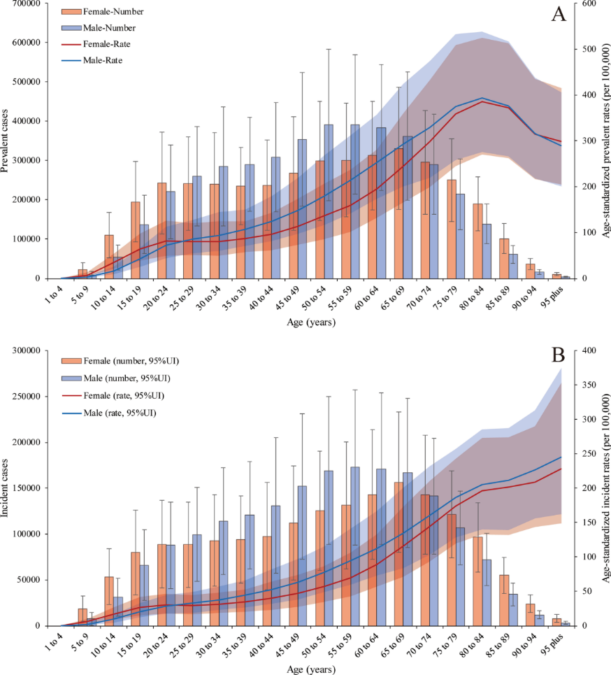
Fig.2 The global, regional and national burden of peptic ulcer disease from 1990 to 2019
Fourth, the intersection of ulcer disease with antithrombotic therapy is a daily dilemma in aging populations, where the cardiologist's antiplatelet agent and the gastroenterologist's ulcer history collide; evidence supports continuing indicated aspirin with PPI or vonoprazan gastroprotection rather than abandoning either, and combination of a COX-2 inhibitor with a PPI offers the strongest protection for very high-risk patients requiring anti-inflammatory therapy [18]. Finally, the field should not lose sight of the global picture: in much of the developing world, H. pylori prevalence remains high, resistance patterns differ, bismuth availability is inconsistent, and the condition's burden, including its downstream contribution to gastric cancer, remains substantial, making eradication strategy a matter of public health as much as individual care. This review's limitations should be acknowledged: as a narrative synthesis it does not recalculate effect sizes, guidelines differ by region, and the therapeutic landscape, particularly around potassium competitive acid blockers and susceptibility guided therapy, is evolving rapidly.
Conclusion
Duodenal ulcer has been transformed over a century from a surgical affliction managed by resection into a medical condition that is, in its H. pylori driven majority, curable with a fortnight of combination therapy. The contemporary clinician's task is nevertheless far from trivial. It demands accurate endoscopic diagnosis, systematic testing for H. pylori in every patient, selection of eradication regimens appropriate to the era of clarithromycin resistance, with optimized bismuth quadruple therapy as the current first line standard, disciplined confirmation of cure, thoughtful use of the most potent acid suppressants including vonoprazan where indicated, scrupulous management of NSAID and antithrombotic exposure, and readiness to recognize and treat the hemorrhage, perforation, penetration, and obstruction that still bring ulcer patients to emergency care. The growing cohort of idiopathic ulcers reminds us that the final chapter of this disease has not been written. Yet the overarching message remains one of medicine's quieter triumphs: for most patients with a duodenal ulcer today, the combination of accurate diagnosis, rational pharmacology, and bacterial eradication offers not merely healing but cure.
References
Marshall BJ, Warren JR. Unidentified curved bacilli in the stomach of patients with gastritis and peptic ulceration. Lancet. 1984;1(8390):1311 to 1315.
Nobel Prize Organization. The Nobel Prize in Physiology or Medicine 2005: Barry J. Marshall and J. Robin Warren. NobelPrize.org; 2005.
Sugawara K, Koizumi S, Horikawa Y, et al. Is the new potent acid inhibitory drug vonoprazan effective for healing idiopathic peptic ulcers? A multicenter observational study in Akita Prefecture, Japan. J Gastroenterol. 2019;54(11):963 to 971.
Kamada T, Satoh K, et al. Evidence based clinical practice guidelines for peptic ulcer disease 2026. J Gastroenterol. 2026.
Chey WD, Howden CW, Moss SF, et al. ACG Clinical Guideline: Treatment of Helicobacter pylori Infection. Am J Gastroenterol. 2024;119(9):1730 to 1753.
Suerbaum S, Michetti P. Helicobacter pylori infection. N Engl J Med. 2002;347(15):1175 to 1186.
Lanas A, Chan FKL. Peptic ulcer disease. Lancet. 2017;390(10094):613 to 624.
Wong GLH, Wong VWS, Chan Y, et al. High incidence of mortality and recurrent bleeding in patients with Helicobacter pylori negative idiopathic bleeding ulcers. Gastroenterology. 2009;137(2):525 to 531.
Schoenfeld P. ACG Guideline on Treatment of Helicobacter pylori. Am J Gastroenterol Evidence Based Reviews. 2024.
Leontiadis GI, Howden CW. The role of proton pump inhibitors in the management of upper gastrointestinal disorders. Gastroenterol Hepatol. 2009;5(3):174 to 176.
Yang Z, et al. Efficacy and safety of vonoprazan versus proton pump inhibitors in the treatment of peptic ulcer disease: a systematic review and network meta-analysis of randomized controlled trials. Front Nutr. 2024;11:1436993.
Bandyopadhyay S, et al. Efficacy of vonoprazan in nonsteroidal anti-inflammatory drug induced ulcer in terms of ulcer recurrence and gastrointestinal bleeding: a systematic review and meta-analysis. 2025.
Ford AC, Delaney BC, Forman D, Moayyedi P. Eradication therapy in Helicobacter pylori positive peptic ulcer disease: systematic review and economic analysis. Am J Gastroenterol. 2004;99(9):1833 to 1855.
University of Minnesota College of Pharmacy. 2024 American College of Gastroenterology H. pylori Guidelines: Curbside Consult. 2025.
Chey WD, Megraud F, Laine L, et al. Vonoprazan triple and dual therapy for Helicobacter pylori infection in the United States and Europe: randomized clinical trial. Gastroenterology. 2022;163(3):608 to 619.
Barkun A, et al. Management of nonvariceal upper gastrointestinal bleeding: guideline recommendations from the American College of Gastroenterology. Am J Gastroenterol. 2021/2024 update.
Sung JJY, Barkun A, Kuipers EJ, et al. Intravenous esomeprazole for prevention of recurrent peptic ulcer bleeding: a randomized trial. Ann Intern Med. 2009;150(7):455 to 464.
Chan FKL, Wong VWS, Suen BY, et al. Combination of a cyclo-oxygenase-2 inhibitor and a proton-pump inhibitor for prevention of recurrent ulcer bleeding in patients at very high risk: a double blind randomised trial. Lancet. 2007;369(9573):1621 to 1626.
Malfertheiner P, Megraud F, Rokkas T, et al. Management of Helicobacter pylori infection: the Maastricht VI/Florence consensus report. Gut. 2022;71(9):1724 to 1762.
Lau JYW, Sung JJY, Hill C, et al. Systematic review of the epidemiology of complicated peptic ulcer disease: incidence, recurrence, risk factors and mortality. Digestion. 2011;84(2):102 to 113.