
The Brain Stem: Anatomy of Body`s Control Hub
1. Zarina Zhamaldinovna Toichieva
2. Kumaravel Komala Sathia
Arulkumaran Poonkodi
Thiyageshwaran Ganesan
Mohamed Hafeez Syed Basha
Jegan Raj Sahaya Arockiaraj
(1. Lecturer, International Medical Faculty, Osh State University, Osh, Kyrgyz Republic
2. Students, International Medical Faculty, Osh State University, Osh, Kyrgyz Republic)
ABSTRACT:
The brain stem represents an integral part of the central nervous system that bridges the cerebrum with the spinal cord, coordinating numerous life-supporting functions. From the standpoint of gross anatomy, the brain stem consists of three parts, namely the midbrain, pons, and medulla oblongata, each having its unique function in sensory, motor, and autonomic systems.
The brain stem contains significant nuclei of cranial nerves, ascending and descending nervous fibers, and the reticular formation that plays a significant role in the regulation of consciousness and sleep-wakefulness cycle. Furthermore, it has an important role in the development of reflex responses and physiological controls such as breathing, heartbeat, and blood pressure. Because of its small size and high level of functional specialization, the brain stem injury may lead to serious neurologic deficits.
KEY WORDS: Brainstem, Midbrain, Pons, Medulla oblongata, Cranial nerves, Reticular formation, Neural pathways, Autonomic regulation, Central nervous system, Neuroanatomy, Sensory pathways, Motor pathways, Brainstem lesions, Vital functions.
INTRODUCTION:
The brainstem is an important component of the central nervous system that acts as a connection between the brain and the spinal cord. In spite of its rather compact nature, it helps to regulate numerous vital life processes such as respiration, heartbeat, and level of consciousness. In addition to its regulatory functions, the brainstem functions as a main nerve pathway that links the brain and other parts of the human body through nerve fibers. It is very important to study the structure of the brainstem due to the fact that any injury to this organ leads to serious neurological problems and even death. The brainstem is made up of three important sections, which include the midbrain, pons, and medulla oblongata.
GENERAL ORGANIZATION OF BRAIN STEM:
It occupies the posterior cranial fossa, positioned anteriorly to the cerebellum. Its superior part continues with the diencephalon and ends inferiorly at the spinal cord. The brainstem has three divisions that include the midbrain, the pons, and the medulla oblongata.
The internal structures of the brainstem include nuclei (groups of cell bodies of neurons), ascending tracts that transmit sensory information, descending tracts that transmit motor information, and the reticular formation network. The brainstem is the origin of most cranial nerves responsible for functions like eye movements and hearing.
A notable characteristic of the brainstem is its function as a conduction pathway. It conducts sensory messages from the body to higher regions in the brain and also conducts motor signals from the brain to the spinal cord.
THE MIDBRAIN [MESENCEPHALON]:
The midbrain is the highest part of the brain stem and lies between the diencephalon and pons. It is rather small in size and consists of several crucial components related to vision, audition, and movement.
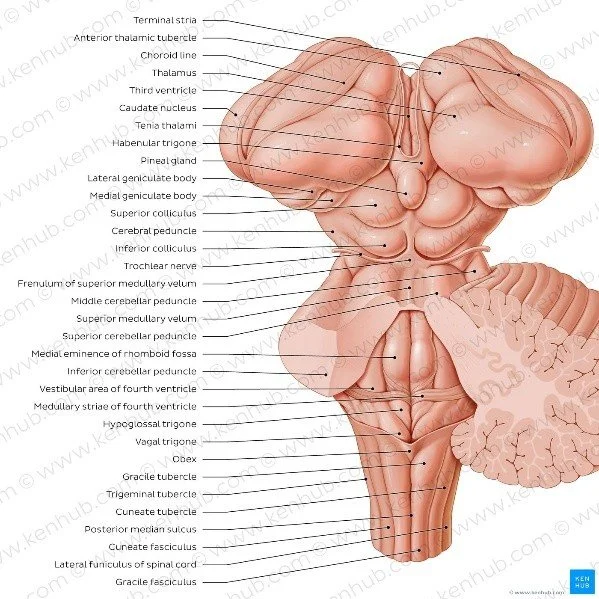
External Features:
The anterior surface of the midbrain is characterized by two large fiber bundles known as the cerebral peduncles. These structures contain descending motor fibers that connect the cerebral cortex to lower brain centers and the spinal cord. On the posterior surface, the midbrain features four rounded elevations called the colliculi. These are divided into:
● Superior colliculi: involved in visual reflexe
● Inferior colliculi: involved in auditory processing
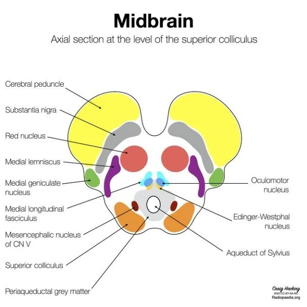
Internal Structure:
Within, the midbrain has three major components:
• Tectum (the posterior section with colliculi)
• Tegmentum (the central area with nuclei and fiber tracts)
• Basis pedunculi (the anterior part with motor fibers)
One of the vital nuclei of the midbrain is the substantia nigra, which has an essential function in movement control. Damage to this nucleus leads to movement abnormalities, such as those seen in Parkinsonian syndrome. Another significant nucleus is the red nucleus.
Cranial Nerves:
The midbrain is associated with two cranial nerves:
● Oculomotor nerve (III)
● Trochlear nerve (IV)
These nerves control eye movements and pupil constriction.
THE PONS:
The pons lies between the midbrain and the medulla oblongata. It acts as a bridge connecting different parts of the nervous system, including the cerebrum and cerebellum.
External Features:
The anterior aspect of the pons is described as a rounded structure. The transverse fibers found here link the cerebellum through the middle cerebellar peduncles. The posterior aspect forms a portion of the floor of the fourth ventricle which is full of cerebrospinal fluid.
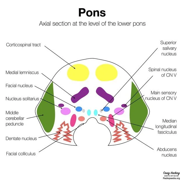
Internal Structure:
The structure of the pons includes two major components:
● Basilar portion (anterior): includes motor tract and nuclei of pons
● Tegmental portion (posterior): includes cranial nerve nuclei and sensory tract
Nuclei of the pons transmit information between the cerebrum and the cerebellum, serving an important function in movement coordination. Reticular formation of the pons regulates sleep and breathing processes.
Cranial Nerves:
The Pons has many nerves associated with it:
● Trigeminal nerve (V) - Facial sensation and chewing
● Abducens nerve (VI) - Eye movements
● Facial nerve (VII) - Facial expressions
● Vestibulocochlear nerve (VIII) - Hearing and equilibrium
THE MEDULLA OBLANGATA:
The medulla oblongata is the lowest part of the brainstem and continues inferiorly as the spinal cord. It is crucial for regulating vital autonomic functions.
External Features:
The anterior surface bears two parallel ridges referred to as the pyramids. They have motor neurons that decussate to the other side of the body at an area referred to as the pyramidal decussation. Lateral to the pyramids are the olives that bear nuclei that assist in motor coordination. The posterior surface helps form the fourth ventricle.
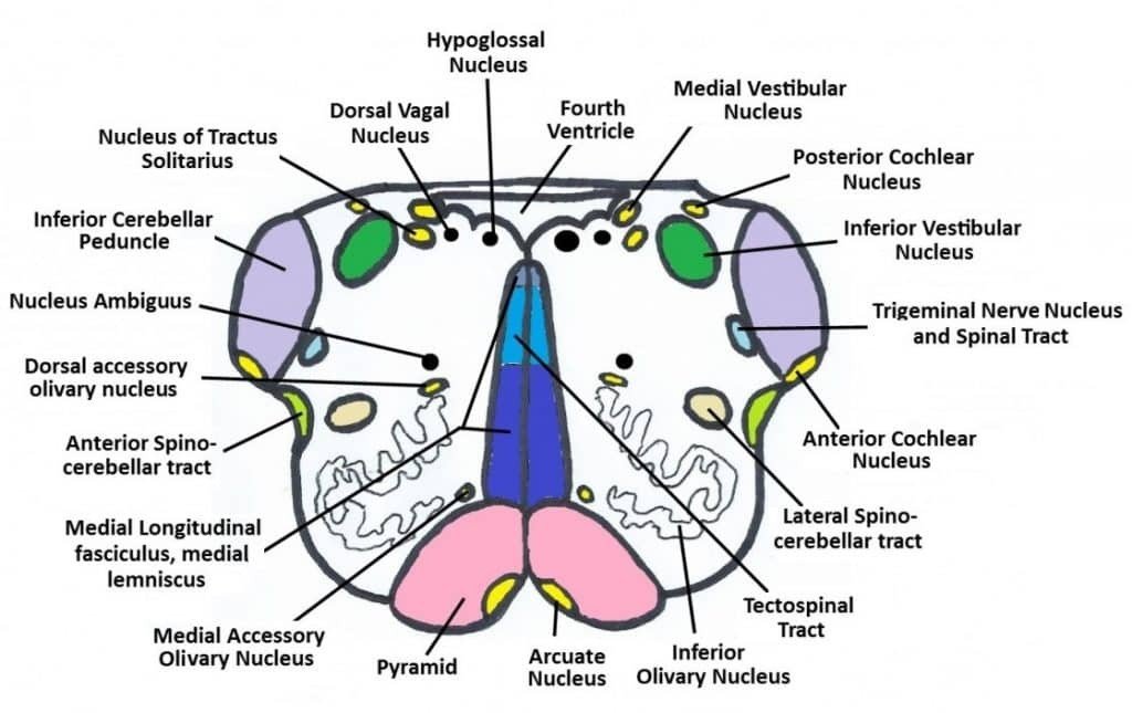
Internal Structure:
Some of the nuclei present in the medulla are:
● Cardiac nucleus: regulates cardiac function
● Respiratory nucleus: regulates respiration
● Vasomotor nucleus: regulates blood pressure
The medulla also houses ascending sensory tracts and descending motor tracts. The reticular formation in the medulla is involved in controlling consciousness and autonomic functions.
Cranial Nerves:
The medulla is related to the following cranial nerves:
• IX – Glossopharyngeal nerve
• X – Vagus nerve
• XI – Accessory nerve
• XII – Hypoglossal nerve
The functions related to these nerves include speech and the regulation of internal organs through autonomic function.
THE RETICULAR FORMATION:
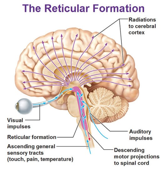
It is described as an elaborate network of neurons spread all through the brainstem without specific borders. It is essential for alertness regulation and control of many other body functions. Reticular Formation Functions are:
● Regulation of Sleep-Wakefulness Circadian Rhythm
● Regulation of Muscle Tone
● Pain Perception Regulation
● Consciousness Maintenance Reticular Formation malfunction leads to loss of consciousness.
ASCENDING & DESCENDING PATHWAYS:
The brainstem serves as a conduit for numerous neural pathways.
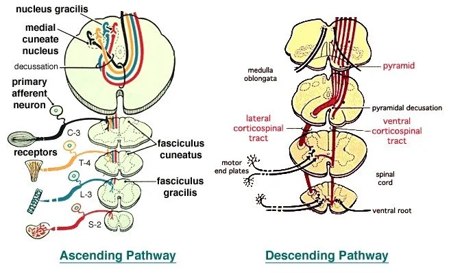
Ascending Pathways:
These pathways carry sensory information from the body to the brain. Examples include:
● Spinothalamic tract (pain and temperature)
● Dorsal column-medial lemniscus pathway (touch and proprioception)
Descending Pathways:
This pathway carries motor instructions from the brain to the spinal cord.
Some examples are:
● Corticospinal tract: voluntary movements
● Reticulospinal tract: posture and muscle tone
The decussation of fibers in the brain stem is responsible for the fact that the brain hemisphere controls the opposite side of the body.
BLOOD SUPPLY OF THE BRAINSTEM:
Blood is mainly supplied to the brainstem via the branches of the vertebral and basilar arteries.
● Midbrain: supplied by branches of posterior cerebral arteries
● Pons: supplied by branches of the basilar artery
● Medulla: supplied by the vertebral arteries and the anterior spinal artery
Strokes affecting any of these areas will lead to brainstem stroke symptoms.
FUNCTIONAL IMPORTANCE OF BRAIN STEM:
Importance of Brain Stem:
Brain stem is very important for survival as it helps regulate various automatic actions. They are:
• Breathing
• Heart rate
• Blood pressure
• Swallowing
Brain stem is involved in various reflex actions like sneezing, coughing, and vomiting. Besides, it is also responsible for postural control and coordination of movements.
CLINICAL SIGNIFICANCE:
A lesion in the brainstem would have a catastrophic effect because the number of important structures is high within a very compact space. The most common medical conditions associated with the brainstem are the following:
● Stroke
● Neoplasms
● Demyelinating conditions
Injury Possible symptoms may be the following:
● Paralysis
● Sensory deficits
● Difficulty swallowing
● Diminished awareness Since cranial nerves originate from the brainstem, lesions affect one side of the body while leaving the other side relatively intact. These are referred to as “crossed signs.”
CONCLUSION:
The brain stem is a crucial and well-organized structure that serves an essential purpose in sustaining life and regulating various neurological activities. The brain stem can be categorized into three different sections, namely the midbrain, the pons, and the medulla oblongata. These structures are responsible for the regulation of the autonomic system, act as pathways for transmitting neural impulses, and contain the cranial nerve nuclei. The knowledge of the brain stem structure is indispensable when diagnosing neurological diseases.
REFERENCES:
Textbooks
1. Gray’s Anatomy Standring, S. (2020). Gray’s Anatomy: The Anatomical Basis of Clinical Practice (42nd ed.). Elsevier.
2. Clinically Oriented Anatomy Moore, K. L., Dalley, A. F., & Agur, A. M. R. (2018). Clinically Oriented Anatomy (8th ed.). Wolters Kluwer.
3. Snell's Clinical Neuroanatomy Snell, R. S. (2019). Clinical Neuroanatomy (8th ed.). Wolters Kluwer.
4. Netter's Atlas of Human Anatomy Netter, F. H. (2018). Atlas of Human Anatomy (7th ed.). Elsevier.
5. Principles of Neural Science Kandel, E. R., Schwartz, J. H., & Jessell, T. M. (2021). Principles of Neural Science (6th ed.). McGraw-Hill.
Websites
1. National Institute of Neurological Disorders and Stroke Brainstem and neurological disorders overview. Available at: https://www.ninds.nih.gov