
Thymus Gland
1. Manas kyzy Uulkan
2. Sivabalan Thirumalaikumar
(1. Lecturer, Dept. of Histology, International Medical Faculty, Osh State University, Osh, Kyrgyz Republic.
2. Student, International Medical Faculty, Osh State University, Osh, Kyrgyz Republic.)
Introduction:
Thymus gland refers to the primary immune system organ responsible for the function of immune response development and coordination. Thymus gland qualifies to be the primary lymphoid organ due to the fact that lymphocytes known as T cells originate and develop immunological capability in this body part. Thymus gland attains peak functions during the fetal and infant stages and during childhood years, and even though the gland undergoes involution during puberty, the immune functions are of great significance throughout life.
The thymus has a special place within human physiology due to its connection between the immune system and the endocrine system. The thymus generates a variety of hormones and cytokines involved in the regulation of the development of immune cells. This prevents a condition whereby the organism fails to distinguish between "self" and "non-self" immune antigens. Malfunctioning of the thymus results in immunodeficiency, autoimmunity, or cancers.
Historical Background:
The thymus was documented in ancient literature, but its function was unknown for a long period of human history. Anatomists in ancient times regarded it as a vestige or a part of a system related to emotions or spirituality. Galen described its anatomy, but its physiological purpose was unknown. In the 19th and early 20th centuries, scientists began linking the thymus with child and immune disorders. The identification of lymphocytes and discoveries within immunology during the mid-20th century finally resolved the question of the thymus’ involvement in the development of the immune system. Experiments showed that the loss of the thymus in baby animals would lead to a lack of immunity, proving it to be a very important organ for immunity.
Embryological Development:
The thymus gland arises from the third pharyngeal pouch. In the sixth week of life, epithelial cells of the endoderm of the third pharyngeal pouch grow to form the thymic primordium. The neural crest cells make up the connective tissue and the capsule of the gland. During development, the thymic primordium migrates from the neck region to the superior mediastinum. Anomalies in this migration process can cause ectopic thymic tissue in the neck region and mediastinum. Before birth, the thymus is grossly large in size compared with other organs. However, it continues to grow very rapidly in early childhood.
Microscopic structure (Histology):
Each thymic lobule has two distinct regions: the cortex and the medulla.
Cortex:
· The cortex comprises a darker-staining outer portion because of the high concentration of immature T lymphocytes-thymocytes-it contains. Supporting it is a framework of epithelial reticular cells. The blood-thymus barrier is one distinctive feature of the cortex. It protects T cells from antigens in the blood and potential self-antigens that might otherwise interfere with T-cell maturation.
Medulla:
· The medulla is the inner lighter-staining region that has fewer lymphocytes and more epithelial reticular cells. A major feature of the medulla is the presence of Hassall's corpuscles, which are concentric, keratinized structures formed by epithelial reticular cells. They provide sites for T-cell differentiation and immune tolerance.
In contrast to the lymph nodes and spleen, lymphoid follicles and germinal centers are absent in the thymus.
“The thymus gland is the chief organ of the lymphoid system, lying in the superior mediastinum immediately behind the breastbone and in front of the heart, along with the great vessels, but descends into the lower neck region in childhood. It occurs as a soft, bilobed structure encased in a thin layer of connective tissue, which in turn boasts a delicate network of septa dividing it into countless lobules. Every lobule is comprised of an outer dark-staining cortex heavily populated by undifferentiated T-lymphocytes simply called thymocytes, together with an inner lighter staining medulla showing fewer lymphocytes, along with characteristic Hassall’s corpsucules composed of concentric layers of epithelial cells. “It is rich in blood vessels but devoid of afferent lymphatic channels, which instead come directly from the bloodstream. The gland attains maximal size about puberty but otherwise enters a phase called involution, within which the whole mass of lymphoid tissue becomes replaced by adipose tissue.””
Blood Supply:
Arterial Supply: It is provided by the internal thoracic artery, inferior thyroid artery, and occasionally, pericardiophrenic artery.
Venous drainage: The thymic veins drain into the left brachiocephalic vein
Lymphatic drainage:
The thymus is devoid of afferent lymphatics and possesses efferent lymphatic vessels. Due to this, the thymus cannot be directly reached by antigens.
Nerve supply:
The thymus has autonomic innervation via the vagus nerve and sympathetic fibers from the cervical and thoracic sympathetic chains.
Organ Function:
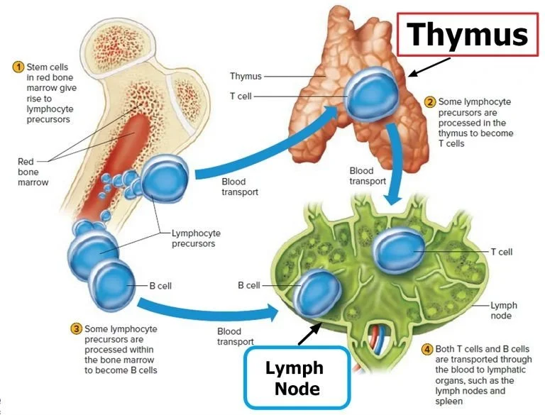
· T-Lymphocyte:The main significance of the thymus is the development of T lymphocytes. Immature cells migrate from the bone marrow to the thymus for differentiation into T cells.
· Positive Selection:Positive selection promotes the ability of T cells to accept self-major histocompatibility complex molecules.
· Negative Selection:It removes self-reactive T cells and thereby suppresses autoimmunity.
· Endocrine Functions:The thymus has several secreted hormones such as thymosin, thymopoietin, thymulin, and thymic humoral factors. These hormones control T cell proliferation, T cell differentiation
Thymic Involution:
Involution of the thymus begins in puberty and continues through adulthood. The functional thymus is secondarily replaced with fatty tissue, but some thymic function is retained. Causes of thymic involution include age, stress, malnutrition, infection, and hormonal changes.
Although involution does occur, it still plays a significant part in immunoregulation, especially in immunologic tolerance.
Role In Immune Tolerance:
The thymus is a crucial part of the mechanism that maintains central immune tolerance. The elimination of self-reactive T cells in the body through the thymus avoids autoimmune diseases. The malfunctioning of this process may cause diseases like systemic lupus erythematosus, rheumatoid arthritis, and type 1 diabetes in humans.
Clinical Significance:
The thymus gland is very important clinically, since it plays an essential role in the development and regulation of the immune system. Disorders in the thymus may be manifested as immunodeficiency disorders, autoimmune diseases, or neoplastic conditions.
1. Congenital Anomalies
· Thymic aplasia or hypoplasia leads to a severe deficiency of T lymphocytes and consequently impairs the cell-mediated immunity.
· DiGeorge syndrome is a classical example, caused by developmental defects of the third and fourth pharyngeal pouches. Patients present with thymic aplasia, recurrent infections, hypocalcemia, and cardiac anomalies.
2. States of Immunodeficiency
· Destruction or absence of the thymus during early life results in severe immunodeficiency. Defective T-cell–mediated immunity causes children to experience viral, fungal, and opportunistic infections repeatedly.
3. Autoimmune Diseases
· Abnormal thymic function may lead to the failure of negative selection of T cells, allowing self-reactive lymphocytes into circulation. This contributes to autoimmune disorders such as:
A)Myasthenia gravis
B)Systemic lupus erythematosus
C)Rheumatoid arthritis
Myasthenia gravis has often been associated with thymic hyperplasia or thymoma, and clinical symptoms have improved following thymectomy.
4. Thymic Tumors
· Thymoma is the most common tumor of the thymus and arises from epithelial cells. It is often associated with myasthenia gravis and other paraneoplastic syndromes.
Conclusion:
The thymus gland plays a crucial role in the development of the immune system. Its importance in the medical field can be gauged by the drastic effects of its malfunctioning, such as immunodeficiency, autoimmunity, and tumors. To treat the immune disorders that occur in the body, it is important to have proper knowledge about the thymus gland.
References:
1)Standring S, editor. Gray’s Anatomy: The Anatomical Basis of Clinical Practice. 42nd ed. London: Elsevier; 2021.
2)Chaurasia BD. Human Anatomy, Volume 2: Thorax and Abdomen. 9th ed. New Delhi: CBS Publishers & Distributors; 2022.
3)Romanes GJ. Cunningham’s Manual of Practical Anatomy, Volume 2. 16th ed. Oxford: Oxford University Press; 2018.
4)Mescher AL. Junqueira’s Basic Histology: Text and Atlas. 16th ed. New York: McGraw-Hill Education; 2021.
5)Young B, O’Dowd G, Woodford P. Wheater’s Functional Histology: A Text and Colour Atlas. 6th ed. Philadelphia: Elsevier; 2020.
6)Murphy K, Weaver C. Janeway’s Immunobiology. 10th ed. New York: Garland Science; 2022.
7)Delves PJ, Martin SJ, Burton DR, Roitt IM. Roitt’s Essential Immunology. 13th ed. Hoboken: Wiley-Blackwell; 2017.
8)Hall JE. Guyton and Hall Textbook of Medical Physiology. 14th ed. Philadelphia: Elsevier; 2021.
9)Kumar V, Abbas AK, Aster JC. Robbins and Cotran Pathologic Basis of Disease. 10th ed. Philadelphia: Elsevier; 2021.
10)Jameson JL, Fauci AS, Kasper DL, Hauser SL, Longo DL, Loscalzo J, editors. Harrison’s Principles of Internal Medicine. 21st ed. New York: McGraw-Hill Education; 2022.