
Clinical Bedside Examination Techniques for the Musculoskeletal and Peripheral Neurological Systems
1. Ranjit Rajesh Gaud
2. Tushar Kumar
3. Pankaj Kumar Rana
(1. Student, Osh State Medical University, Osh, Kyrgyz Republic
2. Student, Osh State Medical University, Osh, Kyrgyz Republic
3. Student, Osh State Medical University, Osh, Kyrgyz Republic)
Abstract
Accurate physical exams are still the basis for diagnosing neurological problems, especially in places where there is not quick access to advanced imaging. This review brings together ten common exams done at the bedside. These include the Straight Leg Raise (SLR) test, Gowers sign, upper and lower limb neurological exams, the Stork (Gillet) test, the FABER (Patrick's) test, the Functional Gait Assessment (FGA), and the developmental milestone exam. The review uses known sources of exams and research on how accurate each test is. Each test is explained in terms of how the patient should be positioned, how the examiner should do the test, how to rate the results, and what they mean. There is also a summary of how reliable and valid each test's where such information is available. The tests reviewed here were used by the author when working under supervision on medicine and orthopedic wards. This review is meant to bring that experience into a clear reference. Looking at all the tests reviewed, single orthopedic special tests, like the Stork test on its own, usually have limited value for diagnosis. Using several tests together or using tools like the FGA that are standardized makes the results more reliable and more helpful in real life. This review ends with the idea that the bedside tests are best used as part of a thinking process during a clinical exam, not as tools that work alone.
Keywords: physical examination, straight leg raise test, neurological examination, sacroiliac joint, FABER test, functional gait assessment, developmental milestones, clinical skills.
1. Introduction
The physical examination remains one of the cost-effective and immediately available diagnostic tools in clinical medicine. In low-resource settings and even in well-equipped hospitals, a carefully performed bedside test can narrow a differential diagnosis before any imaging or laboratory investigation is ordered. This is particularly true for complaints such as low back and hip pain and for the assessment of neurological deficits affecting the limbs, where special orthopedic tests and structured neurological examinations continue to guide referral and further work-up.
This review consolidates a set of bedside examination techniques encountered and applied during training and ward-based patient assessment, spanning orthopedic provocation tests (the Straight Leg Raise test, the FABER test and the Stork test), core components of the neurological examination (tone, power, reflexes, function and sensation for both the upper and lower limb), a validated composite gait instrument (the Functional Gait Assessment) and pediatric developmental screening. The objective is twofold: first to present each test in a reproducible format suitable for clinical reference; and second, to summarize the reliability and validity evidence available for each test so that findings can be interpreted with appropriate clinical caution.
A recurring theme throughout this review is that individual special tests, taken in isolation, frequently show modest diagnostic accuracy, while clusters of tests or standardized multi-item instruments tend to perform considerably better. This distinction has implications for how these tests should be used and reported in clinical practice and is discussed further in Section.
2. Methods
This is a narrative review, not a systematic review or an original research study. The source material came from (a) structured clinical‑examination notes that were gathered during bedside teaching and patient assessment and
(b) the literature on diagnostic accuracy, reliability and validity for each technique, mainly taken from Physiopedia. A peer‑reviewed physiotherapy and medicine reference and from standard clinical‑examination textbooks. Each test was examined for its purpose, how it is performed, the grading or interpretation criteria, and any published reliability data (intra‑rater, inter‑rater or internal consistency) and validity data (concurrent, construct or diagnostic). If a test belongs to a recommended cluster (for example, the sacroiliac test cluster), this is noted explicitly because the performance of a single test and the performance of a cluster are not the same.
No new patient data, statistical analysis or human‑subjects research is reported in this review; all reliability and validity figures cited are taken from published sources and are reproduced here for reference and synthesis.
3. The Straight Leg Raise (SLR) Test
3.1 Background and Purpose
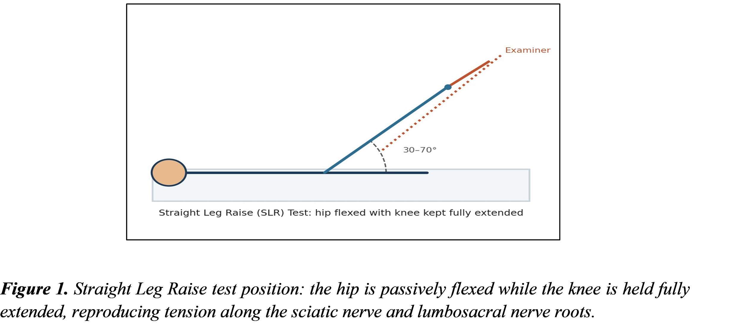
The Straight Leg Raise test is a test used to find disc problems or nerve root irritation in people with low back pain and leg pain. It was first described by Lasègue more than a hundred years ago. That is why it is also sometimes called Lasègues test. This test adds tension to the nerve. Pulls on the nerves coming from the lower spine, especially the L4 through S2 nerve roots. When the leg is lifted, the nerve roots are pulled down and forward. This movement stretches the dura mater. Creates pressure that follows a path: starting at the sciatic foramen, then moving across the sacrum, and finally going through the intervertebral foramen. Pain or tenderness felt during this test shows up near the sciatic notch.
3.2 Technique
The SLR is a test that is done on one leg at a time. The examiner starts with the leg to set a range. The patient lies flat on the back no pillow under the head. The examiner stands on the side of the leg that is being tested. The examiner holds the heel with one hand. Places the hand on the front of the thigh, keeping the knee fully straight. The leg is then lifted by bending at the hip. The knee stays fully extended. The lift continues until the patient feels pain or tightness in the back or the back of the leg.
3.3 Interpretation and Clinical Reasoning
• If the reproduced symptom is mainly back pain the underlying disc herniation is more likely to be central, pressing on the part of the spinal cord. Central prolapses tend to be smaller.
• If the pain is mainly felt in the leg, the problem is more likely to be on the side.
• Problems that are in between these two extremes usually cause pain in both the back and the leg.
• Neurologic pain that happens in both the leg and low back between 30–70 degrees of hip flexion suggest a disc problem that affects the L4–S1 nerve roots.
Clinically, the SLR is best regarded as a screening tool for lumbar radiculopathy rather than a definitive diagnostic test; a positive result should prompt further neurological assessment and, where indicated, imaging correlation.
4. Gowers' Sign
Gowers' sign is an observation that shows weakness in the muscles near the center of the body, especially in the legs. The sign is found by asking the person to stand up from a sitting or crouching position. A positive sign occurs when the person has to use their hands to push up their legs and body to get up because the hip and thigh muscles are not strong enough. This sign is usually linked to conditions that affect the muscles near the center of the body. It is often seen in children with Duchenne muscular dystrophy. When this sign is noticed, it is important to check the muscles and nerves.
5. Upper Limb Neurological Examination
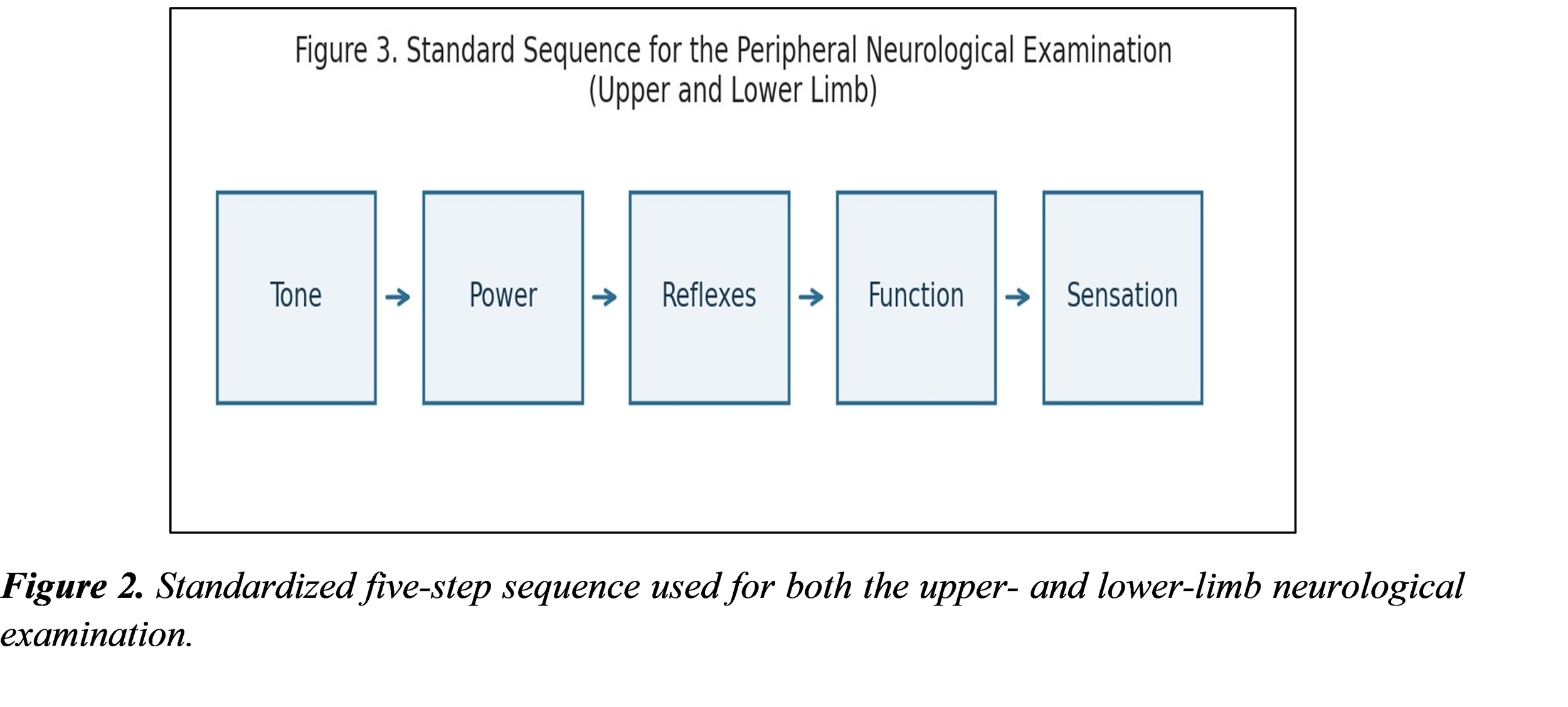
The upper limb examination is used to find signs of nerve problems in the arms and hands. People often come with changes in feeling, like tingling or numbness, and weakness. The weakness can be in bursts, as in conditions that damage nerve insulation, such as multiple sclerosis, or it can be steady, as in motor neurone disease. The limb neurological examination can also spot single nerve problems, like wrist drop or Erb's palsy, and many nerve problems, such as the "glove and stocking" pattern that appears in diabetes mellitus. A thorough upper limb examination looks at both movement and feeling using a set order of steps.
5.1 Preparation
Like any examination, the examiner introduces themselves, confirms the patient’s identity, explains the procedure, and obtains consent before beginning. The upper body is. The examiner looks at the arms for muscle wasting, fasciculations, or asymmetry before proceeding to formal testing.
5.2 Tone and Power
Tone is first checked near the shoulder, looking at how easy it's to move the shoulder without effort. After that, the check moves to the elbow, wrist, and hand joints. Then power is measured at each one by one. The shoulder moves out and in, the elbow and wrist bend and straighten, and the fingers move out and in; even the thumb moves. At each joint, the examiner sees how much force the patient can push against.
5.3 Reflexes
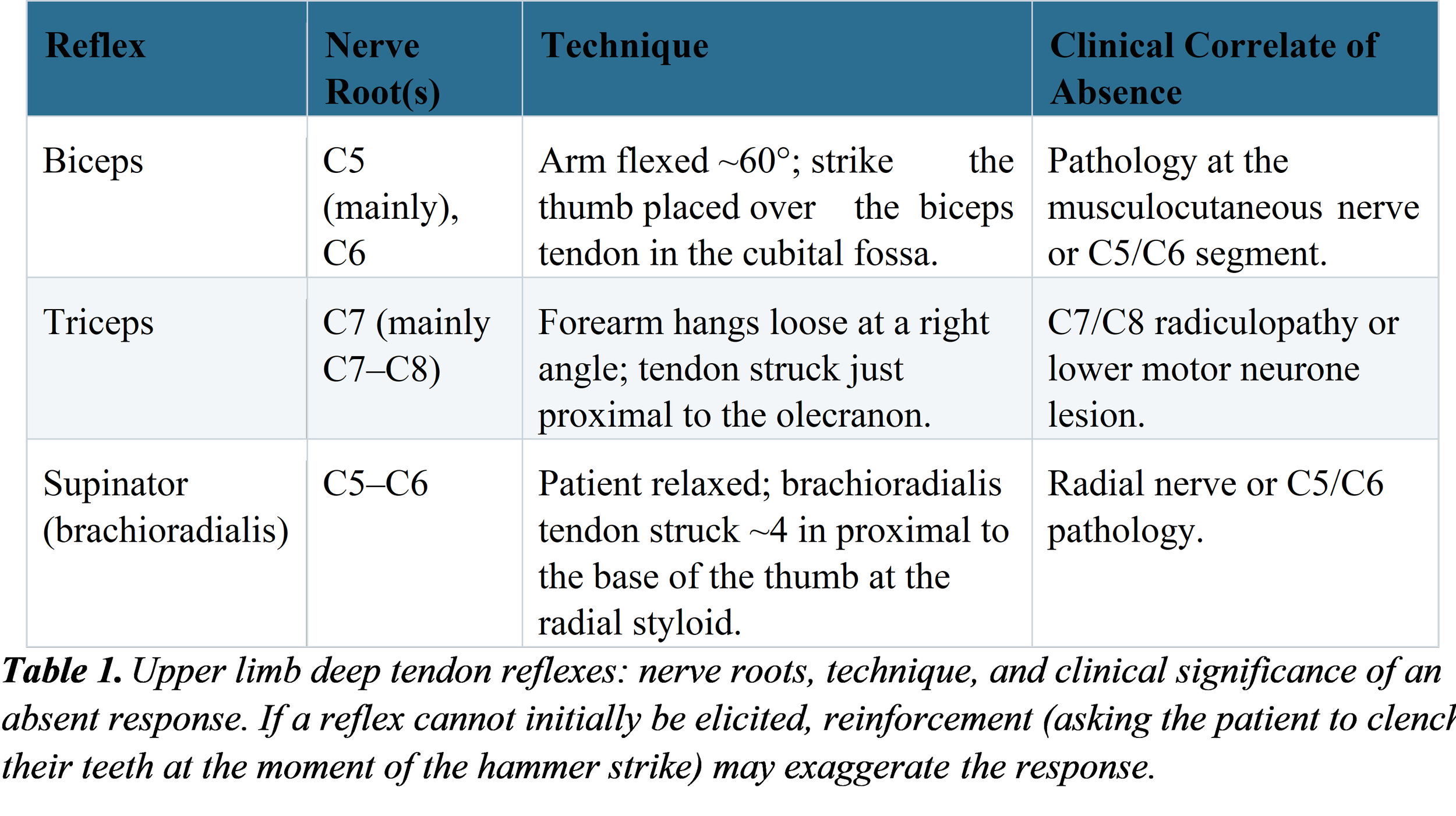
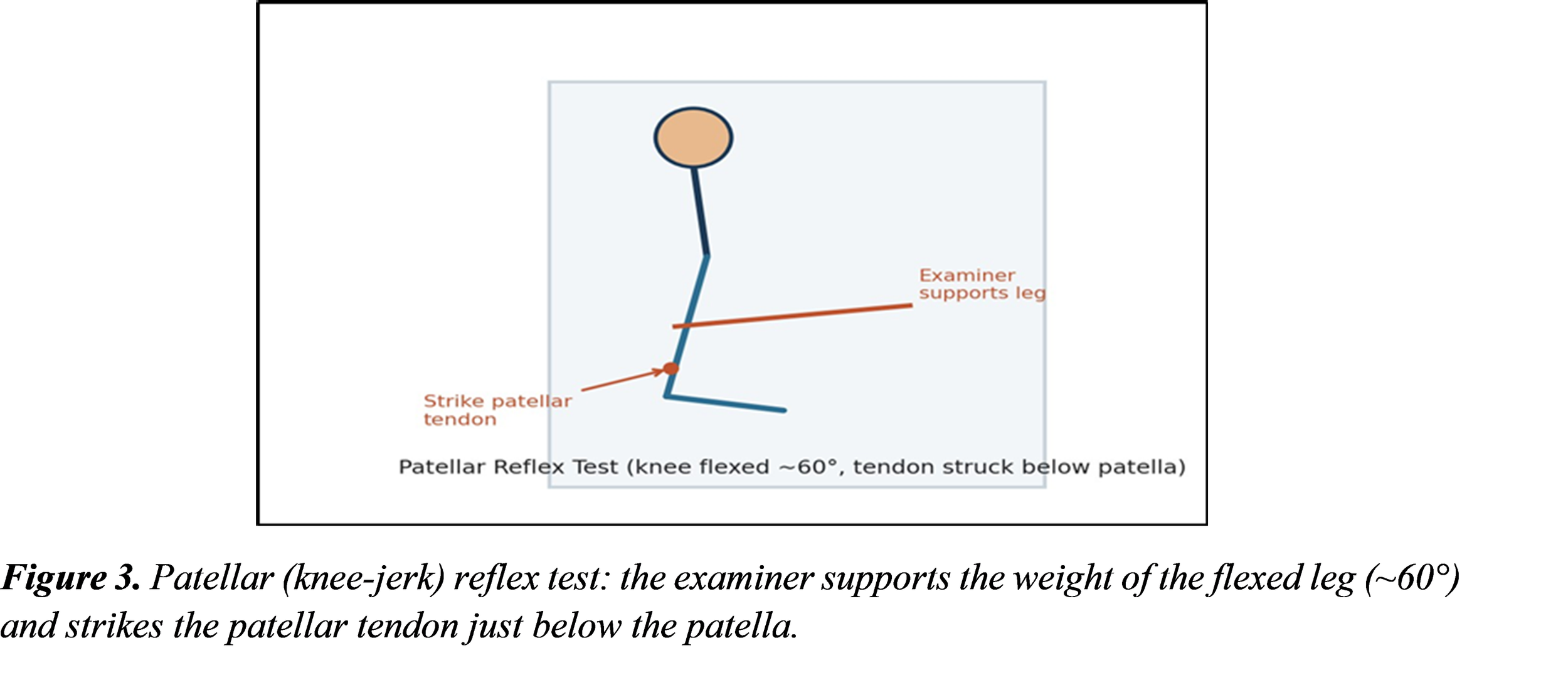
In the limb we routinely check three deep tendon reflexes: the biceps reflex, the triceps reflex and the supinator reflex (also known as brachioradialis reflex). When the muscle contracts strongly, that is a deep tendon reflex. When the muscle contracts weakly or not at all, that is a deep tendon reflex. A brisk deep tendon reflex often points toward upper motor neuron pathology. An absent or reduced deep tendon reflex often points toward a lower motor neuron lesion.
5.4 Function and Sensation
I do an assessment by asking the patient to touch their head with both hands and to pick up a small object, such as a coin, with each hand so I can see how well the patient moves small muscles. After that, I perform testing that includes light touch, pinprick, vibration, and joint position sense. I start touch and pinprick on the sternum so the patient knows what the stimulus should feel like. Then I apply them over each dermatome while the patient keeps their eyes closed. I test vibration with a sounding tuning fork, beginning at the end on the bony prominence at the base of the thumb and moving toward the body: first the radial styloid, then the olecranon only if the first test shows a problem. I examine proprioception by holding the tip of the thumb by its sides, showing "up" and "down" positions while the joint is either straight or bent, and then I ask the patient, with eyes closed, to say what position the joint is in after I move it.
6. Lower Limb Neurological Examination
The limb neurological examination follows the same pattern as the upper limb examination looking at tone, power, reflexes, function and sensation. The limb neurological examination helps to find nerve problems that serve the legs and feet. In cases just like the upper limb examination patients may report paraesthesia, numbness or loss of power. These symptoms can point to mononeuropathies for example foot drop or to polyneuropathies, such, as a glove-and-stocking pattern that often appears in diabetes mellitus.
6.1 Tone
When the patient’s lower body is properly exposed the examiner first looks at the legs for signs of muscle wasting, twitching or unevenness. Tone is checked by rolling the leg across the bed to see how easily it moves, by lifting under the knee to feel for resistance. To test for ankle clonus the examiner turns the patient’s leg outward gently relaxes the ankle and then quickly lifts the foot upward; if a rhythmic movement follows this indicates clonus a sign of a motor neuron problem.
6.2 Power
Power is tested at the hip, knee, ankle and toes. Power is measured at the hip during flexion, extension, abduction and adduction. Power is measured at the knee during flexion and extension. Power is measured at the ankle during dorsiflexion and plantarflexion. Power is measured at the toes during dorsiflexion and plantarflexion. The examiner checks how much Power the patient can generate when the patient pushes against resistance at each joint.
6.3 Reflexes
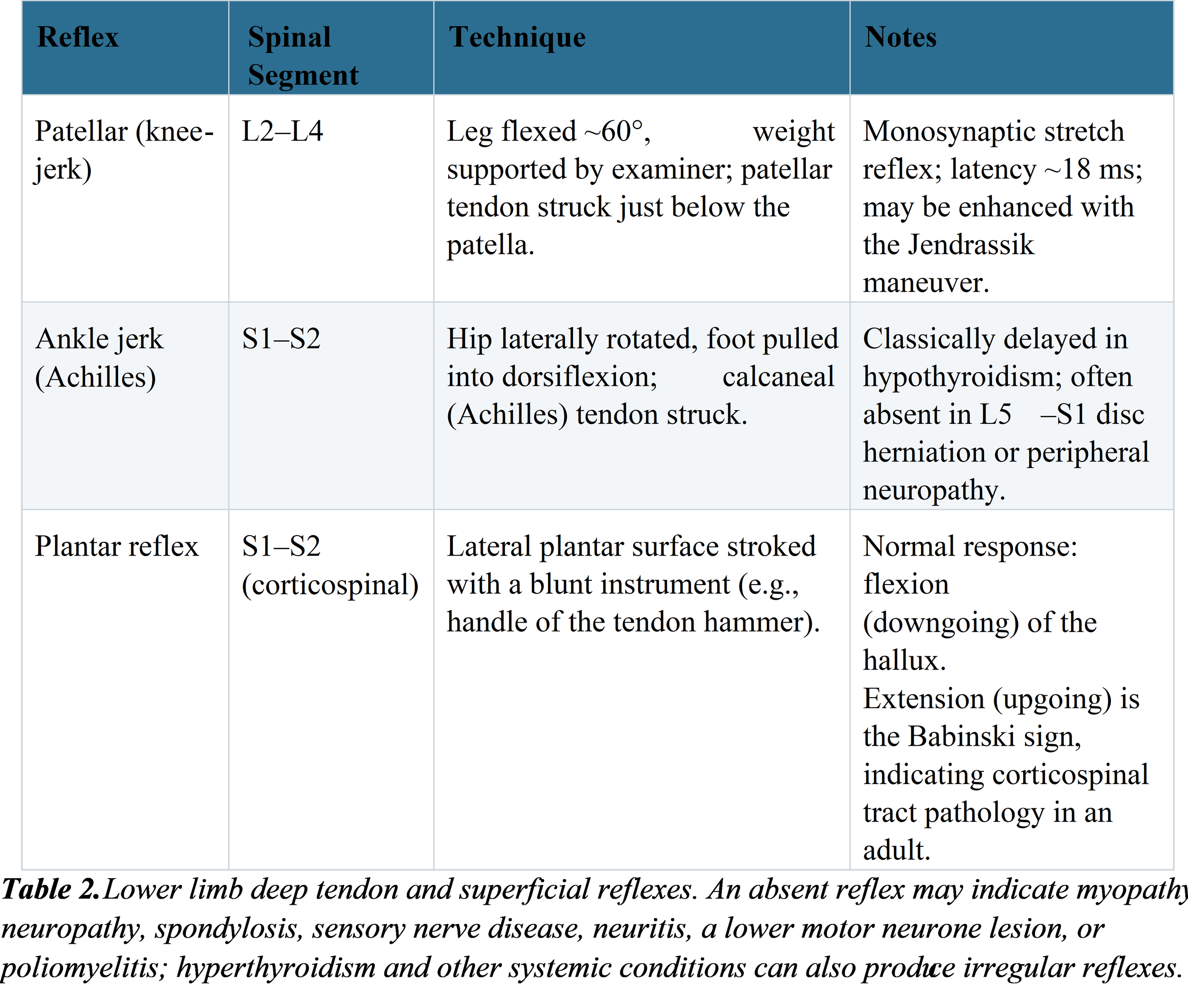
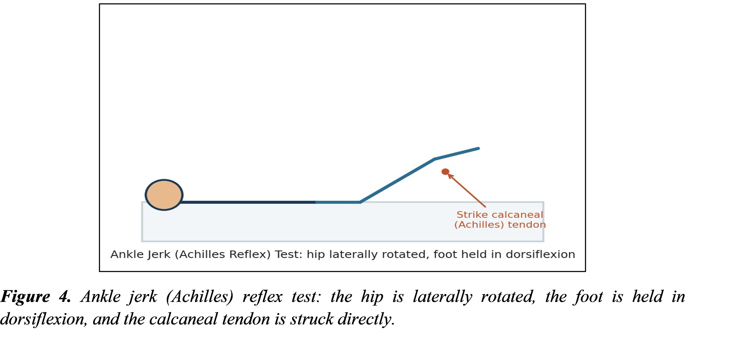
6.4 Function and Sensation
Functional lower-limb assessment includes looking at the way a person walks for any signs. It also includes Romberg's test, where the patient stands with their feet together and eyes closed. If they sway, it suggests a problem with the column, which is related to feeling, rather than a problem with the cerebellum.
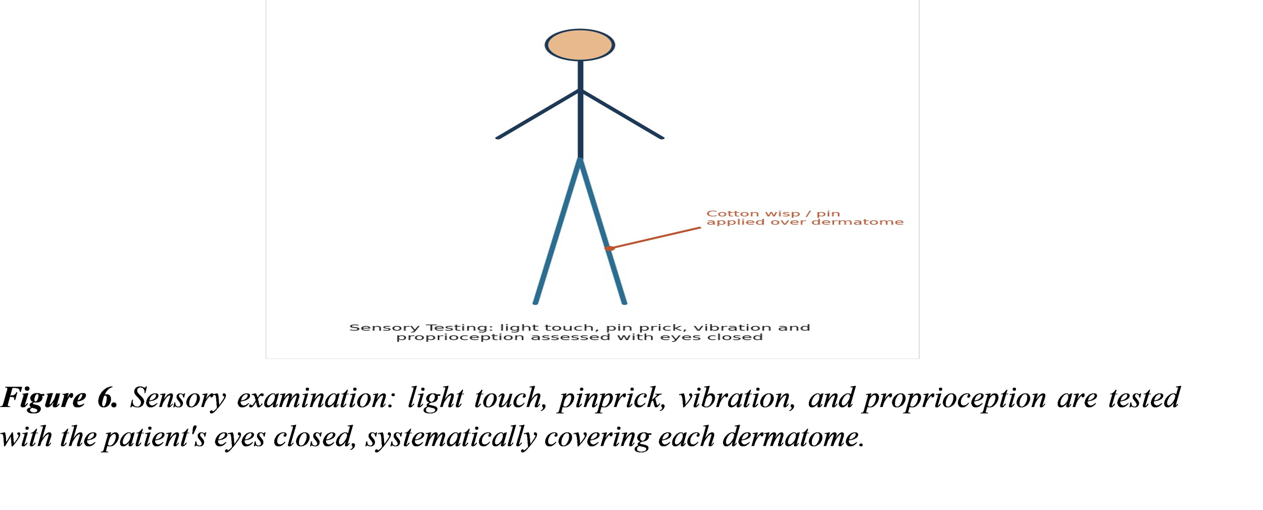
Testing the sense follows the methods as for the upper limb. This includes checking for touch, pinprick, vibration, and proprioception. Vibration is tested starting from the toe and moving up toward the tibial epicondyle and the greater trochanter if the feeling in the feet is not normal. Proprioception is checked at the toe joints.

7. Sacroiliac Joint Assessment: The Stork (Gillet) Test
7.1 Purpose and Technique
The Stork test, also called the Gillet test or March test, is one of the mobility tests used to check the sacroiliac joint for problems. The sacroiliac joint is the part of the body where the spine meets the pelvis. When this joint is not working right, it can cause pain in the back or around the pelvis. The test checks how it moves. The examiner uses their fingers to feel for movement inside the pelvis. One thumb is placed on the bony part at the back of the hip called the superior iliac spine. The other finger is placed near the bottom of the spine on the base. The patient stands up. Then lifts the leg on the side being tested to 90 degrees or more. The examiner watches how the bony part on the hip moves compared to the spine. The same thing is done on the other side, and the two sides are compared.
In a pelvis that works normally, the side being tested moves backward. This makes the bony part on the hip drop. Move down in relation to the spine. The movement should be the same on both sides. If the bony part on the side as the leg is being lifted moves very little, does not move at all, or moves with pain, the test is considered positive. This is seen as a sign that the sacroiliac joint is not moving properly.
7.2 Diagnostic Evidence and Limitations
The evidence that supports the Stork test on its own is limited. The normal range of SIJ motion is small – less than 4 degrees of rotation and up to 1.6 mm of translation – which makes it hard to reliably tell the difference between symptomatic and asymptomatic movement at the bedside. Results from sacroiliac mobility tests, including the Stork test, are not reliable or valid for diagnosing SIJ dysfunction when used alone. Asymmetry in SIJ movement can also be found in people who do not have symptoms. Reliability improves when a group of mobility and provocation tests is used together. One study found that adding the Stork test to an examination of irritation points during testing improved the assessment of SIJ dysfunction. Notably, Hungerford and colleagues found that physical therapists could reliably feel and recognize a pattern of intrapelvic motion on the support side during the Stork test and could reasonably distinguish no relative movement from anterior rotation of the innominate bone during a load‑bearing task. However, further research was recommended to confirm the test’s validity for detecting girdle dysfunction specifically.
A positive Stork test, with other positive sacroiliac mobility tests, suggests likely impairment of SIJ mobility, but it is not diagnostic on its own.
• The recommended SIJ Test Item Cluster includes the distraction test, compression test, thigh thrust test, Gaenslen's test and sacral thrust test.
• Diagnostic discrimination for SIJ pain is strongest when three or more of these five tests are positive, and at least one of the positive results is either the thigh thrust or compression test.
8. The FABER (Patrick's) Test
8.1 Definition and Clinically Relevant Anatomy
The FABER test. Which stands for Flexion, Abduction, and External Rotation. Is also called Patrick’s test or the "figure-4" test. It is a pain-provocation test used to help find problems in the hip, back, or sacroiliac area. The hip joint is a ball-and-socket joint where the head of the femur fits into the acetabulum. This structure allows for both stability and a wide range of motion, including flexion, extension, abduction, adduction, and internal and external rotation. The joint is supported by ligaments inside the capsule and outside it.
8.2 Technique and Interpretation
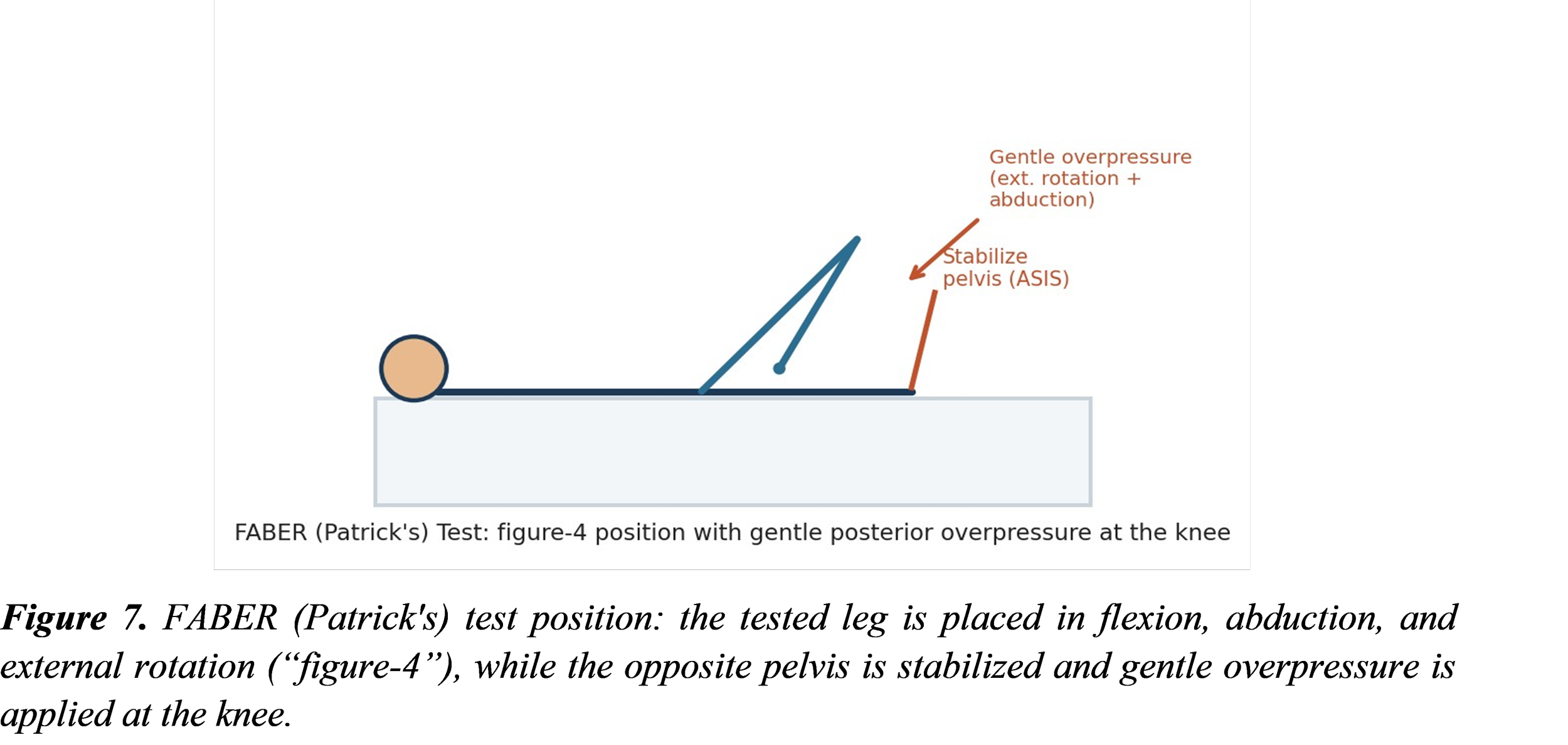
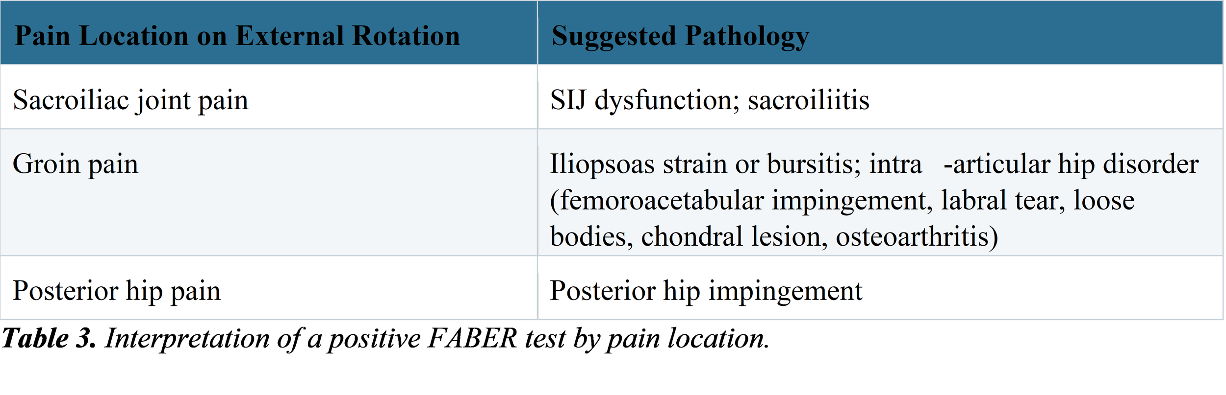
Patient lies supine and test leg is placed in figure‑4 position – hip flexed and abducted. Lateral ankle rests on thigh above knee. Examiner stabilizes side of pelvis at the anterior superior iliac spine. Examiner applies external rotation, abduction, and posterior overpressure to knee on same side until end‑range is reached, sometimes with small oscillations to check for pain. Patient reproduces pain. Shows limited range of motion. A positive test means pain or limited range.
8.3 Diagnostic Evidence
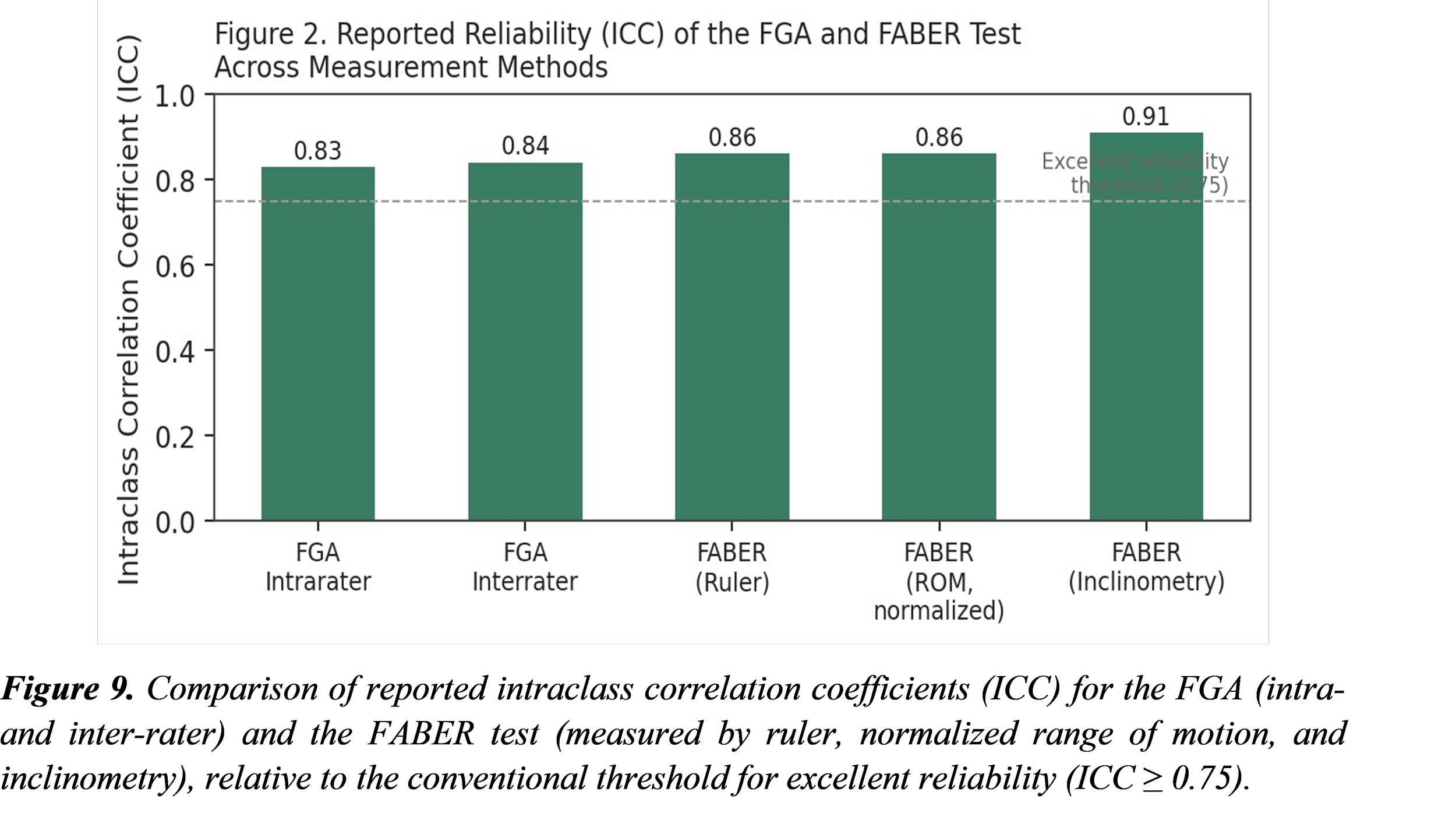
Reliability of the FABER test changes with how it's measured. When the FABER test is measured with a ruler, the FABER test shows intra‑rater reliability (ICC 0.86). The FABER test also shows intra‑rater reliability (ICC 0.86) when the FABER test uses a normalized range of motion. The FABER test is best measured with inclinometry, giving an ICC of 0.91. That score is even higher when the FABER test is done by a clinician. The FABER test can detect hip pathology with a sensitivity of 0.89 when compared to arthroscopy. The FABER test’s positive result is moderately linked to osteoarthritis (r = 0.54). When the FABER test is used to find tears seen on MR arthrography, the FABER test has very high specificity and a positive predictive value of 100 %. However, the FABER test has a sensitivity of 41 % and a low negative predictive value (9 %). That means the FABER test is much better at confirming a tear than at ruling it out. In the literature, the FABER test’s validity is debated. Some authors say the FABER test is a pain‑provocation tool. Others say the FABER test is unreliable when used alone. The main lesson is that physical special tests, like the FABER test, should be backed up by the clinical picture instead of being relied on as a single diagnostic test.
9. Functional Gait Assessment (FGA)
9.1 Overview
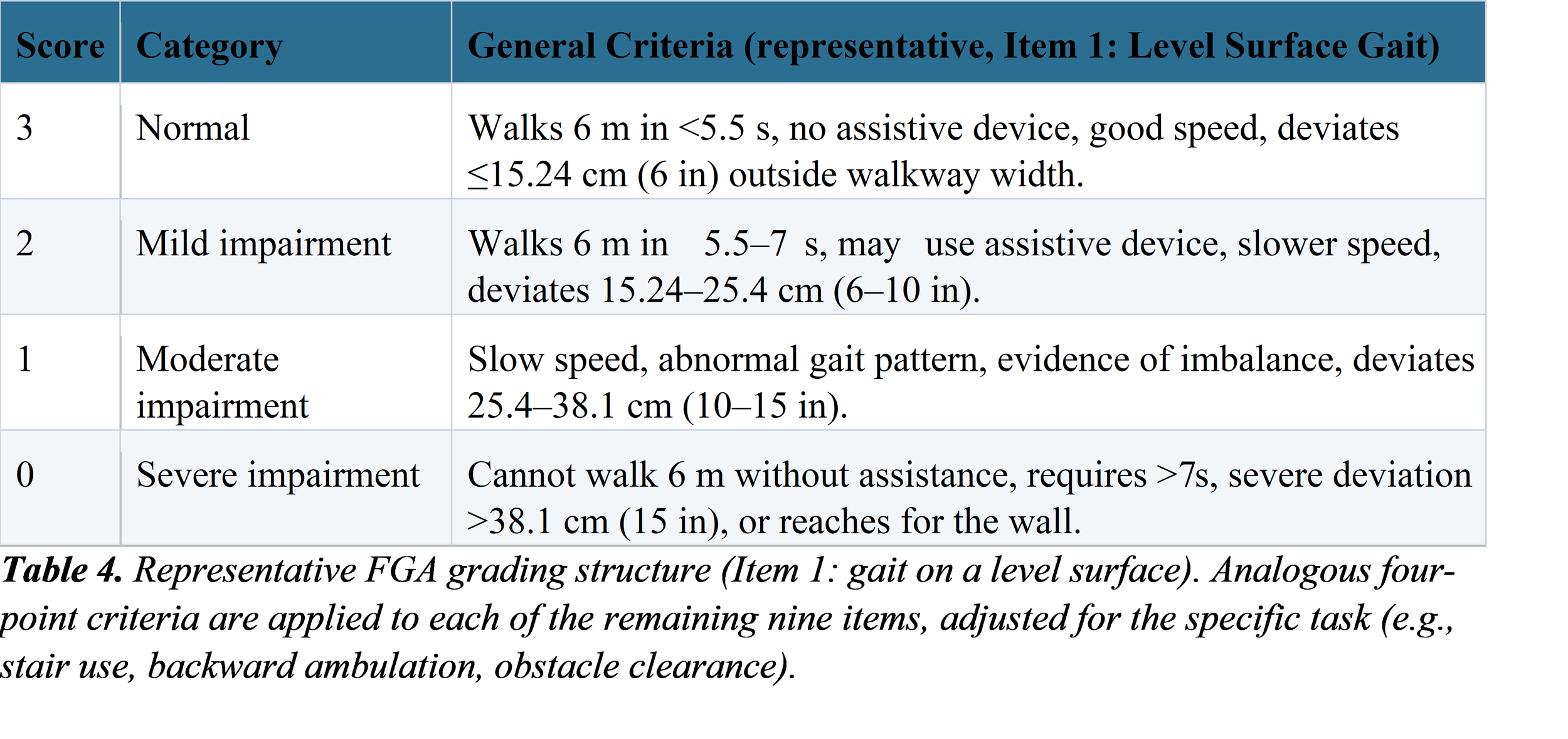
The Functional Gait Assessment or FGA is a version of the Dynamic Gait Index or DGI. It adds complex walking tasks to make it better suited for people with vestibular disorders. The goal is to avoid the ceiling effect that happens with the test, where patients score high even if they still have problems. Three new items were added because they are especially hard for people who have dysfunction. The FGA is used with adults who have Parkinson’s disease, stroke or vestibular issues. It checks how well someone keeps their balance while doing walking activities. The test takes between five and ten minutes to complete. There are ten items in total. Each item gets a score from zero to three—zero means difficulty and three means normal walking. The highest possible total score is thirty, out of thirty.
9.2 Test Items
The ten FGA items are: (1) gait on a level surface (2) change in speed (3) gait with horizontal head turns, (4) gait with vertical head turns, (5) gait and pivot turn, (6) step over obstacle (7) gait with a narrow base of support (8) gait with eyes closed (9) ambulating backwards and (10) steps (stair ascent/descent). Each of the ten FGA items is scored against criteria that describe deviation outside a standardized 30.48 cm (12 in) walkway width. The criteria also cover changes, in speed use of assistive devices and loss of balance.
9.3 Requirements
• A marked, level 6 m (20 ft) walkway, 30.48 cm (12 in) in width
• A stopwatch
• A step or shoe box (for the obstacle item)
• A flight of stairs (for the steps item)
9.4 Reliability and Validity
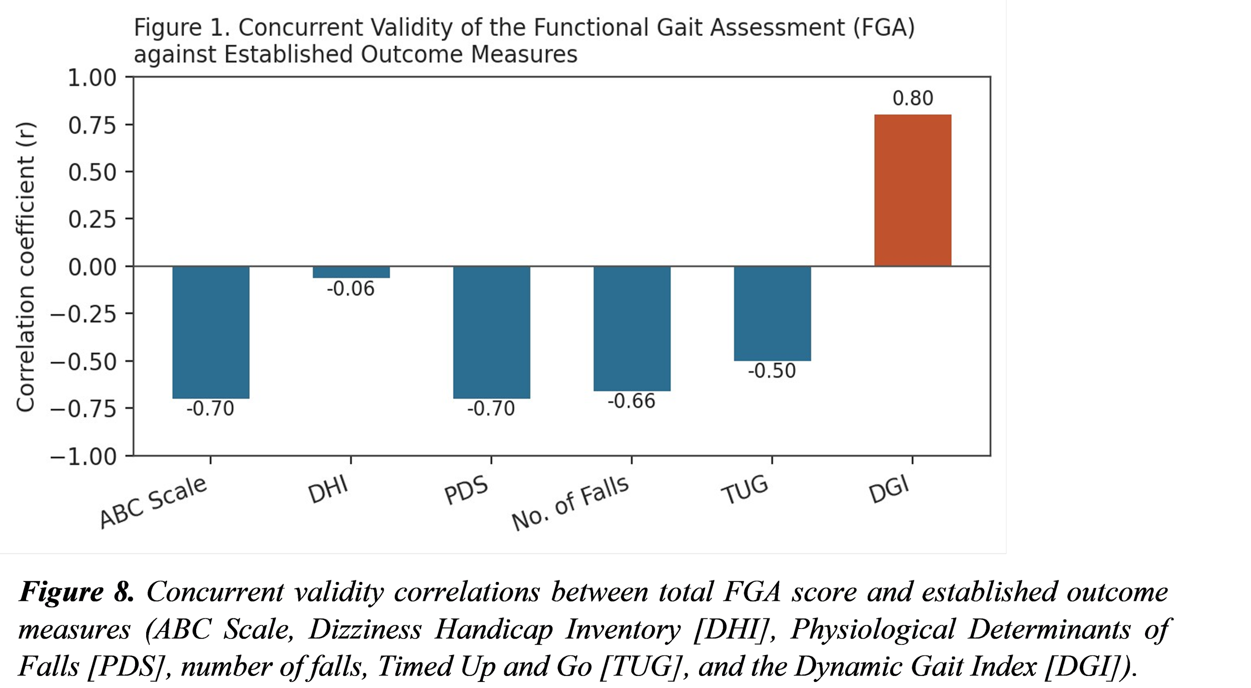
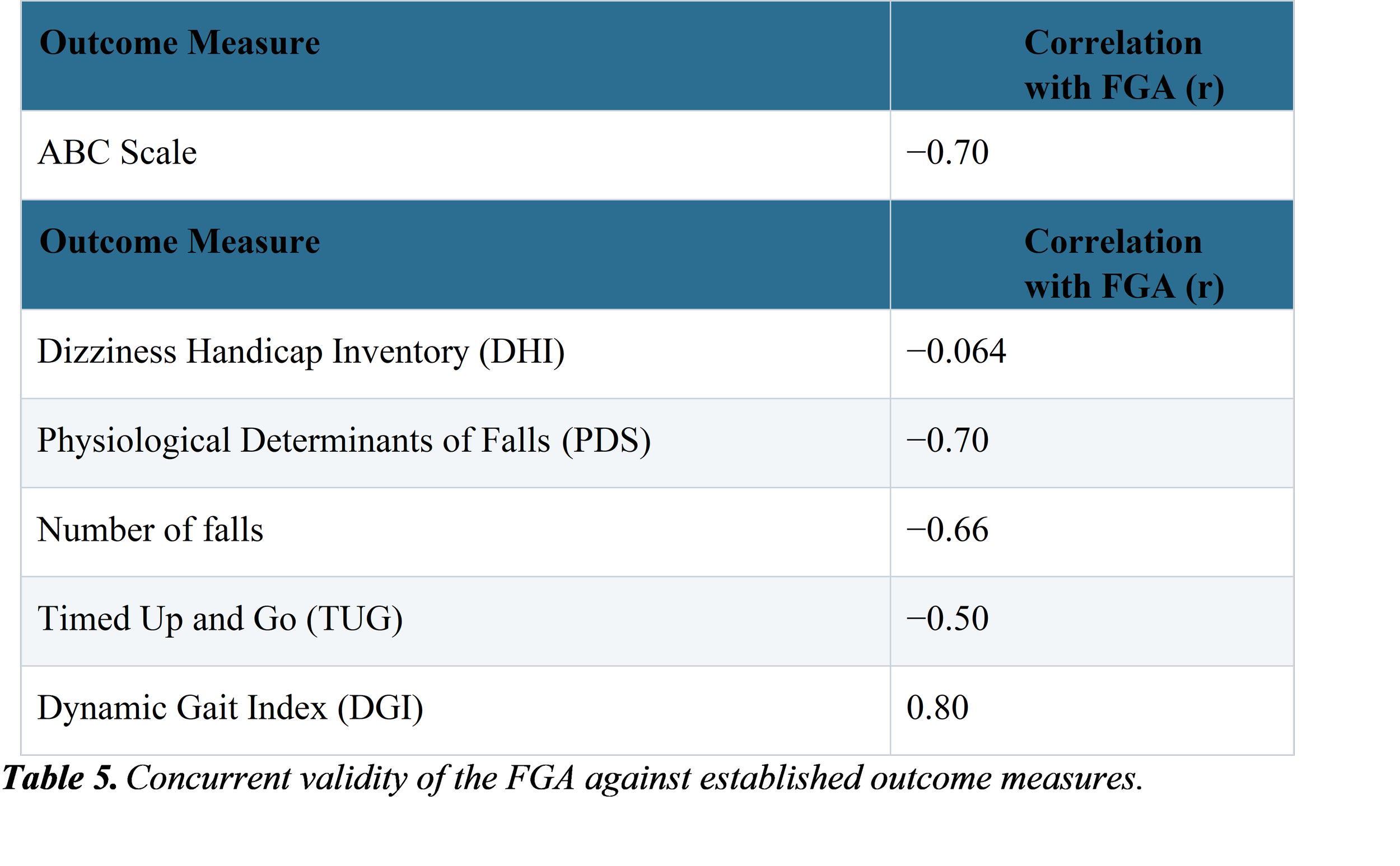
The FGA shows good reliability: the intra‑rater reliability of the total score is ICC 0.83, the inter‑rater reliability is ICC 0.84, and the internal consistency across both trials is Cronbach’s alpha 0.79. The FGA is valid, with several other outcome measures as summarized in Figure 8 and Table 5 below. The FGA is negatively correlated with the Activities‑specific Balance Confidence (ABC) Scale, the Fear of Falling / Physiological Determinants of Falls (PDS) measure, and the number of falls, a finding that fits the idea that a lower FGA, meaning poorer gait function, is linked to lower balance confidence and a greater history of falls. The FGA is positively correlated with the Dynamic Gait Index, from which the FGA was derived.
Taken together with the Dynamic Gait Index and its four-item short form, the FGA shows sufficient validity, responsiveness, and reliability for assessing walking function in patients undergoing stroke rehabilitation, and is generally recommended over the original DGI on the basis of its psychometric properties.
10. Developmental Milestone Examination
10.1 Developmental Delay and Cerebral Palsy Screening
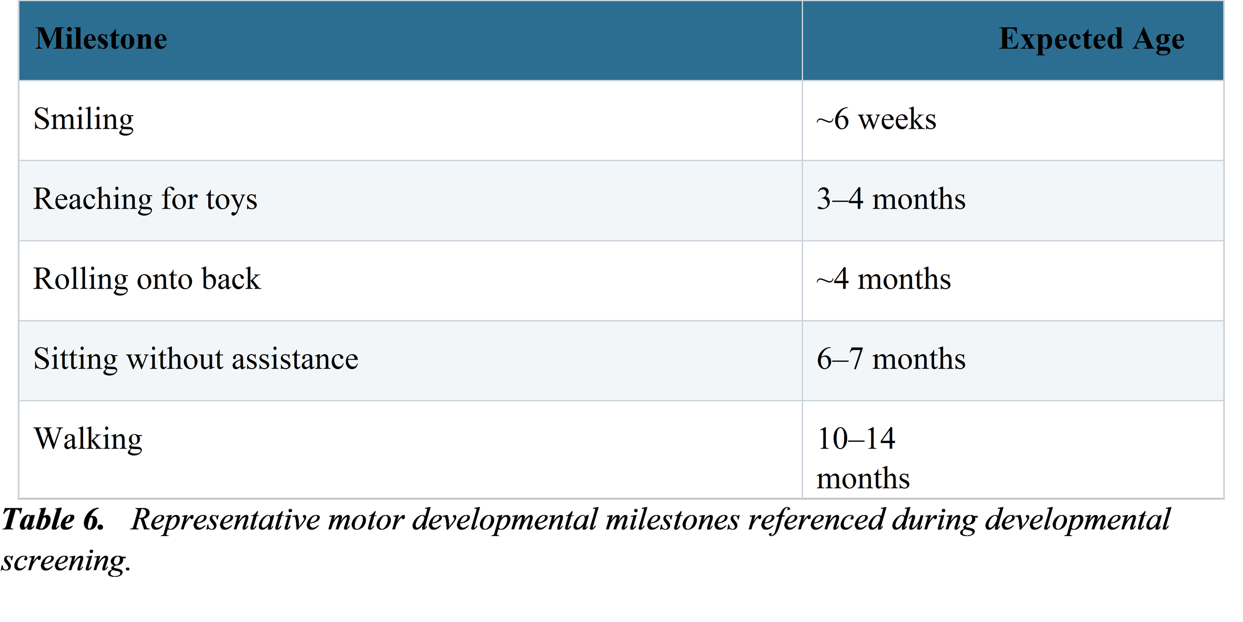
Developmental delay means a child is not reaching expected milestones at the expected time. This can be one of the signs that a child might have cerebral palsy or another developmental disability. It is normal for children to develop at different speeds, and some may reach milestones a little later than others without any issues. When delays happen, especially along with other risk factors, like certain physical findings, imaging results, or medical history, it becomes more serious. These concerns should be taken seriously by caregivers and doctors.
10.2 Evaluation Process
When developmental delay is suspected, evaluation follows a two‑step process. First, developmental screening is carried out. During screening, the clinician talks to the parents and meets the child directly to check the child’s skills, reflexes, and reactions. If developmental delay is found, a detailed developmental evaluation is then carried out. The developmental evaluation is usually done by a specialist such as a pediatrician, a developmental psychologist, or a pediatric neurologist. Additional tests are used during the evaluation to confirm or rule out possible conditions.
Anatomic signs that are important for the developmental evaluation are loose limbs, poor torso support, trouble balancing, and infant reflexes that do not develop or that stay too long. Failure to thrive is usually defined as growth that's below the third to fifth percentile for weight or a sudden sharp drop in the growth rate. Failure to thrive can appear at the time as developmental delay and may be caused by digestive problems, dietary issues or bone and joint problems that make feeding difficult.
10.3 Radiological Correlation
Cerebral palsy comes from an injury or malformation of the brain. Injuries or malformations can be seen with neuroimaging. Cranial ultrasound is a choice for very young infants, but it is less reliable than CT or MRI. MRI requires the infant to stay and that can be a practical problem. About eighty‑three percent of children with palsy have a visible brain injury on imaging. Therefore, looking at imaging helps to confirm what the clinical tests show for palsy.
11. Discussion
The ten techniques I looked at show a trend. When you use a structured, part-based exam or a standard tool made up of several parts like the five-step neurological exam or the ten-item FGA, they are more likely to be reliable. This isn't by chance. A single test, like the Stork test or the FABER test, only does one physical action to find where the pain is coming from. The movement in the SIJ is very small, just a few millimeters and a few degrees. Also, many parts around the hip and pelvis can cause pain in different areas. So one test alone can't tell for sure which part is the problem.
In contrast, the FGA's strength is in combining ten walking tasks into one combined score. This combined score cancels out errors that happen in each task and makes the FGA better at distinguishing differences. The FGA shows reliability between raters and within the same rater, and the FGA has clear links with other tests that measure balance confidence and records of falls. The same logic also informs the recommendation that appears often in the sacroiliac and hip literature. The recommendation says special tests should be read as a group; for example, the five-test SIJ cluster, rather than one by one.
From a clinical-reasoning point of view, the practical takeaway is that one positive orthopedic test, like a Stork test or a positive FABER test, should make you suspicious of the related condition, but it should not be used alone to confirm a diagnosis. These tests are most helpful when combined with the patient’s history, the pattern of pain that comes up during testing, positive findings in the same group of tests, and imaging if needed. On the other hand, standardized tools such as the FGA can be trusted more when used by themselves as outcome measures, especially when checking how a patient improves over time during rehabilitation, through repeated assessments.
An observation that is seen in the neurological examination, the SLR test, and the FABER test is the important role of reinforcement and movement in bringing out subtle signs. For example, asking a patient to clench their teeth can make a weak reflex more obvious. Comparing both sides during the Stork test can show differences. These simple tests done at the bedside have no cost. Improve the chances of finding something important. They do not need tools. This shows that a careful physical examination is still very important when there are advanced imaging options available.
11.1 Limitations
This review is a synthesis rather than a systematic review, and this review does not apply a formal quality‑appraisal framework such as QUADAS‑2 to the underlying studies cited for each test.
This review reproduces reliability and validity figures from literature reviewed instead of recalculating them from primary data, and these figures may differ across patient populations, examiner experience, and measurement method, as illustrated by the range of FABER ICC values obtained with a ruler versus inclinometry.
This review describes the application of these tests as supervised bedside use during training rather than an independently validated diagnostic protocol, and any findings from any single test discussed in this review should always be interpreted within the full clinical context of the patient.
12. Conclusion
This review brings together ten bedside examination techniques. These include musculoskeletal provocation testing, peripheral neurological examination, standardized gait assessment, and pediatric developmental screening. All were applied during patient assessments. The evidence shows a clinical pattern: single special tests have real value but limited diagnostic power. They are most useful when used as part of a structured evaluation. In contrast, standardized composite tools like the Functional Gait Assessment offer reliability and validity. These tools can be trusted more, especially when tracking a patient’s progress over time. A disciplined, accurate physical exam, done with understanding of each test’s limits, remains a vital part of clinical care. It works hand in hand with taking a patient’s history and ordering investigations. This approach is essential in medical practice.
Declarations
Author Contributions
(Ranjit Rajesh Gaud). came up with the idea, wrote the text for the text, made the figures and tables, and put the final version together. (Tushar Kumar) and (Pankaj Kumar Rana) helped gather data and supported the research. Worked on editing the final manuscript. All authors read the paper. Agreed that it was ready to be published.
Conflicts of Interest
The author declares no conflicts of interest.
Funding
This review received no external funding.
Data Availability
No original datasets were generated for this review. All reliability and validity figures cited are reproduced from the previously published sources listed in the References section.
Ethical Approval
Not applicable. This narrative review does not report original human-subjects research; no patient-identifiable data are included.
References
1. Fuller, G. (2019). Neurological Examination Made Easy (6th ed.). Elsevier.
2. Hungerford, B., Gilleard, W., & Hodges, P. (2007). Evidence of altered lumbopelvic muscle recruitment in the presence of sacroiliac joint pain. Spine, 32(14), 1478–1485.
3. Martin, R. L., Kelly, B. T., & Leunig, M. (2008). Diagnostic accuracy of clinical tests for the different types of femoroacetabular impingement: a systematic review. Arthroscopy, 24(9), 1013–1018.
4. Nelson, W. E., Kliegman, R. M., St. Geme, J. W., & Blum, N. J. (Eds.). (2020). Nelson Textbook of Pediatrics (21st ed.).
5. Elsevier.
6. Petty, N. J. (2011). Neuromusculoskeletal Examination and Assessment: A Handbook for Therapists (4th ed.). Churchill Livingstone.
7. Physiopedia contributors. (n.d.). Developmental Milestones. Physiopedia. Retrieved from https://www.physio-pedia.com/Developmental_Milestones
8. Physiopedia contributors. (n.d.). FABER Test. Physiopedia. Retrieved from https://www.physio-pedia.com/FABER_Test
9. Physiopedia contributors. (n.d.). Functional Gait Assessment. Physiopedia. Retrieved from https://www.physio-pedia.com/Functional_Gait_Assessment
10. Physiopedia contributors. (n.d.). Gowers' Sign. Physiopedia. Retrieved from https://www.physio-pedia.com/Gowers_Sign
11. Physiopedia contributors. (n.d.). Lower Limb Neurological Examination. Physiopedia. Retrieved from https://www.physio-pedia.com/Lower_Limb_Neurological_Examination
12. Physiopedia contributors. (n.d.). Stork Test (Gillet Test). Physiopedia. Retrieved from https://www.physio-pedia.com/Stork_Test
13. Physiopedia contributors. (n.d.). Straight Leg Raise Test. Physiopedia. Retrieved from https://www.physio-pedia.com/Straight_Leg_Raise_Test
14. Physiopedia contributors. (n.d.). Upper Limb Neurological Examination. Physiopedia. Retrieved from https://www.physio-pedia.com/Upper_Limb_Neurological_Examination
15. Talley, N. J., & O'Connor, S. (2021). Clinical Examination: A Systematic Guide to Physical Diagnosis (9th ed.). Elsevier.
16. Wrisley, D. M., Marchetti, G. F., Kuharsky, D. K., & Whitney, S. L. (2004). Reliability, internal consistency, and validity of data obtained with the Functional Gait Assessment. Physical Therapy, 84(10), 906–918.