
Cirrhosis of the Liver: Symptomatology and General Principles of Treatment
1. Kebekova Aizat Ashimzhanovna
2. Rishikesh Kumar
3. Talim Khan
(1. Lecturer, International Medical Faculty, Osh State University, Osh, Kyrgyz Republic
2. Student, International Medical Faculty, Osh State University, Osh, Kyrgyz Republic
3. Student, International Medical Faculty, Osh State University, Osh, Kyrgyz Republic)
Abstract
Cirrhosis is the end-stage of chronic liver injury, characterised by diffuse fibrosis, formation of regenerative nodules and distortion of the normal hepatic architecture, leading to portal hypertension and progressive hepatic insufficiency. It is a major cause of morbidity and mortality worldwide. The clinical course is divided into a long, often asymptomatic compensated phase and a decompensated phase marked by ascites, variceal haemorrhage, hepatic encephalopathy and jaundice. This article reviews the clinical manifestations of cirrhosis and the general principles of its management, including aetiological treatment, management of complications, prevention of progression, and liver transplantation.
Keywords: cirrhosis, portal hypertension, ascites, hepatic encephalopathy, variceal bleeding, liver transplantation
1. Introduction
The liver has a remarkable regenerative capacity, but sustained injury from viral, toxic, metabolic or autoimmune causes triggers persistent inflammation and activation of hepatic stellate cells. These cells deposit excess extracellular matrix, producing fibrosis. When fibrosis becomes widespread and the parenchyma is replaced by regenerative nodules, the condition is termed cirrhosis. The process was long considered irreversible, but current evidence shows that removal of the underlying cause (for example, viral eradication or alcohol abstinence) can lead to regression of fibrosis and reduce complications, particularly in early disease.
Cirrhosis is among the leading causes of death from digestive disease globally. Its burden is rising because of the growing prevalence of metabolic dysfunction-associated steatotic liver disease (MASLD), in addition to the continuing impact of chronic hepatitis B and C and alcohol-related liver disease.
2. Aetiology
Chronic liver injury leading to advanced fibrosis and cirrhosis arises from a varied spectrum of etiologies, with chronic alcohol consumption remaining one of the primary drivers globally. Long-term alcohol misuse triggers toxic cellular damage, lipid accumulation, and persistent inflammatory cascades, ultimately resulting in severe metabolic dysfunction and progressive tissue scarring characteristic of alcohol-related liver disease.
Chronic viral hepatitis infections, particularly chronic hepatitis B and C, represent another major cause of global liver morbidity. In cases of hepatitis B, co-infection or superinfection with hepatitis D significantly accelerates the progression toward cirrhosis and hepatocellular carcinoma. These viruses induce chronic immune-mediated destruction of hepatocytes, driving continuous cycles of cell death, regeneration, and fibrotic tissue deposition over several decades.
Metabolic dysfunction-associated steatotic liver disease (MASLD) and its progressive inflammatory counterpart, metabolic dysfunction-associated steatohepatitis (MASH), have emerged as rapidly increasing causes of chronic liver disease. Closely linked with obesity, insulin resistance, type 2 diabetes, and dyslipidemia, MASH promotes hepatic lipotoxicity, oxidative stress, and chronic low-grade inflammation that gradually transforms simple steatosis into bridging fibrosis and irreversible architectural distortion.
Autoimmune and cholestatic pathologies also contribute substantially to systemic liver damage. Autoimmune hepatitis involves T-cell-mediated destruction of hepatic parenchyma, whereas primary biliary cholangitis (PBC) and primary sclerosing cholangitis (PSC) target the biliary tree. PBC leads to autoimmune destruction of small intrahepatic bile ducts, while PSC causes chronic inflammation, concentric fibrosis, and stricturing of both intrahepatic and extrahepatic bile ducts, causing persistent toxic bile acid retention and secondary biliary cirrhosis.
Genetic and metabolic disorders disrupt normal hepatic function through the pathological accumulation of metabolic products or defective protein transport. Hereditary hemochromatosis leads to systemic iron overload with preferential deposition in hepatocytes, Wilson disease causes toxic copper accumulation due to defective biliary copper excretion, and alpha-1 antitrypsin deficiency results in misfolded protein accumulation within the endoplasmic reticulum of liver cells, all of which induce severe oxidative damage and parenchymal injury.
Vascular anomalies that impair hepatic venous drainage can produce chronic passive congestion and ischemic necrosis. Conditions such as Budd–Chiari syndrome, characterized by hepatic vein occlusion, and chronic right-sided heart failure cause severe sinusoidal congestion, central vein dilation, and centrilobular fibrosis, eventually culminating in cardiac cirrhosis if left unmanaged.
Finally, long-term exposure to hepatotoxic drugs and environmental toxins, including chronic administration of medications like methotrexate or amiodarone, can induce subtle, progressive hepatic damage. When a thorough clinical, serological, and histological workup fails to reveal a clear underlying cause, the disease is categorized as cryptogenic cirrhosis, though many such cases are now recognized as late-stage, burnt-out MASH where steatosis has resolved as cirrhosis fully develops.
3. Pathophysiology in Brief
The clinical manifestations of advanced liver disease are fundamentally driven by two primary pathophysiological mechanisms: hepatocellular failure and portal hypertension. Understanding how these two distinct yet overlapping processes develop provides the key to recognizing and managing the vast array of symptoms, physical exam findings, and laboratory abnormalities seen in patients with cirrhosis.
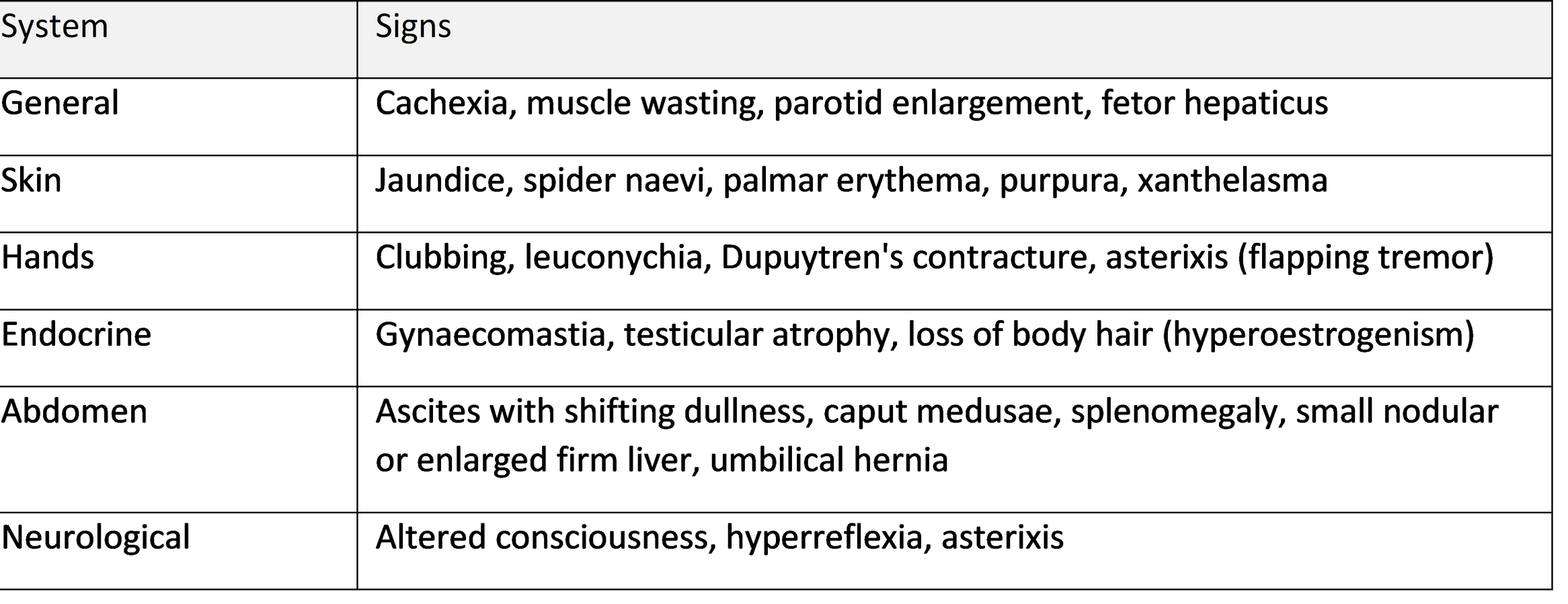
Hepatocellular failure occurs as progressive fibrotic scarring and parenchymal tissue destruction impair the functional capacity of hepatocytes. The resulting loss of synthetic function leads directly to hypoalbuminemia, which lowers plasma oncotic pressure and contributes to peripheral edema and fluid accumulation. Loss of synthetic capacity also manifests as severe coagulopathy due to the diminished production of clotting factors (II, VII, IX, and X), leading to an increased risk of bleeding. Furthermore, impaired detoxifying function causes hyperbilirubinemia, which presents clinically as jaundice and scleral icterus. The liver's inability to metabolize circulating hormones results in hyperestrogenism, yielding findings such as spider angiomas, palmar erythema, gynecomastia, and testicular atrophy. Crucially, the breakdown in detoxifying capacity permits neurotoxic compounds—most notably ammonia—to bypass hepatic processing, resulting in spectrums of cognitive impairment known as hepatic encephalopathy.
Portal hypertension develops due to structural changes within the hepatic architecture, where dense fibrotic bands and regenerating nodules increase intrahepatic vascular resistance to portal blood flow. This structural blockade is compounded by active splanchnic vasodilation driven by local endothelial release of nitric oxide. As portal venous pressure rises above physiological thresholds, the body attempts to decompress the system by opening portosystemic collateral pathways. This leads to the formation of high-risk gastroesophageal varices, caput medusae, and anorectal varices, all of which carry significant risk of life-threatening hemorrhage. Chronic venous congestion within the portal system also leads to congestive splenomegaly, which subsequently causes hypersplenism and secondary sequestration of platelets and leukocytes.
The interplay between these two mechanisms is most evident in the development of ascites and hepatorenal syndrome. Splanchnic arterial vasodilation, triggered by severe portal hypertension, reduces effective arterial blood volume. This systemic circulatory dysfunction activates the renin-angiotensin-aldosterone system (RAAS) and the sympathetic nervous system, promoting intense renal sodium and water retention. Combined with the low oncotic pressure from hepatocellular failure-induced hypoalbuminemia, fluid shifts massively into the peritoneal cavity to form ascites. As intrarenal vasoconstriction progressively worsens in response to persistent arterial hypotension, renal perfusion severely declines, ultimately precipitating hepatorenal syndrome—a functional, life-threatening form of acute kidney injury driven entirely by advanced liver failure.
4. Clinical Stages
The clinical spectrum of cirrhosis is fundamentally divided into compensated and decompensated stages, a distinction that carries profound prognostic implications. In compensated cirrhosis, the liver maintains sufficient functional parenchyma and hemodynamic balance to prevent major systemic complications. Patients in this early phase are frequently asymptomatic, and the diagnosis is often discovered incidentally during routine laboratory testing or abdominal imaging performed for unrelated reasons. Reflecting this stable physiological baseline, the median survival for patients with compensated disease exceeds twelve years.
Decompensated cirrhosis represents a critical transition marked by the clinical emergence of overt manifestations of hepatocellular failure or portal hypertension. A patient is defined as having decompensated disease upon the development of ascites, variceal hemorrhage, hepatic encephalopathy, or jaundice. The onset of any of these major complications signals a sharp breakdown in the body's homeostatic mechanisms, causing median survival to drop precipitously to approximately two years in the absence of liver transplantation.
To systematically quantify disease severity and stratify patient mortality risk, clinicians rely on validated prognostic scoring systems. The Child–Pugh scoring system evaluates five key clinical and laboratory parameters: serum bilirubin, serum albumin, international normalized ratio (INR), and the severity of ascites and hepatic encephalopathy. Based on the cumulative score, patients are categorized into Class A (well-compensated), Class B (significantly compromised), or Class C (severely decompensated), providing a standardized measure of hepatic synthetic function and overall surgical risk.
For organ allocation and transplant prioritization, the Model for End-Stage Liver Disease (MELD) and its variation, MELD-Na, serve as the primary objective tools. Utilizing precise mathematical formulas based on serum bilirubin, INR, serum creatinine, and serum sodium levels, these models accurately predict short-term (90-day) mortality in end-stage liver disease. By relying strictly on objective laboratory values rather than subjective clinical assessments like ascites or encephalopathy grading, the MELD score ensures fair, transparent prioritization for candidates on liver transplant waiting lists.
5. Symptomatology
5.1 Compensated phase
In the early or compensated stages of cirrhosis, symptoms are frequently absent or present in an insidious, non-specific manner. Patients often report generalized systemic complaints such as persistent fatigue, profound weakness, and a noticeable reduction in physical exercise tolerance. These constitutional symptoms stem from underlying metabolic alterations, impaired hepatic glycogen storage, and the systemic inflammatory state associated with ongoing liver injury.
Gastrointestinal and nutritional disturbances are also prominent non-specific manifestations. Patients commonly experience anorexia, nausea, postprandial fullness, early satiety, and progressive weight loss. These features are driven by altered gut hormone signaling, delayed gastric emptying, and passive venous congestion of the splanchnic circulation, which can also present as diffuse abdominal bloating, dyspeptic discomfort, or a vague sensation of heaviness localized to the right upper quadrant due to hepatic enlargement or capsular stretch.
Endocrine dysregulation and metabolic derangements further contribute to subtle systemic complaints. Impaired hepatic clearance of hormones can lead to altered circulating estrogen-to-androgen ratios, resulting in reduced libido and reproductive dysfunction in both male and female patients. Additionally, skeletal muscle cramps are a frequent yet overlooked symptom, arising from secondary electrolyte imbalances, altered peripheral nerve perfusion, and the rapid loss of lean muscle mass characteristic of chronic liver disease.
5.2 Decompensated phase
Symptoms related to hepatocellular failure and portal hypertension:
As liver disease progresses into decompensation, clinical manifestations shift from non-specific complaints to overt signs of organ failure and severe portal hypertension. Jaundice develops when the liver's capacity to conjugate and excrete bilirubin declines, leading to scleral icterus, cutaneous yellowing, and dark, tea-colored urine as conjugated bilirubin spills into the renal filtration system. Pruritus often accompanies this excretory failure, presenting with particular severity in primary cholestatic disorders like primary biliary cholangitis due to the systemic accumulation of bile salts and pruritogenic mediators.
Fluid overload and third-spacing represent major structural complications of decompensation. Severe portal hypertension combined with hypoalbuminemia drives fluid accumulation in the peritoneal cavity, manifesting as progressive abdominal distension, rapid weight gain, and shifting dullness on examination. This systemic fluid retention frequently extends to the lower extremities as dependent ankle edema. In some patients, fluid transudates across diaphragmatic defects into the pleural space—a condition known as hepatic hydrothorax—which can cause significant dyspnea and compromised pulmonary function even with minimal visible ascites.
Decompensation also severely compromises vascular stability and metabolic clearance, leading to high-acuity crises. Rupture of high-pressure gastroesophageal varices presents as catastrophic upper gastrointestinal hemorrhage, manifesting as frank hematemesis or melena. Concurrently, the failure of hepatic detoxification pathways allows neurotoxic metabolites like ammonia to cross the blood-brain barrier, precipitating hepatic encephalopathy. This neurocognitive spectrum ranges from subtle sleep-wake cycle reversal and personality shifts to profound confusion, flapping tremor, lethargy, and eventual hepatic coma.
Hemostatic impairment and systemic inflammatory vulnerability further complicate the clinical course. Reduced hepatic synthesis of clotting factors paired with platelet sequestration from congestive splenomegaly leads to spontaneous coagulopathy, clinically evident as easy bruising, epistaxis, and mucosal gum bleeding. Patients with established ascites face a high risk of spontaneous bacterial peritonitis, which can present insidiously with low-grade fever, diffuse abdominal tenderness, or unexplained worsening of renal function and encephalopathy. Over time, chronic hypermetabolism and systemic inflammation contribute to marked sarcopenia and muscle wasting, severely undermining physical reserve and long-term clinical outcomes.
5.3 Physical signs
5.4 Complications
Ascites represents the most common decompensating event in cirrhosis, arising from the combination of portal hypertension and systemic arterial vasodilation. Increased sinusoidal pressure drives fluid into the peritoneal cavity, while splanchnic vasodilation prompts renal sodium and water retention via activation of the renin-angiotensin-aldosterone system. Spontaneous bacterial peritonitis (SBP) occurs when bacteria, predominantly Gram-negative enteric bacilli like Escherichia coli, translocate across the compromised gut mucosal barrier into this ascitic fluid without an identifiable intra-abdominal surgical source. SBP requires prompt paracentesis for diagnosis, defined by an absolute neutrophil count in the ascitic fluid of 250 cells per cubic millimeter or greater.
Portal hypertension also redirects portal blood flow through collateral pathways, forming gastroesophageal varices and predisposing to portal hypertensive gastropathy. Variceal hemorrhage is a life-threatening emergency caused by increased pressure within thin-walled submucosal veins. Portal hypertensive gastropathy presents as mucosal ectasia and congestion in the gastric body and fundus, leading to chronic slow blood loss and iron deficiency anemia rather than sudden catastrophic bleeding. Management involves non-selective beta-blockers for primary or secondary prophylaxis and endoscopic band ligation during acute bleeding episodes.
Hepatic encephalopathy encompasses a spectrum of neuropsychiatric abnormalities resulting from portosystemic shunting and hepatocellular failure, which prevent the liver from clearing neurotoxins such as ammonia. Clinical severity is classified according to the West Haven criteria across four grades: Grade I manifests as mild confusion, altered attention, or inversion of the sleep-wake cycle; Grade II introduces lethargy, disorientation to time, and clear asterixis (flapping tremor); Grade III presents with somnolence, marked confusion, and disorientation to place; and Grade IV represents full hepatic coma. Treatment relies on non-absorbable disaccharides such as lactulose to trap ammonium in the gut lumen and non-absorbable antibiotics like rifaximin to suppress ammonia-producing colonic microflora.
Functional organ crosstalk failure severely impacts the renal, pulmonary, and cardiovascular systems in advanced disease. Hepatorenal syndrome (HRS) is a form of functional renal failure marked by intense renal vasoconstriction in response to extreme splanchnic vasodilation and arterial hypotension, occurring without structural renal pathology. Pulmonary complications include hepatopulmonary syndrome, driven by intrapulmonary vascular dilations causing arterial hypoxemia that typically worsens in the upright position (platypnea-orthodeoxia), and portopulmonary hypertension, defined by pulmonary arterial hypertension in the setting of portal hypertension. Cirrhotic cardiomyopathy further contributes to systemic circulatory dysfunction, characterized by blunted ventricular responsiveness to stress and altered cardiac electrophysiology.
Additional systemic sequelae and malignant transformation markedly increase morbidity. Hepatocellular carcinoma (HCC) develops at an annual incidence of one to eight percent in cirrhotic livers, driven by chronic inflammation, continuous cell turnover, and fibrotic remodeling, necessitating routine surveillance ultrasound every six months. Vascular complications such as portal vein thrombosis arise from sluggish portal blood flow and altered procoagulant-anticoagulant dynamics. Congestive splenomegaly leads to hypersplenism with secondary sequestration of platelets and leukocytes, while chronic metabolic derangements foster severe protein-calorie malnutrition, sarcopenia, osteoporosis, and an increased susceptibility to systemic bacterial infections.
Acute-on-chronic liver failure (ACLF) represents a distinct syndrome occurring in individuals with pre-existing chronic liver disease or cirrhosis. It is characterized by an acute hepatic decompensation triggered by precipitants such as bacterial infections, active alcohol consumption, or gastrointestinal bleeding. ACLF is clinically defined by the rapid onset of organ failure involving the liver, kidneys, brain, coagulation system, circulation, or respiration, and is associated with intense systemic inflammation and high short-term mortality requiring critical care stabilization or urgent liver transplantation.
5.5 Laboratory and instrumental findings
Laboratory markers in cirrhosis directly reflect the loss of hepatic synthetic capacity, impaired metabolic clearance, and the secondary effects of portal hypertension. Hypoalbuminemia and a prolonged prothrombin time with an elevated International Normalized Ratio (INR) serve as primary indicators of diminished hepatocyte synthetic function. Serum bilirubin rises as the liver's excretory mechanics fail. Thrombocytopenia routinely manifests as the earliest and most common laboratory abnormality, resulting from platelet sequestration within an enlarged spleen due to congestive splenomegaly, as well as reduced hepatic production of thrombopoietin. Transaminase patterns typically shift such that aspartate aminotransferase (AST) exceeds alanine aminotransferase (ALT), reversing the typical ratio seen in acute liver injury. In advanced disease, dilutional hyponatremia frequently develops as a consequence of persistent non-osmotic vasopressin hypersecretion driven by severe splanchnic vasodilation and arterial underfilling.
Abdominal ultrasonography provides essential non-invasive anatomical confirmation of cirrhosis and its associated vascular alterations. Key structural findings include a nodular, heterogeneous liver surface contour, coarse parenchymal echotexture, and selective hypertrophy of the caudate lobe relative to the right lobe. As intrahepatic vascular resistance increases, ultrasound reveals secondary markers of portal hypertension, including a dilated portal vein measuring greater than thirteen millimeters, splenomegaly, recanalization of the umbilical vein, and the presence of free fluid within the peritoneal cavity.
Non-invasive staging modalities have largely replaced routine tissue sampling for quantifying hepatic fibrosis. Vibration-controlled transient elastography (FibroScan) measures liver stiffness in kilopascals, providing a reliable physical metric of fibrotic remodeling. Simple serum-based scoring systems, such as the Fibrosis-4 (FIB-4) index and the AST-to-Platelet Ratio Index (APRI), combine routine laboratory values to stratify patients by risk of advanced fibrosis or cirrhosis without requiring specialized equipment.
Invasive and procedural evaluations are reserved for risk stratification, disease management, and clarifying ambiguous presentations. Upper gastrointestinal endoscopy (esophagogastroduodenoscopy) is routinely performed to screen for and grade gastroesophageal varices and portal hypertensive gastropathy, guiding primary bleeding prophylaxis. While non-invasive testing and imaging often suffice for diagnosis, a percutaneous or transjugular liver biopsy remains the definitive gold standard when the underlying etiology is uncertain, when overlapping liver disorders are suspected, or when clinical, lab, and imaging findings yield discordant results.
6. General Principles of Treatment
The aims of treatment are to treat or remove the cause, slow or reverse fibrosis, prevent and treat complications, improve quality of life, and select patients for liver transplantation.
6.1 Aetiological treatment
Etiology-specific management aims to arrest ongoing liver injury, reverse fibrosis where possible, and prevent progression to decompensation across different chronic liver diseases.
For alcohol-related liver disease, complete and lifelong abstinence from alcohol forms the absolute cornerstone of therapy. Achieving abstinence requires integrated psychological support, structural counseling, and, when indicated, targeted pharmacotherapy for alcohol use disorder to prevent relapse and mitigate secondary inflammatory responses within the hepatic tissue.
In chronic viral hepatitis, antiviral interventions target the specific mechanisms of viral replication. Treatment for chronic hepatitis B relies on long-term administration of potent nucleos(t)ide analogues such as entecavir or tenofovir, which suppress viral load, reduce parenchymal inflammation, and lower the risk of cirrhotic decompensation and hepatocellular carcinoma. For chronic hepatitis C, treatment with oral direct-acting antiviral (DAA) regimens achieves a sustained virological response—effectively curing the infection—in the vast majority of patients across all genotypes.
Management of metabolic dysfunction-associated steatotic liver disease (MASLD) and steatohepatitis (MASH) prioritizes systemic metabolic control. Sustained weight reduction achieved through dietary modification and regular physical activity restores metabolic balance and reduces hepatic steatosis. Optimal clinical management also demands aggressive control of comorbid conditions, including type 2 diabetes mellitus, dyslipidemia, and arterial hypertension, to limit lipotoxicity and fibrotic progression.
Targeted immune and cholestatic therapies address autoimmune pathologies of the liver. Autoimmune hepatitis requires systemic immunosuppression, typically initiating with corticosteroids such as prednisone or budesonide, often combined with or transitioned to azathioprine to maintain biochemical remission. For primary biliary cholangitis, ursodeoxycholic acid serves as first-line therapy, promoting bile flow, reducing toxic intrahepatic bile acid accumulation, and slowing the destruction of small intrahepatic bile ducts.
Metabolic and genetic liver disorders require interventions designed to eliminate accumulated toxins or prevent their cellular uptake. Hereditary hemochromatosis is managed through therapeutic venesection to mobilize excess iron stores from hepatocytes and maintain target serum ferritin levels. In Wilson disease, long-term therapy utilizes copper chelating agents such as penicillamine or trientine to promote urinary excretion of excess copper, or zinc salts to block intestinal copper absorption.
6.2 General measures, diet and lifestyle
• Avoid alcohol and hepatotoxic drugs; avoid NSAIDs and, where possible, nephrotoxic agents.
• Nutrition: adequate energy (about 35 kcal/kg/day) and protein (1.2–1.5 g/kg/day). Protein restriction is not recommended, even in encephalopathy. Small frequent meals and a late-evening snack reduce overnight catabolism.
• Sodium restriction (about 2 g/day or 88 mmol/day) in patients with ascites.
• Vaccination against hepatitis A and B, influenza, pneumococcus and COVID-19.
• Regular physical activity, smoking cessation, and management of coexisting metabolic disease.
6.3 Management of complications
Ascites
• Sodium restriction plus diuretics: spironolactone (with or without furosemide) in a stepwise manner.
• Large-volume paracentesis with intravenous albumin for tense ascites.
• Refractory ascites: repeated paracentesis, TIPS (transjugular intrahepatic portosystemic shunt) in selected patients, and transplant evaluation.
• Spontaneous bacterial peritonitis: diagnostic paracentesis (ascitic neutrophils ≥250/mm³), intravenous third-generation cephalosporin and albumin; secondary prophylaxis thereafter.
Portal hypertension and varices
• Screening endoscopy in cirrhosis, unless non-invasive criteria exclude high-risk varices.
• Primary prophylaxis: non-selective beta-blockers (propranolol, nadolol, or carvedilol) or endoscopic variceal band ligation.
• Acute bleeding: resuscitation with restrictive transfusion (haemoglobin target about 7–8 g/dL), vasoactive drugs (terlipressin or octreotide), antibiotic prophylaxis (ceftriaxone), and endoscopic band ligation within 12 hours. TIPS is considered early in high-risk patients or when bleeding is not controlled.
• Secondary prophylaxis: combination of beta-blocker and band ligation.
Hepatic encephalopathy
• Identify and correct precipitants (gastrointestinal bleeding, infection, constipation, electrolyte disturbance, dehydration, sedatives).
• Lactulose is first-line for treatment and secondary prophylaxis; rifaximin is added to prevent recurrence.
Hepatorenal syndrome
• Withdraw diuretics and nephrotoxins, expand volume with albumin, and give vasoconstrictors (terlipressin, or noradrenaline in intensive care). Liver transplantation is the definitive treatment.
Hepatocellular carcinoma surveillance
• Ultrasound with or without alpha-fetoprotein every 6 months in all patients with cirrhosis.
Other
• Treat coagulopathy only when bleeding or before invasive procedures; manage hyponatraemia with fluid restriction in severe cases; treat pruritus (cholestyramine) and muscle cramps (albumin, baclofen).
6.4 Liver transplantation
Liver transplantation is the only definitive treatment for advanced decompensated cirrhosis. Referral is indicated at the first major decompensation, a MELD score of 15 or more, or when HCC within accepted criteria develops. Contraindications include active uncontrolled sepsis, extrahepatic malignancy, severe cardiopulmonary disease and ongoing substance misuse without commitment to abstinence.
6.5 Palliative care
Patients not eligible for transplantation benefit from early palliative care for symptom control, advance care planning and family support.
7. Prognosis
Prognosis depends on aetiology, ongoing liver injury and the presence of complications. The development of ascites, variceal bleeding or encephalopathy markedly worsens survival. Child–Pugh class C carries a one-year survival of about 45%, compared with approximately 100% in class A at one year. Successful treatment of the underlying cause substantially improves outcomes.
8. Conclusion
Cirrhosis is a systemic disease with a long silent phase and a progressively severe decompensated phase. Early recognition of symptoms and signs, identification of the cause, and timely treatment of both the aetiology and its complications are essential to improve survival. A multidisciplinary approach, regular surveillance for varices and hepatocellular carcinoma, nutritional support, and timely referral for transplantation form the basis of modern management.
References
European Association for the Study of the Liver. EASL Clinical Practice Guidelines for the management of patients with decompensated cirrhosis. J Hepatol. 2018;69(2):406–460.
Tsochatzis EA, Bosch J, Burroughs AK. Liver cirrhosis. Lancet. 2014;383(9930):1749–1761.
de Franchis R, Bosch J, Garcia-Tsao G, et al. Baveno VII – Renewing consensus in portal hypertension. J Hepatol. 2022;76(4):959–974.
Garcia-Tsao G, Abraldes JG, Berzigotti A, Bosch J. Portal hypertensive bleeding in cirrhosis: risk stratification, diagnosis and management. AASLD practice guidance. Hepatology. 2017;65(1):310–335.
Biggins SW, Angeli P, Garcia-Tsao G, et al. Diagnosis, evaluation and management of ascites, spontaneous bacterial peritonitis and hepatorenal syndrome: AASLD practice guidance. Hepatology. 2021;74(2):1014–1048.
Vilstrup H, Amodio P, Bajaj J, et al. Hepatic encephalopathy in chronic liver disease: AASLD/EASL practice guideline. Hepatology. 2014;60(2):715–735.
Ginès P, Krag A, Abraldes JG, Solà E, Fabrellas N, Kamath PS. Liver cirrhosis. Lancet. 2021;398(10308):1359–1376.
Kumar P, Clark M. Kumar and Clark's Clinical Medicine. Elsevier (latest edition), chapter on liver disease.