
Symptomatology and General Principles of Treatment of Chronic Cholecystitis, Gallstone Disease and Dyskinesia of the Gallbladder
1. Kebekova Aizat Ashimzhanovna
2. Khan Arbaj
3. Rathod Adarsh
(1. Lecturer, International Medical Faculty, Osh State University, Osh, Kyrgyz Republic
2. Student, International Medical Faculty, Osh State University, Osh, Kyrgyz Republic
3. Student, International Medical Faculty, Osh State University, Osh, Kyrgyz Republic)
Abstract
Diseases of the gallbladder and biliary tract represent a significant portion of gastrointestinal morbidity, with chronic cholecystitis, cholelithiasis, and functional biliary dyskinesia forming an intertwined spectrum of hepatobiliary pathology. Functional dyskinesia often leads to bile stasis, creating ideal conditions for lithogenesis and the formation of cholesterol or mixed gallstones. In turn, the mechanical irritation of gallstones combined with recurrent low-grade bacterial invasion or chemical irritation triggers chronic structural changes within the gallbladder wall, culminating in chronic calculous cholecystitis.
The etiology of these conditions is multifactorial, driven by metabolic derangements, altered bile composition, impaired organ motility, and structural abnormalities. Biliary dyskinesia primarily stems from autonomic nervous system dysfunction, hormonal imbalances involving cholecystokinin, or intrinsic smooth muscle dysfunction. Gallstone formation relies on the classic triad of bile supersaturation with cholesterol, accelerated nucleation driven by mucin secretion, and gallbladder hypomotility. Chronic cholecystitis integrates these metabolic and mechanical factors with opportunistic secondary infections, typically involving ascending enteric pathogens like Escherichia coli, Klebsiella species, and Enterococci.
Keywords: chronic cholecystitis, cholelithiasis, gallstones, biliary dyskinesia, biliary colic, cholecystectomy, ursodeoxycholic acid.
1. Introduction
Gallstone disease affects roughly 10–20% of adults in Western populations, and its prevalence rises with age. It is more frequent in women, in obesity, and in people with rapid weight loss, diabetes or pregnancy. Chronic cholecystitis is, in the large majority of cases (about 90–95%), associated with gallstones. Gallbladder dyskinesia (functional gallbladder disorder) is a disturbance of the motor function of the gallbladder and biliary sphincters without structural change such as stones or marked inflammation. Knowledge of the symptoms and the principles of management of these three conditions is essential for every general practitioner.
2. Chronic Cholecystitis
2.1 Definition
Chronic cholecystitis is a chronic inflammatory disease of the gallbladder wall, accompanied by disturbed motor and concentrating function of the organ. It follows a relapsing course with periods of exacerbation and remission.
2.2 Classification
The categorization of gallbladder pathology provides a systematic framework for understanding how the organ responds to disease processes. Classification by the presence of stones distinguishes between calculous forms, which make up the vast majority of cases driven by lithogenic bile and mechanical irritation, and acalculous forms, which typically arise from severe critical illness, ischemia, or stasis without the presence of solid concretions.
When evaluating the disease course, clinicians divide cases into mild, moderate, and severe categories, while also tracking whether the patient is currently experiencing a period of remission or an acute exacerbation. This temporal tracking helps guide acute management decisions versus elective surgical planning.
Assessing the functional state of the gallbladder further refines the clinical picture by determining whether the organ retains preserved contractility, exhibits a reduced ejection fraction, or has progressed to a completely non-functioning state where chronic inflammation has rendered it fibrotic and incapable of normal storage and release.
2.3 Aetiology and pathogenesis
Bile stasis serves as a primary driver of gallbladder pathology, triggered by mechanical blockages like gallstones or cystic duct obstruction, as well as structural anomalies such as gallbladder kinks. Motility disruptions, including hypokinetic dyskinesia, physiological states like pregnancy, a sedentary lifestyle, and irregular meal schedules, further impede normal gallbladder emptying, allowing bile to stagnate within the lumen.
Infection frequently complicates stasis when enteric flora—most commonly Escherichia coli, Klebsiella, Enterococcus species, and various anaerobes—invade the gallbladder. These bacteria typically ascend from the duodenum via the biliary tract, though they can also seed the organ through hematogenous or lymphogenous spread during systemic or localized infections.
Pathologic changes in bile composition, specifically supersaturation with cholesterol leading to lithogenic bile, inflict direct damage on the mucosal lining. This chemical damage is often compounded by mechanical irritation from migrating or impacted stones, as well as the abnormal reflux of pancreatic enzymes into the biliary tree when sphincter mechanics are disrupted.
Systemic and metabolic factors significantly lower the threshold for disease development. Conditions such as obesity, dyslipidemia, diabetes mellitus, a hereditary predisposition, and underlying allergic or autoimmune reactions all alter metabolic homeostasis, accelerate bile lithogenicity, and promote persistent inflammatory changes in the gallbladder wall.
2.4 Clinical features (symptomatology)
Pain syndrome. The leading symptom is a dull, aching or pulling pain in the right hypochondrium and epigastrium, often constant or paroxysmal. It typically appears 1–3 hours after fatty, fried or spicy food, eggs, alcohol or cold carbonated drinks, and after physical strain or emotional stress. The pain radiates to the right shoulder, right scapula, right side of the neck and the right half of the back. In hypertonic states or when a stone moves, it may become acute (biliary colic).
Dyspeptic syndrome. Bitter taste in the mouth, belching with bitter or air taste, nausea, occasional vomiting with bile, bloating, flatulence, unstable stool (constipation alternating with diarrhoea) and intolerance of fatty foods. Asthenovegetative syndrome. Irritability, weakness, fatigue, poor sleep, headache. Intoxication and inflammatory syndrome. During exacerbation there may be low-grade fever, malaise and chills.
Physical examination. In remission, the examination may be normal. During periods of remission, the physical examination of a patient with chronic cholecystitis may be entirely unremarkable. However, an acute exacerbation brings out key physical findings localized to the right upper quadrant and distant visceral reflex points. Primary among local signs is focal tenderness directly over the gallbladder point, situated at the intersection of the right costal margin and the lateral border of the rectus abdominis muscle. Kehr's sign reflects this localized sensitivity upon palpation of the gallbladder anatomical projection. Murphy's sign serves as a cardinal physical indicator, elicited when the patient abruptly halts deep inspiration because the inflamed gallbladder descends and hits the examiner's palpating hand. Ortner's (Grekov–Ortner) sign further confirms local peritoneal and hepatic capsule irritation when percussion or light tapping along the right costal arch with the ulnar border of the hand provokes sharp discomfort.
Irritation of the phrenic nerve and thoracic spinal nerve roots produces distinct neuro-reflexive signs. The Georgievsky–Mussy (phrenicus) sign manifests as exquisite tenderness when pressure is applied between the heads of the right sternocleidomastoid muscle, reflecting phrenic nerve hypersensitivity. Posteriorly, Boas' point identifies localized tenderness just to the right of the spine at the level of the tenth to twelfth thoracic vertebrae, while Zakharyin–Head zones describe broad areas of cutaneous hyperaesthesia mapping across the right hypochondrium and right shoulder. General systemic signs such as a heavily coated tongue and subtle scleral subicterus can also present when minor biliary stasis or systemic inflammation accompanies the flare.
2.5 Diagnosis
Laboratory evaluation during an acute flare of chronic cholecystitis provides essential objective measures of inflammation and ductal involvement. A complete blood count typically demonstrates leukocytosis with a left shift alongside an elevated erythrocyte sedimentation rate (ESR), while serum C-reactive protein (CRP) serves as a sensitive marker for tissue reaction. Evaluating liver enzymes—specifically alanine aminotransferase (ALT), aspartate aminotransferase (AST), alkaline phosphatase (ALP), and gamma-glutamyl transferase (GGT)—along with total and direct bilirubin helps rule out choledocholithiasis or ascending cholangitis. Serum amylase and lipase levels are drawn concurrently to detect early secondary pancreatic inflammation, while a full lipid profile assesses underlying metabolic dyslipidemia driving lithogenesis.
Abdominal ultrasonography remains the undisputed first-line diagnostic method due to its accessibility, safety, and high sensitivity. Key sonographic criteria confirming chronic cholecystitis include a thickened gallbladder wall measuring greater than three millimeters, localized wall compaction, structural deformity, and the presence of intraluminal biliary sediment or acoustic shadowing from gallstones. Furthermore, real-time functional assessment using a fatty test meal during ultrasound can reveal significantly impaired gallbladder contractility.
Advanced functional and structural imaging modalities further refine complex or ambiguous cases. Dynamic cholescintigraphy (HIDA scan) provides precise quantitative assessment by measuring the gallbladder ejection fraction to diagnose biliary dyskinesia or cystic duct patency issues. When complications like choledocholithiasis, choledochal sloughing, or tissue necrosis are suspected, magnetic resonance cholangiopancreatography (MRCP), endoscopic ultrasound (EUS), or contrast-enhanced CT scans offer high-resolution visualizations of the pancreaticobiliary tree. Fractional duodenal intubation with bile microscopy and microbial culture has largely fallen out of routine practice due to its invasive nature and the superiority of modern non-invasive imaging.
Establishing a rigorous differential diagnosis is essential, as right upper quadrant pain can mimic numerous gastrointestinal, renal, and cardiac pathologies. Clinicians must distinguish cholecystitis from peptic ulcer disease, chronic pancreatitis, gastroesophageal reflux disease (GERD), and irritable bowel syndrome based on symptom timing relative to meals and targeted diagnostic studies. Additionally, acute processes like acute hepatitis, right-sided nephrolithiasis, and atypical presentations of coronary artery disease must be systematically excluded to prevent catastrophic misdiagnoses.
2.6 Complications
Acute cholecystitis, choledocholithiasis, obstructive jaundice, cholangitis, secondary biliary pancreatitis, pericholecystitis and adhesions, empyema, gallbladder perforation, biliary fistulas, and (in long-standing disease) increased risk of gallbladder carcinoma.
3. Gallstone Disease (Cholelithiasis)
3.1 Definition
Gallstone disease is a metabolic disorder characterised by the formation of stones in the gallbladder (cholecystolithiasis) and/or bile ducts (choledocholithiasis, hepatolithiasis).
3.2 Types of stones
• Cholesterol stones (about 80% in Western countries): yellowish, round or oval, radiolucent.
• Pigment stones (bilirubinate): small, dark; associated with haemolysis (sickle cell disease, thalassaemia), cirrhosis and biliary infection.
• Mixed stones: contain cholesterol, bilirubin and calcium salts; the most common form in many series.
3.3 Pathogenesis and risk factors
The three key mechanisms are supersaturation of bile with cholesterol, nucleation (pronucleating factors, mucin), and gallbladder hypomotility with bile stasis. Classic risk factors are summarised as the "five F": female, fat, fertile (multiparous), forty, fair. Others include rapid weight loss or bariatric surgery, total parenteral nutrition, oestrogen therapy, diabetes mellitus, metabolic syndrome, Crohn's disease or ileal resection, fibrates, ceftriaxone, family history and a sedentary lifestyle.
3.4 Stages of the disease
1. Prelithiasis (physicochemical) stage: lithogenic bile, biliary sludge; no stones, often no symptoms.
2. Stage of latent stone carrying: stones are present but symptoms are absent ("silent stones"). Most patients stay asymptomatic; the risk of developing symptoms is approximately 1–4% per year.
3. Clinical stage: symptomatic disease, with biliary colic or chronic calculous cholecystitis.
4. Stage of complications.
3.5 Clinical features
Biliary colic is the typical manifestation: a sudden, severe, steady pain (not truly colicky) in the epigastrium or right upper quadrant, rising within minutes to a plateau and lasting from 30 minutes to several hours, often at night or after a heavy fatty meal. It radiates to the back, right scapula or shoulder and is accompanied by nausea and vomiting, restlessness (the patient cannot lie still) and sweating. Pain lasting more than 6 hours, fever or jaundice suggests a complication.
Between attacks the patient may be entirely asymptomatic or have the dyspeptic features of chronic cholecystitis.
Complications and their signs:
• Acute cholecystitis: persistent pain for more than 6 hours, fever, leucocytosis, positive Murphy's sign.
• Choledocholithiasis and obstructive jaundice: jaundice, dark urine, pale stools, pruritus.
• Acute cholangitis: Charcot's triad (fever with chills, jaundice, right upper quadrant pain); with hypotension and confusion it becomes Reynolds' pentad, which is a surgical emergency.
• Acute biliary pancreatitis: severe epigastric pain radiating to the back, raised amylase and lipase.
• Less common: Mirizzi syndrome, cholecystoenteric fistula, gallstone ileus, empyema and perforation.
3.6 Diagnosis
Transabdominal ultrasound is the method of choice (sensitivity over 95% for gallbladder stones): mobile hyperechoic foci with posterior acoustic shadowing. Liver tests and bilirubin, together with ductal dilatation on ultrasound, guide the probability of common bile duct stones. MRCP or endoscopic ultrasound is used for intermediate-risk patients; ERCP is therapeutic. Oral cholecystography is now historical. CT is helpful in complications.
4. Dyskinesia of the Gallbladder (Functional Gallbladder Disorder)
4.1 Definition
Biliary dyskinesia is a functional disorder of the motility of the gallbladder, bile ducts and sphincter of Oddi, leading to impaired bile excretion without organic disease (no stones, no marked inflammation). According to the Rome IV criteria, it is classified as a functional gallbladder disorder, and disorders of the sphincter of Oddi are classified separately.
4.2 Aetiology
The cause is mainly neurohumoral dysregulation: neuroses and chronic stress, irregular and unbalanced diet, hormonal and endocrine disorders (hypothyroidism, diabetes, menopause), reduced cholecystokinin response, previous hepatitis or infection, gastro-duodenal diseases, helminthiasis (e.g. giardiasis), and anatomical anomalies. It may be primary, or secondary to other diseases.
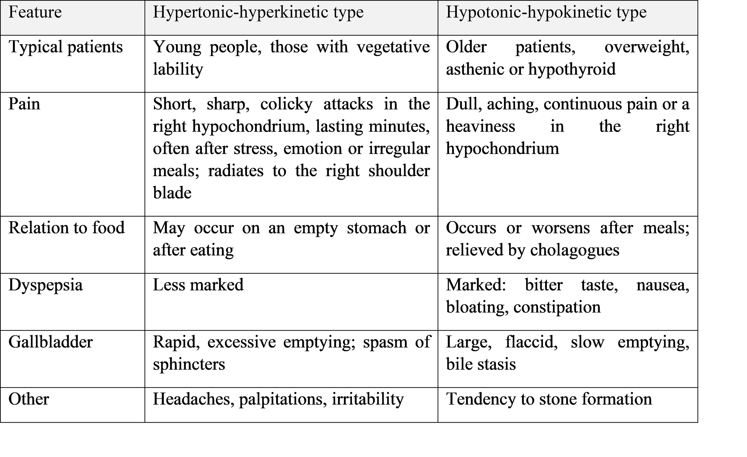
4.3 Types and clinical picture
Rome IV diagnostic criteria for functional gallbladder pain include episodes of pain in the epigastrium and/or right upper quadrant that is steady, builds to a plateau, lasts 30 minutes or longer, recurs at variable intervals, is severe enough to interrupt daily activities or lead to an emergency visit, and is not relieved by bowel movement, posture change or antacids, with absence of structural disease (stones, sludge or other pathology). Supportive features are nausea and vomiting, radiation to the back or right subscapular region and waking from sleep.
4.4 Diagnosis
Diagnosis is by exclusion. It requires normal liver tests, amylase and lipase; a normal ultrasound and upper endoscopy; and impaired gallbladder emptying on cholescintigraphy with CCK stimulation (a gallbladder ejection fraction below about 35–40% is regarded as abnormal). Ultrasound with a test meal (a fatty breakfast) can also assess the contraction volume. Other causes of upper abdominal pain must be excluded first: peptic disease, GERD, pancreatic disease, irritable bowel syndrome and others.
5. General Principles of Treatment
5.1 Chronic cholecystitis
Aims: relieve pain and inflammation, restore bile flow, prevent complications and relapse.
1. Regimen and diet. Frequent small meals (4–6 times a day) at regular times, which promote regular gallbladder emptying. Therapeutic diet No. 5 (Pevzner): limit animal fats, fried, spicy and smoked foods, eggs yolks, alcohol, carbonated and cold drinks; favour boiled, steamed or baked dishes, vegetable oil in moderation, dietary fibre, fruit and low-fat dairy. Weight reduction in obesity (gradual, to avoid triggering stones). Regular physical activity.
2. Antibacterial therapy during exacerbation with signs of infection: drugs that achieve high concentration in bile and are active against enteric flora (e.g. amoxicillin–clavulanate, cephalosporins, fluoroquinolones, with metronidazole when anaerobes are suspected), chosen according to local resistance. Not used routinely in remission.
3. Pain and spasm relief: antispasmodics (drotaverine, mebeverine, otilonium bromide, hyoscine butylbromide); NSAIDs (e.g. diclofenac, ketorolac) for severe pain. Opioids are used cautiously; morphine may increase sphincter of Oddi tone.
4. Choleretics and cholekinetics (only after stones are excluded, and in the absence of obstruction): bile-acid preparations, herbal agents, and prokinetics (domperidone, itopride) for hypomotility.
5. Detoxification during acute exacerbation if needed.
6. Physiotherapy and spa treatment in remission (electrophoresis, UHF, mineral water, peloid therapy), if there are no stones or other contraindications.
7. Surgery: cholecystectomy is indicated in recurrent exacerbations, calculous disease with symptoms, or a non-functioning gallbladder.
5.2 Gallstone disease
Asymptomatic stones: expectant management is the rule; prophylactic cholecystectomy is not recommended, except in specific high-risk situations (porcelain gallbladder, large stones above 3 cm, gallbladder polyps larger than 1 cm, sickle cell disease, some transplant candidates, and certain ethnic groups with high cancer risk).
Symptomatic stones:
• Laparoscopic cholecystectomy is the gold standard, ideally performed early (within about 72 hours of onset in acute cholecystitis, or electively after biliary colic). Open conversion is required in difficult anatomy or complications. Intraoperative cholangiography is used selectively.
• Biliary colic: analgesia (NSAIDs first-line; opioids if needed), antispasmodics, antiemetics, and nil by mouth during the attack, followed by early surgical referral because recurrence is very common.
• Common bile duct stones: endoscopic sphincterotomy and stone extraction (ERCP), followed by cholecystectomy; in cholangitis, urgent biliary drainage and antibiotics.
• Oral litholysis with ursodeoxycholic acid (UDCA), 10 mg/kg per day (often 8–15 mg/kg), in the evening: used only for small (under 10 mm) radiolucent cholesterol stones in patients with a functioning gallbladder and patent cystic duct, who refuse or cannot undergo surgery. Treatment lasts 6–24 months and recurrence after stopping is about 50% within 5 years. UDCA is also used for prevention of stones during rapid weight loss.
• Extracorporeal shock-wave lithotripsy is rarely used today.
• Dietary prevention: regular meals, weight control, adequate fibre, unsaturated fats, coffee in moderate quantity, and physical activity.
5.3 Gallbladder dyskinesia
Treatment depends on the type. Common elements are normalisation of lifestyle, sleep and stress, and treatment of the underlying cause (thyroid disease, giardiasis, gastroduodenal disease).
Hypertonic-hyperkinetic type
• Diet No. 5 with exclusion of strong stimulants of gallbladder contraction (rich broths, egg yolks, concentrated fats, spices, cold and carbonated beverages).
• Antispasmodics (drotaverine, mebeverine, otilonium bromide, hyoscine); nitrates or calcium channel blockers occasionally for sphincter spasm.
• Sedatives and herbal agents (valerian, motherwort), psychotherapy and anxiolytics when indicated.
• Low-mineral water, warm, taken in small sips; thermal physiotherapy.
• Avoid strong cholekinetics.
Hypotonic-hypokinetic type
• Diet No. 5 with increased amounts of vegetable oil, cream, dietary fibre and magnesium-rich foods (to stimulate emptying), and frequent small meals.
• Cholekinetics: magnesium sulphate, sorbitol, xylitol, and high-mineral water at room temperature.
• Choleretics and prokinetics (domperidone, itopride).
• Duodenal tubage ("blind probing") with warm mineral water, magnesium sulphate or sorbitol, once or twice a week in a course.
• Tonic measures: exercise therapy, massage, electrostimulation.
Surgery (cholecystectomy) may be considered for patients with typical biliary pain, a persistently low ejection fraction on CCK-HIDA, and failure of conservative therapy, after careful exclusion of other causes, as results are variable.
6. Prevention and Follow-up
Primary prevention of gallbladder disease centers on modifying underlying metabolic and lifestyle risk factors to maintain normal bile composition and organ motility. Adopting a healthy, balanced diet with regular, scheduled meals helps stimulate consistent gallbladder emptying, preventing the stasis that favors cholesterol crystal nucleation. Sustaining a normal body weight through routine physical activity significantly lowers cholesterol saturation in bile, but weight loss strategies must be managed carefully, as rapid or abrupt weight reduction mobilizes tissue cholesterol and paradoxically increases the risk of stone formation. Additionally, aggressive clinical management of coexisting metabolic disorders, such as dyslipidemia and diabetes mellitus, reduces the metabolic driver behind lithogenic bile production.
Secondary prevention focuses on limiting disease progression, preventing recurrent acute exacerbations, and detecting complications early in patients with established gallbladder pathology. Strict adherence to dietary guidelines—particularly avoiding heavy, fatty, or fried foods—is paired with prophylactic courses of therapy typically scheduled during high-risk spring and autumn seasonal transitions. Routine monitoring via abdominal ultrasonography every six to twelve months allows clinicians to track changes in wall thickness, sludge accumulation, or stone formation over time. Addressing coexisting gastrointestinal conditions, such as irritable bowel syndrome or peptic disease, minimizes shared reflex pathways that trigger biliary symptoms, while structured spa and balneotherapeutic regimens are sometimes integrated into long-term supportive care.
Comprehensive patient education is a critical safeguard to ensure timely intervention before minor flares evolve into life-threatening emergencies. Patients must be explicitly instructed on key red-flag symptoms that warrant immediate medical evaluation rather than home management. These warning signs include high fever accompanied by rigors or chills, visible jaundice of the skin or sclera, continuous epigastric or right upper quadrant pain persisting for more than six hours, persistent vomiting, and dark tea-colored urine. Recognizing these manifestations early enables rapid assessment for acute cholecystitis, choledocholithiasis, or ascending cholangitis, mitigating the severe morbidity associated with delayed care.
7. Conclusion
Chronic cholecystitis, gallstone disease and gallbladder dyskinesia form a continuum of biliary disorders with overlapping clinical features dominated by right upper quadrant pain and dyspepsia. Ultrasound is the cornerstone of diagnosis, and functional tests help to identify dyskinesia. Management combines a regular fractional diet, antispasmodic or prokinetic and choleretic therapy according to the motor type, treatment of infection in exacerbations, and, in symptomatic gallstone disease, laparoscopic cholecystectomy as the definitive treatment. Early recognition and correct treatment prevent serious complications such as cholangitis and pancreatitis.
References
1. European Association for the Study of the Liver (EASL). Clinical Practice Guidelines on the prevention, diagnosis and treatment of gallstones. Journal of Hepatology, 2016; 65(1): 146–181.
2. Cotton PB, Elta GH, Carter CR, Pasricha PJ, Corazziari ES. Gallbladder and Sphincter of Oddi Disorders. Gastroenterology, 2016; 150(6): 1420–1429 (Rome IV).
3. Miura F, et al. Tokyo Guidelines 2018: initial management of acute biliary infection and flowchart for acute cholangitis. Journal of Hepato-Biliary-Pancreatic Sciences, 2018; 25(1): 31–40.
4. Lammert F, Gurusamy K, Ko CW, et al. Gallstones. Nature Reviews Disease Primers, 2016; 2: 16024.
5. Longo DL, Fauci AS, Kasper DL, et al. (eds). Harrison's Principles of Internal Medicine, 21st ed. McGraw-Hill; chapter on disorders of the gallbladder and bile ducts.
6. Kumar P, Clark M. Kumar and Clark's Clinical Medicine, 10th ed. Elsevier; chapter on biliary disease.
7. Ivashkin VT, et al. Clinical recommendations of the Russian Gastroenterological Association on diagnosis and treatment of cholelithiasis (and cholecystitis), latest edition.