
Clinical Evaluation of Gastrointestinal Disorders in Children: Diagnostic Value of History, Physical Examination and Laboratory Tests
1. Altynai Nuralieva
2. Senthilkumar Sharmila
(1. Lecturer, International Medical Faculty, Osh State University, Osh, Kyrgyz Republic
2. Student, International Medical Faculty, Osh State University, Osh, Kyrgyz Republic.)
1.Abstract
Gastrointestinal disorders are a common cause of illness in children and can affect nutrition, growth, development, and quality of life. Their diagnosis can be challenging because symptoms such as abdominal pain, vomiting, diarrhoea, and constipation may occur in several different conditions, while younger children may have difficulty describing their symptoms accurately. This review examines the diagnostic value of clinical history taking, physical examination, and laboratory investigations in the evaluation of gastrointestinal disorders in children. It discusses the role of symptom assessment, feeding and dietary history, growth monitoring, and systematic abdominal examination in establishing an initial diagnosis. The contribution of laboratory investigations, including complete blood count, inflammatory markers, biochemical tests, stool analysis, and disease-specific serology, is also considered. In addition, the review outlines the role of imaging, endoscopy, and histopathological examination when further diagnostic clarification is required. Particular attention is given to the limitations of individual clinical findings and laboratory tests, the influence of age-related differences on symptom presentation, and the importance of recognizing warning signs that require urgent assessment. Overall, an accurate diagnosis depends on integrating clinical findings with appropriately selected investigations rather than relying on a single symptom or test result. A systematic, age-appropriate, and evidence-based approach can improve diagnostic decision-making, reduce unnecessary investigations, and support the timely recognition of serious gastrointestinal conditions in children.
Keywords: Children; gastrointestinal disorders; clinical history; abdominal examination; laboratory investigations; pediatric diagnosis; diagnostic accuracy.
2. Introduction
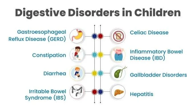
Gastrointestinal (GI) disorders are a common cause of illness in children and may affect their nutrition, growth, development, and overall quality of life. These disorders range from acute conditions, such as gastroenteritis and constipation, to chronic diseases, including celiac disease and inflammatory bowel disease. Although many gastrointestinal symptoms in children are caused by self-limiting conditions, some may indicate serious underlying diseases that require prompt medical attention. Therefore, the early recognition and accurate diagnosis of gastrointestinal disorders are essential components of pediatric healthcare [1,2].
Children with gastrointestinal disorders may present with a variety of symptoms, including abdominal pain, vomiting, diarrhea, constipation, abdominal distension, gastrointestinal bleeding, and poor weight gain. However, identifying the underlying cause can be challenging because these symptoms are often nonspecific and may occur in several different conditions. In addition, young children may have difficulty describing the location, severity, duration, and nature of their symptoms. Consequently, clinicians must carefully interpret information provided by both the child and their caregivers to develop an appropriate initial diagnosis [1,3].
The clinical evaluation of gastrointestinal disorders in children begins with a detailed medical history and a systematic physical examination. History taking provides important information about the onset and progression of symptoms, feeding practices, dietary habits, bowel movements, hydration, medication use, previous illnesses, and family history. A thorough abdominal examination, including inspection, auscultation, percussion, and palpation, helps identify findings such as abdominal distension, tenderness, altered bowel sounds, organ enlargement, and palpable masses. These clinical findings can guide the selection of further investigations and help identify children who require urgent assessment [1,3].
Laboratory investigations play an important supporting role when the clinical presentation suggests a specific disease or when the severity of illness needs to be assessed. Depending on the suspected diagnosis, investigations may include a complete blood count, inflammatory markers, serum electrolytes, liver function tests, celiac disease serology, and selected stool tests. However, laboratory testing should not be performed routinely in every child with gastrointestinal symptoms. For example, uncomplicated acute gastroenteritis can often be diagnosed clinically, and laboratory investigations may be unnecessary unless specific clinical indications are present. Selecting investigations according to the child’s symptoms and clinical condition can improve diagnostic efficiency and reduce unnecessary testing [2,4].
The interpretation of clinical findings also requires consideration of the child’s age and developmental stage. Infants, young children, school-aged children, and adolescents differ in their ability to communicate symptoms, their nutritional requirements, and their vulnerability to dehydration and growth disturbances. Furthermore, gastrointestinal symptoms may overlap with conditions affecting other body systems, making a careful and comprehensive assessment important. Serious conditions, such as acute appendicitis or intestinal obstruction, must be recognized promptly because delayed diagnosis can lead to complications [1,3].
Although clinical history, physical examination, and laboratory investigations are widely used in pediatric practice, their diagnostic value depends on the clinical context and the suspected condition. No single symptom, examination finding, or laboratory result is sufficient to establish every gastrointestinal diagnosis. Instead, clinicians must integrate information from these different sources and select additional investigations when necessary. This approach supports more accurate diagnosis, appropriate use of healthcare resources, and timely identification of children who need specialist assessment or emergency treatment [1–4].
Therefore, this review aims to critically examine the diagnostic value of clinical history taking, abdominal physical examination, and laboratory investigations in children presenting with gastrointestinal symptoms. It will discuss how these methods contribute to the assessment of common pediatric gastrointestinal disorders, evaluate their strengths and limitations, and highlight clinical warning signs that require urgent evaluation. By bringing together relevant research and clinical guidelines, this review seeks to support a systematic, evidence-based approach to the early diagnosis and management of gastrointestinal disorders in children.
3. Anatomical and Physiological Features of the Pediatric Gastrointestinal System
3.1. Development and Age-Related Characteristics of the Digestive System
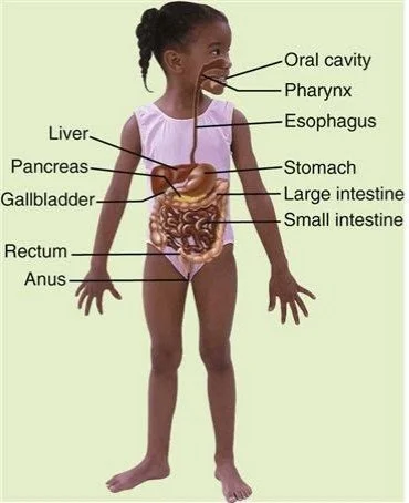
The gastrointestinal (GI) system plays an essential role in the growth, development, and maintenance of health in children. It is responsible for the digestion of food, absorption of nutrients and water, and elimination of waste products. Although the basic structure of the gastrointestinal tract is similar in children and adults, its anatomical features and physiological functions change considerably during childhood. These changes are particularly important during infancy and early childhood, when the body has high nutritional requirements and the digestive system continues to mature [5,6].
The gastrointestinal tract includes the mouth, esophagus, stomach, small intestine, large intestine, and rectum. The liver, pancreas, and gallbladder support digestion by producing or storing substances required for the breakdown and absorption of nutrients. The small intestine is the principal site of nutrient digestion and absorption, while the large intestine is mainly responsible for absorbing remaining water and electrolytes and forming stool [5].
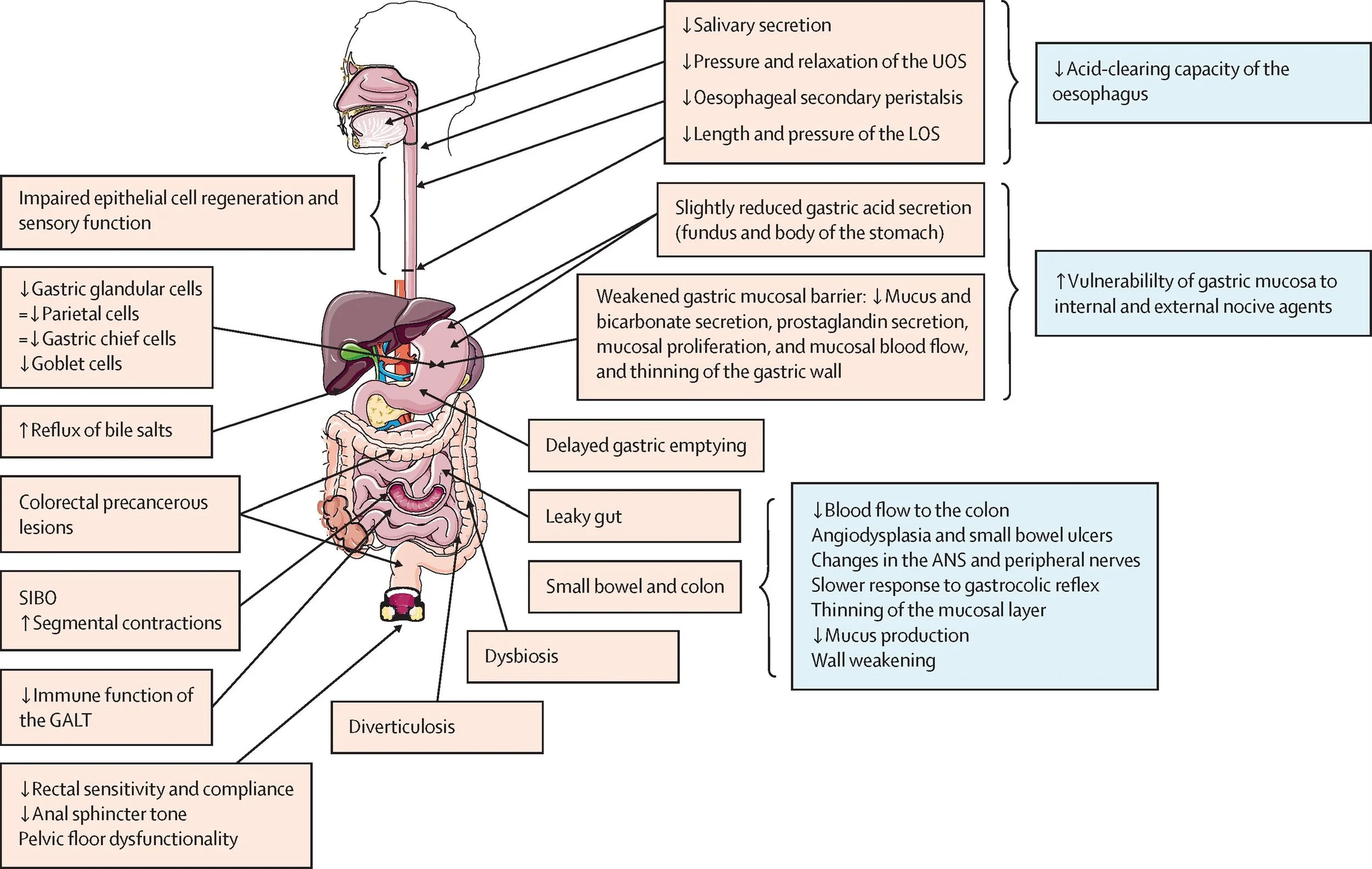
During fetal development, the gastrointestinal tract develops from the primitive gut tube. Its major structures form before birth, but structural development does not mean that every digestive function is fully mature at birth. After birth, the digestive system continues to adapt to feeding, intestinal microorganisms, and the changing nutritional requirements of the growing child. The development of intestinal motility, digestive enzyme activity, mucosal barrier function, and nutrient transport occurs at different rates [6,7].
These developmental differences are clinically significant. For example, premature infants may experience feeding intolerance because of immature gastrointestinal motility and coordination. Infants may also have greater difficulty maintaining fluid balance during episodes of diarrhea or vomiting because they have relatively high fluid requirements and limited physiological reserves. Therefore, the child’s age, gestational history, feeding pattern, and growth should be considered when evaluating gastrointestinal symptoms [6,7].
3.2. Digestive Function and Intestinal Absorption
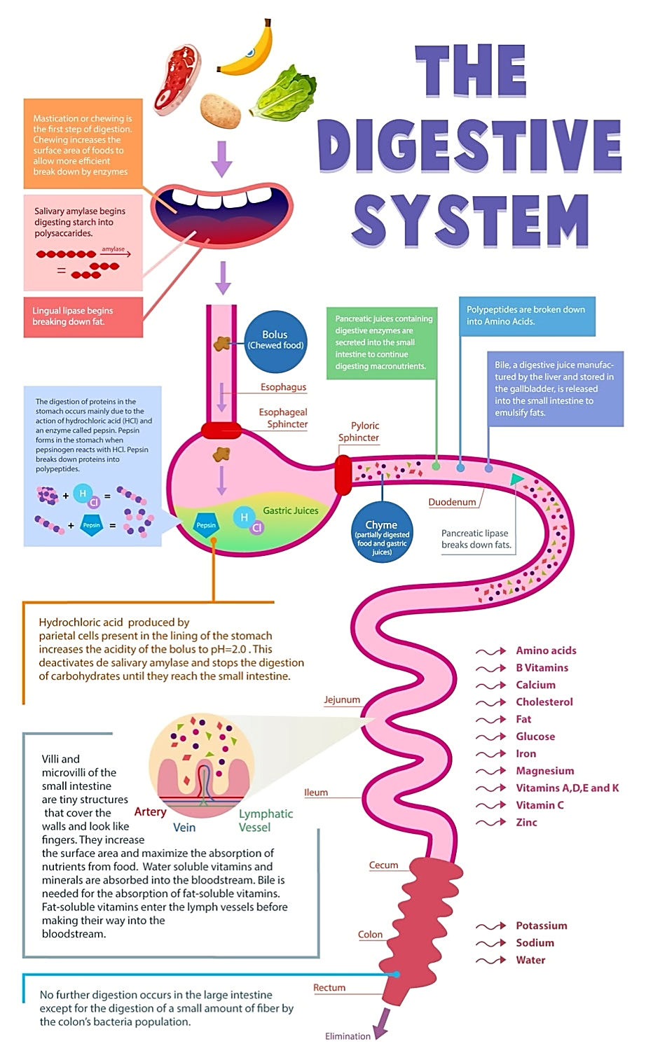
Digestion involves the mechanical and chemical breakdown of food into smaller components that can be absorbed and used by the body. Mechanical digestion begins in the mouth, where food is broken down by chewing. Chemical digestion involves enzymes and digestive secretions that help break down carbohydrates, proteins, and fats. The stomach contributes to protein digestion, while the pancreas and small intestine provide enzymes that support the digestion of different nutrients [5].
The small intestine has a specialized inner surface containing folds, villi, and microscopic microvilli. These structures increase the surface area available for absorption. Carbohydrates are ultimately absorbed mainly as simple sugars, proteins as amino acids and small peptides, and fats largely as fatty acids and monoglycerides. Most absorbed nutrients enter the bloodstream, while many long-chain dietary fats are transported initially through the lymphatic system. Water, vitamins, minerals, and electrolytes are also absorbed through processes that vary according to the substance involved [5,8].
The large intestine has an important role in fluid and electrolyte balance. It absorbs much of the water and sodium that remain after small-intestinal digestion and absorption. Intestinal bacteria also ferment some undigested carbohydrates and produce short-chain fatty acids, which can contribute to colonic health. Disturbances in these processes may lead to diarrhea, constipation, abdominal discomfort, or nutritional problems [5,8].
Digestive and absorptive functions develop throughout infancy and childhood. Breast milk and infant formula provide nutrients in forms suitable for early life, while the introduction of complementary foods gradually expands the range of nutrients consumed. Some digestive processes, including the handling of certain fats and carbohydrates, differ with developmental stage. For this reason, feeding history and the relationship between symptoms and particular foods may provide useful clues when investigating suspected food intolerance or malabsorption [6,7].
For example, persistent diarrhea, abdominal distension, poor weight gain, and nutritional deficiencies may suggest impaired intestinal absorption. In such cases, the clinician may consider conditions such as celiac disease or other intestinal disorders, guided by the child’s history, examination, and appropriate investigations. However, these symptoms are not specific to a single disease, and diagnosis requires a broader clinical assessment [5,8].
3.3. Differences Between Infants, Young Children, and Adolescents
Although there is considerable variation between individuals, several age-related differences are important in pediatric gastrointestinal assessment.
3.3.1. Infants
Infancy is a period of rapid growth and ongoing functional maturation of the gastrointestinal system. Feeding depends on coordinated sucking, swallowing, and intestinal activity. Gastrointestinal motility and some digestive processes differ from those in older children, and premature infants may have more pronounced functional immaturity [6,7].
Regurgitation is common during infancy and may occur because of developmental features of the gastroesophageal junction and feeding patterns. However, persistent or forceful vomiting, bilious vomiting, blood in vomit or stool, marked abdominal distension, and poor weight gain require careful assessment rather than being assumed to be normal infant behavior.
Infants cannot describe abdominal pain or other symptoms clearly. Their illness may instead appear as irritability, persistent crying, refusal to feed, lethargy, changes in stool, or reduced urine output. A careful feeding history, assessment of hydration, and review of weight gain are therefore particularly important. Vomiting and diarrhea may lead to dehydration relatively quickly in infants, making early recognition essential [6,9].
3.3.2. Young Children
In young children, the gastrointestinal system continues to develop while dietary patterns, mobility, and toilet habits change. The transition to solid foods and the development of toilet training may influence bowel habits. Functional constipation is common in this age group and may be associated with painful defecation, stool withholding, and changes in dietary or toileting routines.
Young children may describe pain imprecisely or have difficulty identifying its location. They may show abdominal discomfort through changes in behavior, reduced appetite, crying, or avoidance of usual activities. As a result, clinicians should obtain information from caregivers while also allowing the child to communicate in an age-appropriate way.
Growth monitoring remains important. Persistent abdominal symptoms accompanied by weight loss, poor weight gain, reduced appetite, or delayed growth may indicate an underlying condition rather than an isolated functional problem. The clinical assessment should therefore include dietary history, stool patterns, hydration, and growth measurements [5,9].
3.3.3. School-Aged Children and Adolescents
As children grow older, they generally become better able to describe the location, severity, duration, and character of their symptoms. This can make history taking more precise, although symptom descriptions still need to be interpreted alongside examination findings.
During adolescence, growth spurts, changing dietary habits, stress, and social factors may influence gastrointestinal symptoms. Functional abdominal pain and constipation remain relevant, while inflammatory bowel disease and celiac disease are important considerations in children with persistent symptoms or signs of impaired growth or nutrition. Adolescents may also be reluctant to discuss bowel habits, eating patterns, or other sensitive concerns, so a respectful and confidential approach, appropriate to their age and clinical setting, can improve the quality of the history [5,9].
The ability to communicate more clearly does not eliminate the risk of serious disease. Persistent localized pain, gastrointestinal bleeding, unexplained weight loss, fever, or growth faltering should be investigated according to the clinical presentation, regardless of age.
3.4. Clinical Importance of Age-Related Differences in Diagnosing Disease
Understanding the anatomical and physiological characteristics of the pediatric gastrointestinal system helps clinicians distinguish common developmental patterns from findings that may indicate disease. The same symptom can have different implications depending on the child’s age, feeding history, growth, and overall condition. Consequently, pediatric gastrointestinal assessment should be adapted to the individual child rather than based solely on symptoms commonly observed in adults [5,6,9].
First, age influences the interpretation of symptoms. Regurgitation may be common in an otherwise healthy, thriving infant, whereas persistent vomiting associated with poor growth requires further evaluation. Similarly, constipation in a young child may be related to stool withholding, but constipation accompanied by significant abdominal distension, vomiting, or other concerning findings requires assessment for an underlying cause.
Second, age affects the risk and consequences of dehydration. Infants and young children may deteriorate more rapidly during significant fluid loss. Assessment should therefore include general appearance, fluid intake, urine output, mucous membranes, circulation, and other clinical signs of hydration. Laboratory tests may be needed when dehydration is severe, the diagnosis is uncertain, or electrolyte disturbances are suspected [6,9].
Third, growth and nutritional status provide important diagnostic information. Weight, length or height, and growth trajectories can help identify children with prolonged disease or malabsorption. A single measurement may be less informative than changes over time. Poor growth associated with chronic diarrhea, abdominal pain, or nutritional deficiencies should prompt consideration of conditions such as celiac disease and inflammatory bowel disease, among other possible causes [5,9].
Fourth, developmental anatomy and communication affect physical examination. Infants and young children may have a more prominent abdomen and may be unable to cooperate fully with examination. Clinicians should adapt their approach to the child’s developmental stage, observe behavior and general appearance, and assess abdominal tenderness, distension, bowel sounds, and organ enlargement in context. When necessary, caregivers can help reassure the child and provide information about changes from normal behavior.
Finally, age helps guide the selection of investigations. Not every child with abdominal pain, vomiting, or diarrhea requires extensive testing. The decision to request blood tests, stool studies, ultrasound, or other investigations should be based on the history, physical examination, severity and duration of symptoms, and suspected diagnosis. An age-appropriate, evidence-based approach can reduce unnecessary investigations while helping identify serious conditions without delay [5,9].
In summary, the pediatric gastrointestinal system undergoes important structural and functional changes from birth through adolescence. These changes influence digestion, absorption, fluid balance, symptom presentation, and the interpretation of clinical findings. A clear understanding of these age-related characteristics provides the foundation for systematic history taking, accurate abdominal examination, and appropriate selection of diagnostic investigations in children.
4. Clinical History Taking in Children
Clinical history taking is one of the most important steps in evaluating gastrointestinal disorders in children. It helps clinicians understand the nature of the presenting symptoms, identify possible causes, assess the severity of illness, and determine whether further investigations are required. In many cases, a carefully obtained history provides valuable diagnostic clues before laboratory tests or imaging studies are performed. However, because gastrointestinal symptoms are often nonspecific, the history should always be interpreted alongside the physical examination and the child’s overall clinical condition [10,11].
Pediatric history taking differs from adult assessment because children may not be able to describe their symptoms clearly, particularly during infancy and early childhood. Parents and caregivers therefore provide essential information about feeding, bowel movements, behavior, fluid intake, and changes in normal activity. As children become older, their own descriptions of symptoms become increasingly important. A systematic and age-appropriate approach helps clinicians obtain reliable information while reducing the risk of overlooking significant disease [10,12].
4.1. Presenting Complaints and History of the Current Illness
The initial assessment should identify the main complaint and establish when the symptoms began, how they have progressed, and how they affect the child’s daily activities. Common presenting complaints include abdominal pain, vomiting, diarrhea, constipation, abdominal distension, poor appetite, and gastrointestinal bleeding. Clinicians should determine whether the symptoms are acute, recurrent, or persistent and whether they occur independently or together.
The history of the current illness should explore the onset, duration, frequency, severity, progression, and possible triggers of each symptom. Associated features such as fever, lethargy, reduced appetite, urinary symptoms, skin changes, and weight loss may help distinguish gastrointestinal disease from other medical conditions. Recent travel, exposure to individuals with similar symptoms, changes in diet, and possible ingestion of contaminated food or foreign objects may also be relevant.
The sequence in which symptoms develop is particularly important. For example, vomiting followed by diarrhea may be consistent with acute gastroenteritis, whereas vomiting without diarrhea should prompt consideration of other causes according to the child’s age and clinical presentation. Similarly, abdominal pain that becomes progressively more severe or localized may indicate a condition requiring urgent assessment [10,11].
A clear history should also document any treatment already given, including oral rehydration solutions, prescribed medicines, over-the-counter products, and home remedies. The clinician should establish whether these measures improved the symptoms and whether the child has previously sought medical care for the same problem.
4.2. Assessment of Abdominal Pain
Abdominal pain is a frequent reason for pediatric consultation, but its cause can range from a temporary functional disorder to an acute surgical condition. A detailed pain history helps narrow the differential diagnosis and identify children who require further investigation or urgent referral [10,11].
The assessment should include the following characteristics:
● Location: Determine where the pain began and where it is felt now. Pain may be generalized or localized to a particular region. Asking the child to point to the painful area may be helpful when developmentally appropriate.
● Onset and duration: Establish whether the pain started suddenly or gradually, how long it has lasted, and whether similar episodes have occurred previously.
● Character: Ask whether the pain is cramping, sharp, dull, burning, or intermittent. Younger children may describe pain through behavior rather than words.
● Severity: Assess the effect of pain on sleep, eating, walking, playing, and normal activity. A numerical pain scale or age-appropriate faces scale may help older children communicate its intensity.
● Progression and timing: Determine whether the pain is improving, worsening, continuous, or episodic and whether it occurs before or after meals, during the night, or in relation to defecation.
● Radiation and associated symptoms: Ask whether the pain spreads to another area and whether it occurs with vomiting, fever, diarrhea, constipation, urinary symptoms, or gastrointestinal bleeding.
● Aggravating and relieving factors: Explore whether eating, movement, passing stool, urination, or particular foods influence the symptoms.
These characteristics help guide clinical reasoning, but no single feature establishes a diagnosis. For example, pain associated with defecation may occur in constipation or disorders of gut–brain interaction, while progressive localized pain with guarding may raise concern for an acute inflammatory or surgical condition. The interpretation must take account of the child’s age, examination findings, and clinical course [10,11].
4.3. Vomiting, Diarrhea, Constipation, and Abdominal Distension
4.3.1. Vomiting
The history of vomiting should establish its onset, frequency, duration, approximate volume, appearance, and relationship to feeding. Clinicians should ask whether the vomit contains blood or bile and whether it is forceful, persistent, or associated with abdominal pain or distension.
Bilious vomiting, which is typically green, is an important warning sign because it may indicate intestinal obstruction. Blood in vomit, persistent forceful vomiting, or vomiting accompanied by marked abdominal distension also requires prompt clinical assessment. In infants, it is important to distinguish ordinary regurgitation from true vomiting and to determine whether feeding, hydration, and growth are affected [10,13].
4.3.2. Diarrhea
The assessment of diarrhea should include the number of stools per day, stool consistency, duration, and any change from the child’s normal bowel pattern. The presence of blood, mucus, or unusually watery stools should be documented. Associated fever, vomiting, abdominal pain, recent travel, contaminated food or water exposure, and contact with people who have similar symptoms may help identify a possible infectious cause.
Fluid intake and urine output must be assessed because diarrhea can cause dehydration, particularly in infants and young children. Reduced urination, inability to drink adequately, unusual sleepiness, and worsening general condition are important concerns. Persistent or recurrent diarrhea accompanied by poor growth, weight loss, or nutritional deficiencies may require investigation for malabsorption, celiac disease, inflammatory bowel disease, or other underlying conditions [12,13].
4.3.3. Constipation
A history of constipation should explore stool frequency, stool consistency, painful defecation, excessive straining, and the presence of very large stools or stool withholding. Clinicians should ask whether the child avoids using the toilet, experiences pain during bowel movements, or has episodes of fecal soiling. Dietary habits, fluid intake, physical activity, toilet routines, and previous treatment should also be reviewed.
Constipation in children is often functional, meaning that it occurs without an identifiable structural or biochemical disease. However, the clinician should look for warning features such as onset during the neonatal period, delayed passage of the first stool after birth, persistent vomiting, significant abdominal distension, blood in the stool without an obvious anal cause, or poor growth. These findings may justify further assessment for an underlying disorder [10,11].
4.3.4. Abdominal Distension
Abdominal distension may result from gas, constipation, changes in intestinal motility, fluid accumulation, or intestinal obstruction. The history should establish when the distension began, whether it is persistent or intermittent, and whether it is associated with pain, vomiting, changes in stool passage, feeding difficulties, or weight changes.
In infants, persistent distension associated with feeding intolerance, vomiting, or delayed passage of stool requires careful assessment. In older children, progressive distension with severe pain, bilious vomiting, or inability to pass stool or gas may indicate obstruction and requires urgent evaluation [10,13].
4.4. Feeding History, Dietary Habits, and Hydration
Feeding history is particularly important in infants and young children because nutrition is closely linked to growth and gastrointestinal health. Clinicians should ask whether the child is breastfed, formula-fed, or receiving complementary foods, as appropriate for age. The assessment should include feeding frequency, approximate quantity, preparation practices for formula, feeding difficulties, and any recent changes in diet.
For older children and adolescents, dietary history should explore meal patterns, fluid intake, food variety, fiber intake, and the relationship between particular foods and symptoms. Restrictive eating, avoidance of certain food groups, and the use of special diets may also be relevant when assessing nutritional deficiencies or suspected food-related disorders.
When symptoms appear to follow the consumption of particular foods, clinicians should investigate the timing and consistency of the association rather than assuming that the food is responsible. For example, symptoms after dairy consumption may warrant assessment for lactose intolerance in the appropriate clinical context, while persistent gastrointestinal symptoms associated with poor growth may raise suspicion of celiac disease. A careful dietary history helps determine whether targeted testing is appropriate [11,12].
Hydration assessment is essential in children with vomiting, diarrhea, or reduced oral intake. Clinicians should ask how much fluid the child has consumed, whether fluids can be retained, and whether urine output has decreased. In infants, the number of wet diapers can provide useful information. These details help estimate the risk of dehydration and determine the urgency of physical assessment and treatment [12,13].
4.5. Growth History and Weight Changes
Growth assessment is an essential component of pediatric gastrointestinal evaluation. Unlike adults, children require sufficient energy and nutrients not only to maintain normal body functions but also to support physical growth and development. Persistent gastrointestinal disease may interfere with nutrient intake, digestion, absorption, or utilization, leading to poor weight gain, weight loss, or reduced linear growth.
The history should include birth weight where available, previous weight and height measurements, appetite, feeding difficulties, and any concerns about growth raised by caregivers or healthcare professionals. Growth measurements should be compared with appropriate age- and sex-specific growth charts. Changes over time are generally more informative than a single measurement.
Poor weight gain or weight loss associated with chronic diarrhea, recurrent abdominal pain, persistent vomiting, or fatigue may indicate an underlying organic disorder. Conditions such as celiac disease and inflammatory bowel disease should be considered when supported by the overall clinical presentation. Nevertheless, growth changes can have multiple causes, so they should not be attributed to gastrointestinal disease without further assessment [11,14].
A normal growth pattern can be reassuring, but it does not completely exclude gastrointestinal pathology. Growth history should therefore be interpreted together with symptom duration, physical findings, dietary intake, and other relevant clinical information.
4.6. Birth History, Previous Illnesses, Medication Use, and Family History
A complete pediatric history should include relevant information from pregnancy, birth, the neonatal period, and previous medical care. Prematurity, neonatal complications, feeding difficulties, and delayed passage of the first stool may be important in selected children, particularly when gastrointestinal symptoms began early in life.
Previous illnesses, hospital admissions, abdominal surgery, and recurrent episodes of similar symptoms should also be documented. A history of chronic medical conditions may influence the likely causes of gastrointestinal symptoms and the selection of investigations. Recent infections and contact with ill family members may support an infectious explanation, although alternative diagnoses should still be considered when symptoms are atypical or severe [10,12].
Medication history is equally important. Clinicians should document prescribed medicines, non-prescription drugs, supplements, recent antibiotic use, and known allergies. Some medications can cause nausea, abdominal discomfort, diarrhea, or constipation, while antibiotics may alter the intestinal microbiota and increase the risk of antibiotic-associated diarrhea. The timing of symptoms in relation to medication use may help identify a possible adverse effect.
Family history should explore gastrointestinal disorders such as celiac disease, inflammatory bowel disease, recurrent gastrointestinal problems, and relevant liver disorders. A family history of these conditions may increase clinical suspicion in a child with compatible symptoms, but it does not establish the diagnosis.
For adolescents, additional aspects of the medical and psychosocial history may be relevant, including menstrual symptoms, eating patterns, stress, school attendance, and other factors affecting gastrointestinal health. Sensitive questions should be asked respectfully and in a manner appropriate to the adolescent’s age and circumstances [10,11].
4.7. Age-Appropriate Communication with Children and Caregivers
Effective communication is essential for obtaining a reliable history in pediatric practice. The approach should be adapted to the child’s developmental stage, language ability, emotional state, and level of understanding. In infants and very young children, most historical information is obtained from parents or caregivers, who can describe changes in feeding, stool patterns, sleep, crying, and behavior. However, caregiver observations should be interpreted alongside the child’s examination and clinical findings [10,12].
School-aged children can often describe their symptoms directly when questions are simple, clear, and non-leading. Open-ended questions, such as “Can you tell me what happens when your stomach hurts?”, allow the child to explain the experience in their own words. More focused questions can then clarify the location, duration, severity, and associated symptoms.
Adolescents should be treated as active participants in their healthcare. Where appropriate, part of the consultation should take place privately, with confidentiality explained according to local laws and safeguarding requirements. This may help adolescents discuss sensitive matters such as eating behaviors, medication or substance use, menstrual history, and psychosocial stressors. Caregiver information remains valuable, but the adolescent’s own account should also be considered.
Communication should remain supportive and non-judgmental. Clinicians should avoid suggesting an answer, dismissing symptoms because the child appears well, or assuming that recurrent abdominal pain is necessarily psychological. When the history is incomplete or the diagnosis remains uncertain, reassessment and additional information may be needed.
4.8. Clinical Value of a Systematic Pediatric History
A systematic history provides the foundation for selecting appropriate physical examinations and investigations. It helps distinguish acute from chronic symptoms, identify possible infectious or dietary triggers, assess hydration and nutritional status, and recognize features that may indicate serious disease. It also helps clinicians avoid unnecessary testing when the history and examination support a straightforward diagnosis.
However, history taking has limitations. Young children may be unable to describe symptoms accurately, caregivers may not observe every episode, and early manifestations of different diseases may overlap. Therefore, historical information should not be used in isolation. The findings must be integrated with the physical examination, growth assessment, and targeted investigations when indicated [10,11].
Particular attention should be paid to bilious vomiting, gastrointestinal bleeding, severe or worsening abdominal pain, significant distension, persistent vomiting, dehydration, unexplained weight loss, and growth faltering. These features do not all indicate the same diagnosis, but they may signal a need for urgent assessment or further investigation.
In conclusion, careful, age-appropriate history taking is a central component of the clinical evaluation of gastrointestinal disorders in children. A structured assessment of presenting complaints, abdominal pain, bowel symptoms, feeding, hydration, growth, medical background, medication use, and family history helps establish a clinically useful differential diagnosis. When combined with physical examination and appropriately selected investigations, it supports earlier recognition of serious disease and more effective, individualized pediatric care.
5. Physical Examination of the Abdomen
Physical examination of the abdomen is an essential component of the clinical evaluation of gastrointestinal disorders in children. It helps clinicians identify abnormalities, assess the severity of illness, and determine whether further investigations are necessary. When combined with a detailed clinical history, abdominal examination can provide important clues to conditions such as gastroenteritis, constipation, appendicitis, intestinal obstruction, and hepatosplenomegaly. However, physical findings should always be interpreted in the context of the child’s age, symptoms, and general clinical condition [15,16].
5.1. General Assessment
The abdominal examination should begin with an overall assessment of the child’s appearance, behavior, hydration, nutritional status, and vital signs. Before examining the abdomen, the clinician should observe whether the child appears comfortable, restless, unusually sleepy, pale, or acutely unwell. A child who avoids movement, cries continuously, or appears significantly distressed may be experiencing severe pain. Infants and young children may not be able to explain their symptoms verbally, so changes in behavior and activity can provide valuable diagnostic information [15,16].
Vital signs, including temperature, heart rate, respiratory rate, and blood pressure when appropriate, help assess the severity of illness. Fever may accompany an infectious or inflammatory condition, while an increased heart rate may occur with pain, fever, dehydration, or circulatory compromise. The clinician should also assess hydration by examining the mucous membranes, eyes, peripheral circulation, capillary refill, and urine output history. Reduced urine output, prolonged capillary refill, and an altered level of consciousness may indicate significant dehydration or circulatory impairment. Nutritional assessment should include the child’s weight and growth history, because poor weight gain or weight loss may suggest a chronic gastrointestinal disorder or impaired nutrient absorption [15,17].
5.2. Inspection of the Abdomen
Inspection is the first specific step of the abdominal examination. The child should be positioned comfortably, usually lying on the back, with sufficient exposure of the abdomen while maintaining privacy and warmth. In younger children, examination may be easier while they sit on a caregiver’s lap. The clinician should observe the overall shape and symmetry of the abdomen and look for distension, scars, bruising, skin discoloration, prominent veins, visible masses, and abnormal movements [15,16].
Abdominal distension may occur because of intestinal gas, constipation, obstruction, organ enlargement, or fluid accumulation within the abdominal cavity. The child’s age must be considered because a mildly prominent abdomen can be normal in some toddlers. Visible intestinal movements may occasionally be seen in thin children, but prominent or unusual peristaltic waves associated with vomiting and distension may suggest an intestinal motility problem or obstruction. Scars may indicate previous abdominal surgery, while bruising or skin discoloration may suggest trauma or another underlying condition. The umbilicus and groin regions should also be examined for abnormalities such as umbilical or inguinal hernias [15,16].
Inspection alone cannot establish the diagnosis, but it helps direct the remaining examination. For example, marked distension combined with persistent vomiting and inability to pass stool or gas raises concern for intestinal obstruction and requires urgent assessment. The findings should therefore be interpreted together with the child’s symptoms, vital signs, and general appearance.
5.3. Auscultation and the Clinical Significance of Bowel Sounds
Auscultation involves listening to bowel sounds using a stethoscope. It is generally performed after inspection and before percussion and palpation because touching the abdomen may alter intestinal activity. The clinician listens across the abdominal regions and assesses the presence, frequency, and character of the sounds. Normal bowel sounds result from the movement of gas and fluid through the intestines during intestinal contractions [15,18].
Increased bowel sounds may occur with diarrhea or increased intestinal activity, while reduced bowel sounds may be associated with decreased motility, ileus, or some cases of intestinal obstruction. High-pitched sounds can occasionally occur in mechanical obstruction. However, bowel sounds vary naturally, and neither increased nor decreased sounds are sufficiently reliable to establish a diagnosis independently. Their significance depends on the associated findings, particularly abdominal pain, vomiting, distension, and tenderness [15,18].
Very quiet or apparently absent bowel sounds should be interpreted cautiously because listening technique and examination duration can influence the findings. When abnormal bowel sounds occur alongside a rigid abdomen, severe pain, bilious vomiting, or signs of systemic illness, further urgent evaluation may be necessary. Auscultation therefore contributes to the overall clinical assessment but should not be used alone to exclude or confirm serious abdominal disease.
5.4. Percussion of the Abdomen
Percussion involves gently tapping the abdominal wall and interpreting the sounds produced by the underlying structures. It helps clinicians assess whether an area mainly contains gas, fluid, or relatively solid tissue. A tympanic sound is commonly heard over gas-filled intestinal loops, whereas dullness may occur over solid organs, such as the liver, or over areas containing fluid or a mass [18,19].
Percussion can help estimate liver size and identify changes in the expected pattern of abdominal resonance. A larger area of dullness may suggest organ enlargement, a mass, or fluid accumulation, although further examination and, when indicated, imaging are needed to determine the cause. In selected cases, specialized percussion techniques, such as assessing shifting dullness, may help evaluate suspected ascites. These techniques can be difficult to interpret in children and may be less useful when the amount of fluid is small [18,19].
Percussion may also reveal discomfort when the abdominal wall or underlying tissues are inflamed. Tenderness elicited by percussion can contribute to the assessment of peritoneal irritation, particularly when it occurs with guarding or severe localized pain. Nevertheless, percussion findings are not diagnostic on their own and should be interpreted alongside inspection, auscultation, palpation, and the child’s clinical history.
5.5. Palpation of the Abdomen
Palpation is an important part of abdominal examination because it helps identify tenderness, muscle guarding, rigidity, masses, and enlarged abdominal organs. The examination should be gentle and adapted to the child’s age, anxiety, and level of pain. It is often helpful to begin with the least painful region and examine the area of greatest discomfort last. A caregiver’s reassurance and distraction techniques may improve cooperation in infants and young children [15,16].
Light palpation is used initially to assess superficial tenderness, muscle tension, and obvious abnormalities. Deeper palpation may then be performed, when appropriate, to assess deeper masses and abdominal organs. Localized tenderness may indicate inflammation in the affected region, while generalized tenderness may occur in several conditions, including gastroenteritis and more widespread abdominal inflammation. Involuntary guarding, rigidity, or marked pain with gentle movement may raise concern for peritoneal irritation and requires careful clinical assessment [15,16].
The clinician should also assess for palpable masses and fecal loading when indicated. A mass may arise from an enlarged organ, an accumulation of stool, a distended bladder, or another abdominal condition. Its location, size, consistency, mobility, and associated tenderness should be documented. Palpation findings can help guide further investigations, but a mass generally requires additional assessment to establish its nature.
Rebound tenderness, which refers to increased pain when pressure is released, may indicate peritoneal irritation. However, it can be uncomfortable and should not be elicited repeatedly or unnecessarily in children. Other clinical findings, including the child’s willingness to move, localized tenderness, and guarding, may also provide useful information. The examination should be stopped or modified if the child experiences significant distress, and urgent medical assessment should not be delayed when serious disease is suspected [15,16].
5.6. Assessment of the Liver, Spleen, and Other Abdominal Findings
Assessment of the liver and spleen is an important part of the pediatric abdominal examination. The liver is examined mainly in the right upper abdomen, while the spleen is assessed in the left upper abdomen. Palpation and percussion may help estimate organ size and identify enlargement. However, normal findings vary with age and body habitus, and a liver edge may be palpable below the right costal margin in some healthy children. Therefore, palpability alone does not necessarily indicate disease [19,20].
An enlarged liver, known as hepatomegaly, may be associated with infections, liver inflammation, metabolic disorders, congestion, or other systemic conditions. Depending on the clinical presentation, associated findings may include jaundice, abdominal discomfort, poor growth, or changes in urine and stool color. Splenomegaly, or enlargement of the spleen, may occur in certain infections, hematological disorders, and systemic diseases. When either organ appears enlarged, the clinician should consider the child’s history, general examination findings, and the need for laboratory investigations or abdominal ultrasonography [19,20].
The examination should also include assessment for other relevant abnormalities, such as an abdominal mass, distended bladder, hernia, or signs of fluid accumulation. Depending on the child’s symptoms, examination beyond the abdomen may be necessary to identify alternative or associated causes of abdominal pain. For example, urinary tract disorders, respiratory infections, and genital conditions can sometimes present with abdominal symptoms. A complete clinical assessment therefore extends beyond the abdominal examination when the history or findings suggest another source of illness [15,16].
5.7. Recognition of Signs Requiring Urgent Medical Evaluation
Recognizing warning signs during abdominal examination is essential because some pediatric gastrointestinal disorders require immediate treatment. Severe or worsening abdominal pain, involuntary guarding, abdominal rigidity, marked distension, and significant tenderness may indicate a serious inflammatory or surgical condition. These findings become particularly concerning when accompanied by persistent vomiting, fever, abnormal vital signs, or a deteriorating general condition [15,16].
Bilious vomiting, which is typically green, is an important warning sign because it may indicate intestinal obstruction. A markedly distended abdomen accompanied by vomiting and an inability to pass stool or gas also raises concern for obstruction. Gastrointestinal bleeding, severe dehydration, signs of shock, and altered consciousness require urgent assessment. In infants, persistent vomiting, poor feeding, unusual lethargy, and abdominal distension may be important signs of serious illness even when the child cannot communicate pain clearly [15,17].
Suspected appendicitis is another important consideration, particularly when pain becomes localized, movement worsens discomfort, or examination reveals localized tenderness and guarding. However, appendicitis may present atypically in younger children, and no single examination finding is sufficient to rule it in or out. Repeated clinical assessment, appropriate investigations, and timely surgical consultation may be required when suspicion remains [15,16].
When these warning signs are present, clinicians should prioritize stabilization and urgent medical assessment rather than delay care to complete every component of a routine examination. The urgency of further investigations depends on the child’s overall condition and the suspected diagnosis.
5.8. Clinical Value and Limitations of Abdominal Examination
A systematic abdominal examination provides valuable information about the location and severity of gastrointestinal disease and helps determine which investigations are most appropriate. Inspection may reveal distension or visible abnormalities, auscultation provides information about bowel activity, percussion helps assess abdominal resonance and organ size, and palpation identifies tenderness, guarding, masses, and organ enlargement. Together, these findings help clinicians develop and refine a differential diagnosis [15,18].
Nevertheless, the accuracy of abdominal examination depends on the child’s age, cooperation, the examiner’s experience, and the stage of the illness. Young children may be frightened or unable to describe pain accurately, while early disease may produce subtle or nonspecific findings. Normal bowel sounds or the absence of marked tenderness do not exclude every serious condition. For this reason, physical examination must be interpreted alongside the clinical history and, when indicated, laboratory investigations and imaging [15,16].
In conclusion, abdominal examination is a fundamental part of the assessment of gastrointestinal disorders in children. A structured evaluation of general appearance, vital signs, hydration, abdominal inspection, auscultation, percussion, palpation, and organ size can identify important diagnostic clues and warning signs. When performed carefully and interpreted in the context of the child’s age and clinical presentation, it supports appropriate investigation selection, timely recognition of serious disease, and effective pediatric care.
6. Laboratory Investigations in Pediatric Gastrointestinal Disorders
Laboratory investigations are an important part of the diagnostic evaluation of gastrointestinal disorders in children. They provide additional information about infection, inflammation, blood loss, dehydration, nutritional deficiencies, and abnormalities affecting organs such as the liver and pancreas. When interpreted alongside clinical history and physical examination, laboratory results can help clinicians narrow the differential diagnosis, assess disease severity, and determine whether further investigations are necessary [21,22].
However, laboratory testing should be guided by the child’s symptoms, age, clinical condition, and suspected diagnosis. Not every child with abdominal pain, vomiting, diarrhea, or constipation requires a complete set of investigations. For example, a child with uncomplicated acute gastroenteritis who has no signs of significant dehydration may not need routine blood tests. In contrast, children with persistent symptoms, gastrointestinal bleeding, poor growth, severe dehydration, or suspected inflammatory or surgical disease may require more extensive evaluation. A targeted approach helps reduce unnecessary testing while ensuring that important conditions are not overlooked [21,22].
6.1. Complete Blood Count
A complete blood count (CBC) is a commonly used blood test that measures different types of blood cells, including red blood cells, white blood cells, and platelets. It provides useful information when gastrointestinal disease is associated with infection, inflammation, blood loss, or nutritional deficiency. Although the CBC cannot identify every gastrointestinal disorder, it may reveal abnormalities that support a suspected diagnosis and indicate the need for further assessment [21,23].
The hemoglobin concentration and red blood cell indices help identify anemia, which may occur in children with chronic gastrointestinal bleeding, iron deficiency, or impaired nutrient absorption. Iron deficiency may be associated with conditions such as celiac disease and inflammatory bowel disease, particularly when symptoms are persistent or growth is affected. A reduced mean corpuscular volume may support iron deficiency, although additional tests, including iron studies, may be required to determine the cause of anemia. Anemia should therefore be interpreted in relation to the child’s diet, growth pattern, clinical history, and other laboratory findings [23].
The white blood cell count may increase during infection or inflammation, while the platelet count may rise in some inflammatory conditions, including inflammatory bowel disease. However, these changes are not specific to gastrointestinal disease. A normal white blood cell count does not exclude appendicitis or another serious abdominal condition, and an elevated count does not establish a bacterial infection. The CBC is therefore most useful when its results are considered together with the clinical presentation and other investigations [21,24].
6.2. Inflammatory Markers: C-Reactive Protein and Erythrocyte Sedimentation Rate
C-reactive protein (CRP) and erythrocyte sedimentation rate (ESR) are blood tests used to assess systemic inflammation. CRP is a protein produced mainly by the liver in response to inflammatory signals and can increase relatively quickly after inflammation begins. ESR measures the rate at which red blood cells settle in a tube over a specified period. It is influenced by inflammation as well as factors such as anemia and certain changes in blood proteins. Both tests can support the assessment of suspected inflammatory or infectious disease, but neither identifies the cause of inflammation by itself [21,24].
In pediatric gastrointestinal practice, CRP and ESR may be requested when inflammatory bowel disease, significant intestinal infection, appendicitis, or another inflammatory condition is suspected. Persistently elevated inflammatory markers, particularly when accompanied by diarrhea, blood in the stool, abdominal pain, anemia, or poor growth, may increase concern about an underlying inflammatory disorder. These tests can also help assess disease activity in selected patients with an established diagnosis [24,25].
The limitations of inflammatory markers are important. CRP may remain normal during the early stages of some illnesses, while ESR may increase more slowly and remain elevated after the initial inflammatory process has improved. Mild or localized disease may also occur without substantial changes in either marker. Consequently, normal CRP and ESR results cannot reliably exclude every serious gastrointestinal condition, including early appendicitis or inflammatory bowel disease. Their diagnostic value depends on the duration of symptoms, the suspected disease, and the findings from the clinical examination and other investigations [24,25].
6.3. Electrolytes, Glucose, and Renal Function
Measurements of serum electrolytes, glucose, and renal function are particularly useful when a child presents with significant vomiting, diarrhea, poor fluid intake, dehydration, or systemic illness. Persistent gastrointestinal fluid loss can disturb the balance of sodium, potassium, chloride, and bicarbonate in the body. These disturbances may affect circulation, muscle function, and neurological activity, especially in infants and young children [22,26].
Serum sodium helps assess abnormalities in fluid balance, while potassium is important for normal muscle and cardiac function. Bicarbonate may decrease during significant diarrheal illness because of bicarbonate loss in stool and other physiological changes. Severe vomiting may contribute to different acid–base disturbances, depending on its duration and associated fluid losses. The interpretation of these results requires consideration of the child’s hydration status, symptoms, and the type of fluid loss involved [22,26].
Blood urea nitrogen, where routinely measured, and serum creatinine are used to assess renal function. They may become abnormal when dehydration reduces blood flow to the kidneys or when another renal problem is present. Blood glucose is useful in children who are seriously unwell, have altered consciousness, or have symptoms suggesting a metabolic disorder. For example, abdominal pain and vomiting accompanied by excessive thirst, frequent urination, and abnormal breathing may indicate diabetic ketoacidosis rather than a primary gastrointestinal disorder.
These investigations are not required routinely for every child with mild gastroenteritis. They are more appropriate when there is significant dehydration, suspected electrolyte disturbance, severe illness, or a need for intravenous fluid treatment. Current guidance recommends selecting biochemical tests according to the clinical severity and specific risks rather than performing them automatically in all children with vomiting or diarrhea [22,26].
6.4. Liver Function Tests and Bilirubin
Liver function investigations are useful when the clinical history or examination suggests hepatobiliary disease or a systemic condition affecting the liver. The commonly requested tests include alanine aminotransferase (ALT), aspartate aminotransferase (AST), alkaline phosphatase (ALP), gamma-glutamyl transferase (GGT), bilirubin, and, in selected cases, albumin and coagulation tests. These measurements provide different types of information and should not be interpreted as interchangeable indicators of liver function [21,24].
ALT and AST are enzymes that may increase when liver cells are injured, although AST can also originate from tissues outside the liver. ALP and GGT can provide information about possible cholestasis or disorders affecting bile flow. ALP requires particular care in children because bone growth can produce higher values than those commonly seen in adults. Results must therefore be interpreted using appropriate pediatric reference ranges. Bilirubin testing helps assess jaundice and abnormalities in bilirubin processing or excretion. Albumin and coagulation measurements may provide additional information about the liver’s protein production when significant liver disease is suspected [21,24].
In children with abdominal pain, jaundice, dark urine, pale stools, hepatomegaly, or suspected gallbladder disease, liver investigations may help guide further assessment. Abnormal results can occur in viral hepatitis, biliary obstruction, some metabolic disorders, and selected complications of inflammatory bowel disease. However, mildly abnormal liver enzymes may also arise from infections or conditions outside the gastrointestinal system. Laboratory findings should therefore be interpreted together with the child’s symptoms and, when appropriate, abdominal ultrasonography or other targeted investigations [21,24].
6.5. Pancreatic Enzymes
Serum lipase and amylase are used when acute pancreatitis is suspected. Pancreatitis may present with significant upper abdominal pain, often accompanied by vomiting and tenderness. In children, possible causes include biliary disease, certain medications, metabolic abnormalities, trauma, infections, and genetic or structural conditions. The diagnosis requires an appropriate clinical assessment rather than relying on abdominal pain alone [27].
Lipase is generally preferred because it is more specific to the pancreas than amylase and usually remains elevated for longer after the onset of acute pancreatitis. Amylase may also increase in conditions unrelated to the pancreas, which limits its specificity. In pediatric practice, acute pancreatitis is generally diagnosed when at least two of the following three criteria are met: characteristic abdominal pain, serum lipase or amylase at least three times the upper limit of normal, and imaging findings consistent with pancreatitis. The criteria must be applied in the context of the clinical presentation and relevant pediatric guidance [27].
Pancreatic enzymes should not be measured routinely in every child with abdominal pain. They are most useful when the symptoms or examination findings suggest pancreatic disease. If pancreatitis is suspected, the clinician may also assess electrolytes, glucose, renal function, liver tests, calcium, and triglycerides to identify complications or possible causes. Imaging may be required to assess the pancreas and biliary system, depending on the clinical circumstances [27].
6.6. Stool Examination, Stool Culture, and Pathogen Testing
Stool investigations can help identify infectious causes of diarrhea and provide evidence of intestinal disease. Depending on the clinical presentation, testing may include stool microscopy, bacterial culture, molecular pathogen panels, antigen testing, or selected tests for parasites. The appropriate method depends on the suspected organism, local laboratory availability, exposure history, and duration and severity of symptoms [22,28].
Stool microbiological testing is not routinely necessary for every child with acute, uncomplicated diarrhea. It becomes more important when diarrhea is bloody or mucoid, the child is severely unwell or immunocompromised, symptoms persist, recent travel or a specific exposure raises concern, or an outbreak is suspected. Stool testing may also be appropriate when identifying a pathogen would affect treatment, infection-control measures, or public health action [22,28].
Bacterial culture can identify certain bacterial pathogens and may allow additional antimicrobial susceptibility testing. Molecular tests can detect genetic material from several pathogens and may provide results more quickly, depending on the laboratory. However, a positive molecular result does not always prove that the detected organism is responsible for the current symptoms, because some organisms may be present without causing active disease. Results should therefore be interpreted alongside the clinical history and local epidemiology [28].
Stool examination for parasites may be appropriate in children with prolonged diarrhea, relevant travel or environmental exposure, or suspected parasitic infection. Testing for specific organisms, such as Giardia, may be particularly useful when the symptoms and exposure history are compatible. The selection of stool tests should remain targeted because broad testing in children with short-lived, uncomplicated illness may have limited diagnostic value.
6.7. Fecal Occult Blood and Fecal Calprotectin
Fecal occult blood testing is used to detect small amounts of blood in stool that may not be visible to the naked eye. It can provide useful information when gastrointestinal bleeding is suspected, although its usefulness depends on the clinical question and the type of test used. A positive result does not identify the source of bleeding or establish a specific diagnosis. The result must be interpreted alongside the child’s symptoms, examination findings, and other investigations [21,29].
Fecal calprotectin is a protein released mainly by neutrophils during intestinal inflammation. Measuring it in stool provides a non-invasive way to assess whether inflammation within the gastrointestinal tract may be present. It is particularly useful when inflammatory bowel disease is being considered in a child with persistent abdominal pain, chronic diarrhea, blood in the stool, or poor growth. A low result can make active intestinal inflammation less likely in an appropriate clinical setting, whereas an elevated result may support the need for further evaluation, including specialist assessment and possible endoscopy [25,29].
Fecal calprotectin is sensitive to intestinal inflammation but is not specific to inflammatory bowel disease. Levels can also increase during gastrointestinal infections and in other inflammatory conditions. Age is another important consideration because healthy infants and younger children may have higher concentrations than older children and adults. Therefore, age-appropriate reference ranges and the clinical context should be considered when interpreting results. An elevated value alone does not confirm inflammatory bowel disease, and a normal value should not override strong clinical evidence of serious disease [25,29].
6.8. Celiac Disease Serology
Celiac disease is an immune-mediated disorder triggered by gluten in genetically susceptible individuals. It can damage the lining of the small intestine and interfere with nutrient absorption. Children may present with chronic diarrhea, abdominal distension, abdominal pain, poor weight gain, iron-deficiency anemia, or reduced growth. Some children have few gastrointestinal symptoms and are identified because of associated conditions or a family history of the disease [30].
Serological testing is an important initial step when celiac disease is suspected. The preferred initial tests are generally immunoglobulin A antibodies against tissue transglutaminase (tTG-IgA) together with measurement of total serum IgA. Total IgA is important because IgA deficiency can produce a falsely negative tTG-IgA result. If IgA deficiency is present, an appropriate IgG-based test, such as tTG-IgG or deamidated gliadin peptide IgG, may be considered according to clinical guidance [30].
Serological results must be interpreted in relation to the child’s symptoms, risk factors, and dietary history. Testing should generally be performed while the child is consuming gluten, because reducing or removing gluten before testing can lower antibody levels and make the results less reliable. A positive screening result does not automatically establish the diagnosis in every child. Depending on antibody concentrations and the applicable pediatric guideline, confirmation may require specialist assessment and, in some cases, duodenal biopsy. Selected children may qualify for a no-biopsy diagnostic pathway under specific criteria established by specialist guidance [30].
6.9. Urinalysis and Other Tests for Alternative Causes of Abdominal Symptoms
Abdominal symptoms in children do not always originate from the gastrointestinal tract. Urinary tract infection, renal disorders, metabolic disturbances, and reproductive or pelvic conditions can produce abdominal pain, vomiting, or changes in appetite. Urinalysis is therefore a useful investigation when the history or examination suggests a urinary cause or when the presentation is unclear. It can detect findings such as leukocytes, nitrites, blood, glucose, ketones, or protein, although abnormal results require interpretation in the clinical context [21,24].
When a urinary tract infection is suspected, urine culture may be needed to confirm the diagnosis and identify the causative organism. The collection method is important because contamination can lead to misleading results, particularly in young children. In a child with vomiting, abdominal pain, excessive thirst, or frequent urination, measurement of blood glucose and urine or blood ketones may be necessary to assess for diabetes-related metabolic complications.
Additional investigations should be selected according to the child’s age, symptoms, and possible alternative diagnoses. In adolescents with lower abdominal pain, pregnancy testing may be appropriate when biologically possible and clinically indicated, with consent, confidentiality, and safeguarding considerations observed according to local requirements. Other tests may be needed when a respiratory, urinary, metabolic, or reproductive condition is suspected. This broader diagnostic approach reduces the risk of attributing every episode of abdominal pain to a primary gastrointestinal disorder [21,24].
6.10. Clinical Interpretation and Limitations of Laboratory Investigations
The diagnostic value of laboratory investigations depends on the clinical question they are intended to answer. The CBC may identify anemia or changes associated with inflammation, CRP and ESR may support the assessment of inflammatory disease, and electrolyte measurements may reveal complications of fluid loss. Stool tests can help identify selected infections or intestinal inflammation, while celiac serology supports the investigation of suspected celiac disease. These tests provide complementary information rather than independent, definitive answers in every clinical situation [21,24,25].
An important limitation is that abnormal laboratory results are not always specific to a single disease. For example, an elevated CRP may occur in different inflammatory or infectious conditions, while anemia may arise from nutritional deficiency, blood loss, chronic inflammation, or other causes. Conversely, normal results do not reliably exclude every serious disorder. Some children with inflammatory bowel disease may have normal routine blood markers, particularly when disease activity is mild [25].
Laboratory investigations should therefore be selected according to the history and physical examination, interpreted using appropriate pediatric reference ranges, and reviewed alongside the child’s growth and overall condition. If symptoms persist or warning signs develop despite initially reassuring results, reassessment and additional investigations may be necessary. In children with suspected acute surgical disease, appropriate referral and treatment should not be delayed while waiting for laboratory results [21,24].
In conclusion, laboratory investigations contribute significantly to the evaluation of pediatric gastrointestinal disorders by identifying possible inflammation, infection, blood loss, nutritional problems, organ dysfunction, and complications of dehydration. Their greatest diagnostic value is achieved when tests are chosen for a clear clinical reason and interpreted together with the history and physical examination. A targeted, evidence-based approach supports accurate diagnosis, avoids unnecessary testing, and helps identify children who require further investigation or urgent specialist care.
7. Additional Diagnostic Methods
Additional diagnostic methods are used when clinical history, physical examination, and laboratory investigations do not provide sufficient information to establish a diagnosis or when a specific gastrointestinal disorder requires confirmation. These methods include imaging techniques, gastrointestinal endoscopy, and histopathological examination. The choice of investigation depends on the child’s age, symptoms, suspected disease, clinical severity, and the information needed to guide treatment. In pediatric practice, the benefits of an investigation must be balanced against potential risks, including radiation exposure, sedation, discomfort, and unnecessary follow-up procedures [31,32].
7.1. Abdominal Ultrasonography
Abdominal ultrasonography is one of the most useful initial imaging methods in pediatric gastrointestinal practice. It uses high-frequency sound waves to produce images of abdominal organs without exposing the child to ionizing radiation. It is generally painless, widely used, and often does not require sedation. Ultrasound can evaluate the liver, gallbladder, bile ducts, kidneys, spleen, and selected intestinal abnormalities. It may also identify abdominal fluid, enlarged lymph nodes, masses, and changes in the intestinal wall [31].
Ultrasonography is particularly valuable when appendicitis, intussusception, pyloric stenosis, gallbladder disease, or other selected abdominal conditions are suspected. In children with suspected appendicitis, ultrasound is generally recommended as the initial imaging examination when appropriate expertise is available. If the result is inconclusive and clinical suspicion remains, further assessment with magnetic resonance imaging (MRI), computed tomography (CT), or a selected repeat examination may be required, depending on local protocols and the child’s condition [31,32].
Despite its advantages, ultrasound has limitations. Its accuracy depends on the operator’s experience, the equipment used, the child’s body habitus, and the condition being investigated. Gas within the intestine may obscure underlying structures, and the appendix may not always be visualized. Therefore, a normal or inconclusive ultrasound does not necessarily exclude disease when the clinical findings remain concerning. The results must be interpreted together with the history, physical examination, and other investigations [31,32].
7.2. Abdominal Radiography
Abdominal radiography uses X-rays to produce images of the abdominal cavity. Although it provides less detail about soft tissues than ultrasound, CT, or MRI, it remains useful in selected clinical situations. It may help identify abnormal gas patterns, dilated bowel loops, certain foreign bodies, and signs suggesting intestinal obstruction or perforation. In specific neonatal conditions, such as suspected necrotizing enterocolitis, abdominal radiography may also contribute to the initial assessment [31].
Radiography should not be requested routinely for every child with abdominal pain or constipation. For example, uncomplicated functional constipation is usually diagnosed from the clinical history and physical examination, and routine abdominal X-rays are generally unnecessary. Imaging may be considered when an alternative diagnosis or complication is suspected and the result is expected to influence management. Because radiography involves ionizing radiation, its use should be justified and the examination should follow pediatric radiation-protection principles [31,32].
7.3. Computed Tomography and Magnetic Resonance Imaging
Computed tomography provides detailed cross-sectional images of the abdomen and pelvis. It can help identify intestinal obstruction, abscesses, perforation, complicated inflammatory disease, trauma-related injuries, and other conditions that may not be adequately evaluated using initial investigations. CT is also useful when rapid diagnosis is necessary and other imaging methods are unavailable, unsuitable, or inconclusive. However, it exposes children to ionizing radiation, and intravenous contrast may be required for some indications. Its use should therefore be based on a clear clinical need, with pediatric-appropriate imaging protocols [31,32].
Magnetic resonance imaging uses magnetic fields and radio waves to produce detailed images without ionizing radiation. It is particularly useful for evaluating selected intestinal, hepatobiliary, and inflammatory conditions. Magnetic resonance enterography, for example, can assess the small intestine and help determine the extent and complications of Crohn’s disease. MRI may also be used to investigate suspected appendicitis when ultrasound is inconclusive and MRI is available. However, examinations may take longer than CT, and some younger children may require sedation or additional support to remain still during scanning [31,32].
The choice between CT and MRI depends on the suspected condition, urgency, availability, and the child’s ability to tolerate the examination. When either method can provide the required information, MRI may be preferred in selected circumstances to avoid radiation exposure. Nevertheless, CT may be more appropriate when rapid imaging is essential or MRI is not readily available. The objective is to obtain reliable diagnostic information while minimizing unnecessary risks [31,32].
7.4. Upper Gastrointestinal Endoscopy and Colonoscopy
Gastrointestinal endoscopy allows direct visualization of the inner surface of the digestive tract using a flexible instrument equipped with a camera. Upper gastrointestinal endoscopy, also known as esophagogastroduodenoscopy, is used to examine the esophagus, stomach, and duodenum. It may be indicated when a child has persistent upper gastrointestinal symptoms, unexplained gastrointestinal bleeding, suspected mucosal disease, or findings suggesting celiac disease or another disorder requiring direct examination and tissue sampling [33].
Colonoscopy allows examination of the rectum and colon and, when appropriate, the terminal ileum. It is particularly useful in children with suspected inflammatory bowel disease, persistent lower gastrointestinal bleeding, or other conditions affecting the large intestine. During endoscopy, clinicians can identify abnormalities such as ulceration, inflammation, erosions, narrowing, and abnormal mucosal patterns. Biopsies can also be obtained to investigate conditions that cannot be diagnosed reliably from visual appearance alone [33,34].
Although endoscopy provides valuable diagnostic information, it is an invasive procedure and requires appropriate preparation, consent, and procedural planning. Depending on the procedure and the child’s condition, sedation or anesthesia may be required. Potential risks include bleeding, perforation, and complications related to sedation, although serious complications are uncommon when procedures are appropriately performed. Endoscopy should therefore be reserved for situations in which its expected diagnostic or therapeutic benefit justifies these risks [33].
7.5. Biopsy and Histopathological Examination
A biopsy involves collecting a small sample of tissue for microscopic examination. In pediatric gastroenterology, biopsies are commonly obtained during upper gastrointestinal endoscopy or colonoscopy when a suspected disorder requires confirmation. Histopathological examination allows specialists to assess tissue structure, inflammation, cellular changes, and damage to the intestinal lining. This information can provide diagnostic evidence that is not available from symptoms, blood tests, or imaging alone [33,34].
In celiac disease, duodenal biopsies may demonstrate characteristic changes in the small-intestinal mucosa, including villous atrophy and increased intraepithelial lymphocytes. However, biopsy requirements depend on the diagnostic pathway being followed. Selected children may receive a diagnosis without biopsy when they meet specific criteria established in pediatric guidelines. In suspected inflammatory bowel disease, tissue samples from the upper and lower gastrointestinal tract help characterize inflammation and distinguish between possible disease types. The diagnosis is based on the combined findings from clinical assessment, endoscopy, histology, laboratory tests, and appropriate imaging [34].
Biopsy findings must be interpreted in the clinical context because similar tissue changes may occur in different conditions, and sampling may not always capture every affected area. Appropriate specimen collection, labeling, and interpretation by an experienced pathologist are therefore important. Histopathology is most valuable when the clinical question is clearly defined and the results are integrated with the other diagnostic findings.
7.6. Selecting Investigations According to Age, Symptoms, and Suspected Diagnosis
The selection of additional diagnostic methods should follow a targeted, evidence-based approach. Children with mild, short-lived gastrointestinal symptoms and no warning signs may not require imaging or endoscopy. In contrast, persistent symptoms, abnormal growth, gastrointestinal bleeding, suspected obstruction, or evidence of significant inflammation may justify further investigation. The clinician should first determine which diagnosis needs to be confirmed or excluded and then choose the method most likely to answer that question [31,32].
Age is particularly important in pediatric diagnostic decision-making. Ultrasound is commonly preferred for several abdominal conditions in children because it avoids ionizing radiation. MRI may be appropriate for selected inflammatory or intestinal disorders, while CT may be necessary when rapid diagnosis is required. Endoscopy and biopsy are more appropriate when direct examination of the gastrointestinal mucosa or tissue confirmation is needed. In infants, special attention should be given to symptoms such as bilious vomiting, persistent forceful vomiting, and significant abdominal distension because these may indicate conditions requiring urgent assessment [31,32].
The availability of specialist services and local diagnostic resources also influences the choice of investigation. When a preferred examination is unavailable, clinicians should consider the safest appropriate alternative and seek specialist advice when necessary. Importantly, further testing should not delay stabilization, emergency referral, or surgical assessment in a child with signs of a serious acute abdominal condition.
In summary, imaging, endoscopy, and histopathological examination provide complementary information in the evaluation of pediatric gastrointestinal disorders. Their appropriate use depends on the clinical question, the child’s age and condition, and the limitations of each technique. A targeted diagnostic strategy helps improve diagnostic accuracy while reducing unnecessary procedures and avoidable risks.
8. Diagnostic Approach to Common Gastrointestinal Disorders in Children
The diagnosis of gastrointestinal disorders in children requires a systematic approach that combines clinical history, physical examination, laboratory investigations, and additional diagnostic methods when indicated. Although many gastrointestinal symptoms overlap between different conditions, the duration and pattern of symptoms, associated clinical findings, and the child’s age can help guide the diagnostic process. The purpose of the initial assessment is not only to identify the most likely diagnosis but also to recognize serious conditions that require urgent treatment [35,36].
8.1. Acute Gastroenteritis and Dehydration
Acute gastroenteritis is a common cause of vomiting and diarrhea in children and is frequently associated with infectious agents. The clinical history should establish the onset and duration of symptoms, stool frequency and consistency, vomiting episodes, fever, abdominal discomfort, fluid intake, and urine output. Recent contact with individuals experiencing similar symptoms, travel, and possible exposure to contaminated food or water may help identify a likely infectious cause. Physical examination should focus on the child’s general appearance, vital signs, hydration status, abdominal findings, and ability to tolerate oral fluids [35].
In uncomplicated cases, acute gastroenteritis is usually diagnosed clinically, and routine laboratory testing is not necessary. However, blood tests may be required when significant dehydration, electrolyte disturbance, renal impairment, or severe systemic illness is suspected. Stool microbiological testing may be indicated in selected children, particularly when diarrhea is bloody, illness is severe or persistent, an outbreak is suspected, or identifying the causative organism would influence treatment. Management decisions depend largely on the severity of dehydration and the child’s clinical condition [35,36].
Dehydration should be assessed using the history of fluid loss and intake together with examination findings such as general appearance, mucous membranes, peripheral circulation, capillary refill, and urine output. Reduced urine output, lethargy, poor peripheral perfusion, or altered consciousness may indicate significant illness. Early recognition is particularly important in infants and young children because they can develop clinically important fluid and electrolyte disturbances. The diagnosis and management of gastroenteritis should therefore focus on assessing severity, identifying warning signs, and selecting investigations only when clinically necessary [35,36].
8.2. Constipation and Functional Abdominal Pain
Constipation is common in childhood and is frequently functional, meaning that no underlying structural or biochemical disease is identified. The history should explore stool frequency, stool consistency, painful defecation, straining, stool withholding, large stools, and fecal soiling. Dietary patterns, fluid intake, physical activity, toilet routines, and previous treatment may provide additional information. Physical examination should include assessment of abdominal distension, tenderness, growth, and any findings suggesting an alternative diagnosis [37].
Functional constipation is generally diagnosed using characteristic symptoms and established clinical criteria. Routine blood tests or abdominal radiography are not necessary in every child with a typical presentation. Additional investigations may be considered when symptoms are atypical, treatment is unsuccessful, or warning signs suggest an underlying disorder. Delayed passage of the first stool after birth, constipation beginning in the neonatal period, significant abdominal distension, persistent vomiting, or poor growth should prompt further evaluation [37].
Functional abdominal pain refers to recurrent or persistent abdominal pain in which symptoms are not adequately explained by another identifiable disease after an appropriate assessment. The diagnostic process should include the duration and pattern of pain, its relationship to meals and defecation, associated bowel symptoms, school and daily functioning, and relevant psychosocial factors. Physical examination and growth assessment are important for identifying warning signs, including gastrointestinal bleeding, unexplained weight loss, persistent fever, or growth faltering. Laboratory tests and imaging should be selected according to the clinical presentation rather than ordered routinely for every child with recurrent pain [38].
A functional diagnosis does not mean that the child’s symptoms are imaginary or unimportant. Disorders of gut–brain interaction involve interactions between intestinal function, sensory processing, and the nervous system. Appropriate diagnosis requires careful assessment, explanation, and follow-up, with further investigation when symptoms change or new warning signs appear [38].
8.3. Appendicitis and Other Acute Abdominal Conditions
Acute appendicitis is an important cause of abdominal pain requiring prompt diagnosis because delayed treatment may lead to perforation and other complications. Children may present with abdominal pain that initially occurs around the umbilicus and later localizes to the right lower abdomen, although the presentation varies considerably. Associated symptoms may include reduced appetite, nausea, vomiting, and fever. Physical examination should assess localized tenderness, guarding, pain with movement, and the child’s general condition [39].
A complete blood count and inflammatory markers may support the assessment, but normal results do not reliably exclude appendicitis, particularly early in the illness. Abdominal ultrasound is generally the preferred initial imaging method in children when appropriate expertise is available. If ultrasound is inconclusive and clinical suspicion remains, MRI or CT may be considered according to availability, urgency, and the child’s condition. Clinical reassessment is important when symptoms persist or progress despite initially inconclusive findings [39,40].
Other acute abdominal conditions, including intestinal obstruction and intussusception, should also be considered when the clinical presentation is compatible. Bilious vomiting, progressive abdominal distension, severe or intermittent pain, gastrointestinal bleeding, and inability to pass stool or gas may indicate a serious underlying condition. Ultrasound is particularly useful in suspected intussusception, while abdominal radiography may contribute to the evaluation of suspected obstruction in selected cases. Urgent pediatric or surgical assessment is required when the history and examination suggest an acute surgical abdomen [39,40].
8.4. Gastroesophageal Reflux Disease
Gastroesophageal reflux occurs when stomach contents move back into the esophagus. It is common in infants and often resolves as the child grows. Gastroesophageal reflux disease (GERD), however, refers to reflux that causes troublesome symptoms or complications. The distinction is important because uncomplicated regurgitation in a thriving infant does not automatically indicate a disease requiring investigation or medication [41].
The diagnostic history should assess regurgitation or vomiting, feeding difficulties, pain associated with feeding, irritability, swallowing problems, and growth. In older children and adolescents, typical symptoms may include heartburn, upper abdominal discomfort, or regurgitation. Physical examination should include assessment of hydration, nutritional status, and growth. Persistent vomiting, bilious vomiting, gastrointestinal bleeding, swallowing difficulties, or poor weight gain should prompt evaluation for alternative or more serious conditions [41].
GERD is usually diagnosed clinically when symptoms are typical and no warning signs are present. Routine imaging and endoscopy are not necessary for uncomplicated reflux. However, upper gastrointestinal endoscopy with biopsy may be considered when complications or another mucosal disorder are suspected, while pH-impedance monitoring may be useful in selected cases when the relationship between symptoms and reflux needs clarification. The choice of investigation should be guided by the clinical question and relevant pediatric recommendations [41].
8.5. Celiac Disease
Celiac disease is an immune-mediated disorder triggered by gluten in genetically susceptible individuals. It can damage the small-intestinal mucosa and interfere with nutrient absorption. Children may present with chronic diarrhea, abdominal distension, recurrent abdominal pain, poor appetite, iron-deficiency anemia, or poor growth. Some children have few gastrointestinal symptoms, and the disease may be suspected because of unexplained nutritional abnormalities, associated conditions, or a family history of celiac disease [42].
The initial diagnostic assessment includes a detailed history, physical examination, growth review, and appropriate serological testing. Tissue transglutaminase IgA antibodies, together with total serum IgA, are generally recommended for initial screening. If IgA deficiency is present, suitable IgG-based tests may be required. Testing should usually be performed while the child is consuming gluten because a gluten-free diet before testing may produce misleading results [42].
A positive serological result should be interpreted according to the child’s clinical presentation and established diagnostic criteria. Some children require upper gastrointestinal endoscopy with duodenal biopsy, while selected children may qualify for a specialist-guided no-biopsy pathway when specific criteria are fulfilled. The diagnostic process should follow current pediatric guidelines rather than relying on symptoms or a single antibody result alone [42].
8.6. Inflammatory Bowel Disease
Inflammatory bowel disease (IBD) includes Crohn’s disease and ulcerative colitis, which are chronic inflammatory conditions of the gastrointestinal tract. Children may present with persistent diarrhea, blood in the stool, abdominal pain, fatigue, anemia, reduced appetite, weight loss, or impaired growth. Crohn’s disease can affect different parts of the gastrointestinal tract, whereas ulcerative colitis primarily affects the colon. However, symptoms can overlap, and the distinction requires a comprehensive diagnostic assessment [43].
The initial evaluation commonly includes a complete blood count, inflammatory markers, albumin, and other tests selected according to the presentation. Stool investigations may be required to exclude infectious causes of diarrhea, while fecal calprotectin can help identify intestinal inflammation and support decisions about further assessment. Nevertheless, fecal calprotectin is not specific to IBD, and results must be interpreted in relation to age, symptoms, and possible infections [43].
Confirmation and classification of pediatric IBD usually require upper gastrointestinal endoscopy and ileocolonoscopy with biopsies, together with small-bowel imaging when indicated. MRI enterography is particularly useful for assessing small-intestinal involvement and complications of Crohn’s disease while avoiding ionizing radiation. The final diagnosis is based on the combined clinical, biochemical, endoscopic, histological, and imaging findings. Early recognition is important because untreated inflammation may interfere with growth, nutrition, puberty, and overall quality of life [43,44].
8.7. Hepatobiliary Disorders
Hepatobiliary disorders affect the liver, gallbladder, and bile ducts. Their clinical presentation varies according to the underlying condition and may include abdominal pain, jaundice, dark urine, pale stools, itching, nausea, or hepatomegaly. Some children have few symptoms, and abnormal liver tests may be the first indication of disease. The history should explore symptom duration, medication use, infections, relevant family history, and other factors that may contribute to liver or biliary abnormalities [45].
The initial laboratory assessment may include ALT, AST, bilirubin, ALP, GGT, albumin, and coagulation tests, depending on the suspected disorder. Results should be interpreted using age-appropriate reference ranges because some values, particularly ALP, can differ from adult levels during normal growth. Abdominal ultrasound is often the initial imaging examination when gallbladder disease, biliary obstruction, or structural liver abnormalities are suspected. Additional investigations may be necessary when the findings suggest hepatitis, metabolic disease, or another specific liver condition [45].
The diagnostic approach must consider the child’s age and the urgency of the presentation. For example, persistent jaundice in an infant, particularly when associated with pale stools or dark urine, requires prompt evaluation for cholestasis and possible biliary disease. Severe abdominal pain with fever and jaundice may also indicate a condition requiring urgent assessment. The interpretation of clinical findings, liver tests, and imaging together helps determine whether specialist referral and additional investigations are necessary [45].
8.8. Integrating the Diagnostic Approach
The evaluation of common pediatric gastrointestinal disorders demonstrates that no single diagnostic method is sufficient for every clinical situation. Acute gastroenteritis may often be diagnosed from the history and examination, whereas suspected appendicitis may require imaging and repeated clinical assessment. Celiac disease requires appropriate serological testing and, in selected cases, intestinal biopsy, while inflammatory bowel disease generally requires a combination of laboratory, endoscopic, histological, and imaging findings [35,39,42–44].
A systematic approach should begin by identifying the presenting symptoms, assessing the child’s general condition, and looking for warning signs. Investigations should then be selected according to the most likely diagnoses and the clinical information needed to confirm or exclude them. When the findings are inconclusive, reassessment and specialist consultation may be appropriate rather than assuming that the initial diagnosis is correct.
In conclusion, an age-appropriate and evidence-based diagnostic approach supports the early identification of common gastrointestinal disorders while reducing unnecessary investigations. Combining history, physical examination, targeted laboratory testing, imaging, and endoscopy when indicated allows clinicians to make more informed decisions, recognize serious disease promptly, and provide appropriate care for children.
9. Diagnostic Value and Limitations of Clinical Examination and Laboratory Tests
9.1. Contribution of History Taking to the Initial Diagnosis
History taking is the first and one of the most important steps in evaluating gastrointestinal disorders in children. Information about the onset, duration, location, and severity of symptoms helps clinicians identify possible causes and decide which investigations are necessary. Associated symptoms, such as vomiting, diarrhoea, fever, blood in the stool, weight loss, and changes in appetite, provide additional diagnostic clues. A detailed history can also help distinguish acute conditions, such as gastroenteritis and appendicitis, from chronic disorders, such as functional abdominal pain and inflammatory bowel disease. However, the accuracy of the history depends on the child's age, communication skills, and the caregiver's observations. Therefore, history taking should guide the initial diagnosis rather than serve as the sole basis for a final conclusion.
9.2. Diagnostic Value of Abdominal Examination Findings
Physical examination provides important information about the possible cause and severity of gastrointestinal symptoms. Abdominal tenderness, guarding, distension, abnormal bowel sounds, and palpable masses may indicate different underlying conditions. For example, localized tenderness in the right lower abdomen may raise suspicion of appendicitis, while abdominal distension with vomiting may suggest intestinal obstruction. Nevertheless, individual examination findings are not always specific to one disease. Their reliability may also be affected by pain, fear, crying, and difficulty cooperating during examination. Repeated assessment and interpretation of findings alongside the child's overall condition can improve diagnostic accuracy.
9.3. Sensitivity and Specificity of Selected Laboratory Tests
Laboratory investigations support clinical assessment by identifying inflammation, infection, anaemia, dehydration, and possible organ dysfunction. Their diagnostic performance varies according to the disease, the timing of testing, and the selected threshold. For example, a systematic review involving 34,839 children found that a white blood cell count of at least 10,000 cells/µL had a sensitivity of approximately 85% for appendicitis, while an absolute neutrophil count of at least 7,500 cells/µL had a sensitivity of approximately 90%. Combining white blood cell count and C-reactive protein (CRP) improved sensitivity further in the studies analysed. However, these results do not mean that either test can independently confirm or exclude appendicitis.
9.4. Limitations of Laboratory Tests in Isolation
Although laboratory tests provide valuable diagnostic information, abnormal results are not always specific to a particular gastrointestinal disorder. Elevated white blood cell counts and CRP levels may occur in several inflammatory or infectious conditions, while normal results may be seen during the early stages of disease. Similarly, stool tests and biochemical investigations must be interpreted according to the child's symptoms and suspected diagnosis. Relying exclusively on laboratory results may therefore lead to false reassurance, unnecessary investigations, or delayed diagnosis.
9.5. Importance of Combining Clinical Findings with Investigations
The most reliable diagnostic approach combines history taking, physical examination, and appropriately selected investigations. Clinical findings establish the initial level of suspicion, while laboratory tests and imaging help refine the diagnosis. For example, suspected appendicitis is assessed using the pattern of abdominal pain, examination findings, inflammatory markers, and imaging when indicated. Evidence shows that no single symptom, physical sign, or laboratory test can reliably confirm or exclude appendicitis in every child. Integrating these findings supports better clinical decisions and reduces the risk of unnecessary testing or missed disease.
9.6. Age-Related Challenges and Risk of Diagnostic Errors
Diagnosing gastrointestinal disorders can be particularly challenging in infants and young children because they may be unable to describe their symptoms accurately. Irritability, poor feeding, vomiting, or unusual behaviour may be the only early signs of illness. Furthermore, abdominal examination may be difficult when a child is frightened or uncooperative, and normal laboratory values can vary with age. These factors increase the risk of delayed recognition or misdiagnosis. Careful observation, age-appropriate interpretation of test results, repeated clinical assessment, and timely referral when serious disease is suspected are therefore essential for safe pediatric care.
10. Red Flags and Indications for Urgent Assessment
Early recognition of warning signs is essential in children presenting with gastrointestinal symptoms because some conditions can progress rapidly and require emergency treatment. Although vomiting, diarrhoea, and abdominal pain are common in childhood, certain features may indicate intestinal obstruction, significant bleeding, severe dehydration, or an acute surgical condition. Identifying these signs helps clinicians prioritise assessment and initiate timely management.
10.1. Bilious Vomiting
Bilious vomiting, which typically appears green, is an important warning sign of possible intestinal obstruction, including malrotation with volvulus. This condition may compromise the blood supply to the intestine and requires urgent assessment. Bilious vomiting should therefore not be assumed to be ordinary gastroenteritis, particularly in infants and young children.
10.2. Gastrointestinal Bleeding
Blood in vomit or stool requires careful evaluation because it may result from conditions ranging from mucosal inflammation to significant gastrointestinal bleeding. Haematemesis may indicate bleeding from the upper gastrointestinal tract, while bloody stools may occur in intestinal infection, inflammatory bowel disease, or conditions such as intussusception. The amount of blood, the child's general condition, and associated symptoms help determine the urgency of assessment. Significant bleeding or bleeding accompanied by pallor, weakness, or altered consciousness requires emergency care.
10.3. Severe or Localised Abdominal Pain
Severe, persistent, or localised abdominal pain may indicate appendicitis or another urgent abdominal condition. Guarding, rebound tenderness, abdominal rigidity, or pain that worsens with movement increases concern for inflammation of the peritoneum or an acute surgical abdomen. Because abdominal pain can have several causes in children, careful examination and repeated assessment are important when the diagnosis remains uncertain.
10.4. Significant Abdominal Distension
Marked abdominal distension, especially when associated with vomiting, severe pain, or reduced passage of stool and gas, may suggest intestinal obstruction or impaired bowel movement. Absent or unusually high-pitched bowel sounds can provide additional clues. These findings require prompt clinical assessment because delayed recognition of obstruction may lead to serious complications.
10.5. Severe Dehydration or Altered Consciousness
Children can develop dehydration rapidly because of fluid loss from vomiting and diarrhoea. Reduced urine output, dry mucous membranes, sunken eyes, tachycardia, and prolonged capillary refill may indicate increasing dehydration. Lethargy, reduced responsiveness, cold extremities, or decreased consciousness are particularly concerning because they may indicate severe dehydration, shock, or another serious illness. Such findings require immediate assessment and appropriate resuscitation when indicated.
10.6. Persistent Vomiting, Weight Loss, or Growth Faltering
Persistent or recurrent vomiting, unexplained weight loss, and poor growth may indicate an underlying disorder rather than a short-lived gastrointestinal infection. Possible causes include malabsorption, coeliac disease, chronic inflammation, and gastrointestinal obstruction. These symptoms warrant further investigation, particularly when accompanied by feeding difficulties, chronic diarrhoea, blood in the stool, or persistent abdominal pain. The urgency depends on the child's overall condition and the severity of associated findings.
10.7. Signs of Intestinal Obstruction or an Acute Surgical Abdomen
A combination of bilious vomiting, significant abdominal distension, severe pain, guarding, or failure to pass stool and gas should raise suspicion of intestinal obstruction or another acute surgical condition. Other important possibilities include intussusception, appendicitis, and an incarcerated hernia. When these conditions are suspected, urgent surgical assessment and appropriate imaging may be necessary. Diagnostic investigations should not delay emergency stabilisation or specialist assessment in a critically unwell child.
11. Conclusion
The evaluation of gastrointestinal disorders in children requires a careful and systematic approach because common symptoms may be associated with a wide range of conditions, from self-limiting illnesses to serious diseases requiring urgent treatment. A detailed clinical history and thorough physical examination remain the foundation of the diagnostic process. Information about symptom onset, progression, feeding habits, bowel movements, hydration, medication use, family history, and growth helps clinicians identify possible causes and determine the need for further investigations.
Laboratory tests provide valuable additional information about inflammation, infection, blood loss, dehydration, nutritional deficiencies, and specific gastrointestinal diseases. However, their findings must be interpreted in the context of the child’s symptoms and examination because normal results do not exclude every serious condition, and abnormal results may have several possible explanations. Imaging, endoscopy, and histopathological examination can provide further diagnostic evidence when clinically indicated, but these methods should be selected carefully to avoid unnecessary procedures and potential risks.
Age-related differences are also important, as infants and young children may express illness through changes in feeding, behavior, or activity rather than clearly described symptoms. Particular attention should be given to warning signs such as bilious vomiting, gastrointestinal bleeding, severe or localized abdominal pain, significant abdominal distension, severe dehydration, persistent vomiting, unexplained weight loss, and growth faltering. Early recognition of these features can help prevent delays in diagnosis and treatment.
In conclusion, no single symptom, physical finding, or laboratory investigation is sufficient to diagnose every pediatric gastrointestinal disorder. The most effective approach combines clinical judgment with targeted investigations, repeated assessment when necessary, and timely specialist referral for concerning presentations. Applying these principles can improve diagnostic accuracy, promote the appropriate use of healthcare resources, and support safer, more individualized care for children with gastrointestinal symptoms.
References
1. Buel KL, Wilcox J, Mingo PT. Acute abdominal pain in children: evaluation and management. Am Fam Physician. 2024;110(6):621–631. Available from: https://www.aafp.org/afp/2024/1200/acute-abdominal-pain-children
2. National Institute for Health and Care Excellence (NICE). Diarrhoea and vomiting caused by gastroenteritis in under 5s: diagnosis and management. Clinical guideline CG84. Available from: https://www.nice.org.uk/guidance/cg84
3. Farrell P, Farrell L, Farrell MK. The approach to management of a child with chronic abdominal pain. Curr Treat Options Pediatr. 2024;10:64–78. doi:10.1007/s40746-023-00288-9
4. World Health Organization. Guideline on management of pneumonia and diarrhoea in children up to 10 years of age. Geneva: WHO; 2024. Available from: https://www.who.int/publications/i/item/9789240103412
5. Kang E, Mencin AA. Normal structure and function of the gastrointestinal tract. In: Rudolph’s Pediatrics. 24th ed. McGraw Hill Medical. Available from: https://accesspediatrics.mhmedical.com/content.aspx?bookid=3469§ionid=299420643
6. Indrio F, Neu J, Pettoello-Mantovani M, et al. Development of the gastrointestinal tract in newborns as a challenge for an appropriate nutrition: a narrative review. Nutrients. 2022;14(7):1405. doi:10.3390/nu14071405
7. Wollmer E, Ungell AL, Nicolas JM, Klein S. Review of paediatric gastrointestinal physiology relevant to the absorption of orally administered medicines. Adv Drug Deliv Rev. 2022;181:114084. doi:10.1016/j.addr.2021.114084
8. Fish EM, Shumway KM, Burns B. Physiology, small bowel. In: StatPearls Internet. Treasure Island (FL): StatPearls Publishing. Available from: https://www.ncbi.nlm.nih.gov/books/NBK532263/
9. Edge D, Davies K. Biological basis of child health 8: development of the gastrointestinal system and associated childhood conditions. Nurs Child Young People. 2021;33(3):32–41. doi:10.7748/ncyp.2020.e1101
10. Gijsbers CF, Kneepkens CMF, Büller HA. Acute abdominal pain in children. Pediatr Gastroenterol Hepatol Nutr. 2013;16(4):219–224. doi:10.5223/pghn.2013.16.4.219
11. Children’s Health Queensland Hospital and Health Service. Gastroenteritis: emergency management in children. Available from: https://www.childrens.health.qld.gov.au/for-health-professionals/queensland-paediatric-emergency-care-qpec/queensland-paediatric-clinical-guidelines/gastroenteritis
12. Royal Children’s Hospital Melbourne. Clinical practice guideline: gastroenteritis. Available from: https://www.rch.org.au/clinicalguide/guideline_index/Gastroenteritis/
13. NSW Health Agency for Clinical Innovation. Abdominal assessment: paediatric. Available from: https://aci.health.nsw.gov.au/ecat/paediatric/assessment/abdominal
14. Royal Children’s Hospital Melbourne. Clinical practice guideline: dehydration. Available from: https://www.rch.org.au/clinicalguide/guideline_index/Dehydration/
15. Ferguson CM. Inspection, auscultation, palpation, percussion of abdomen. In: Walker HK, Hall WD, Hurst JW, editors. Clinical Methods: The History, Physical, and Laboratory Examinations. 3rd ed. Boston: Butterworths; 1990. Available from: https://www.ncbi.nlm.nih.gov/books/NBK420/
16. Abdominal examination. In: StatPearls Internet. Treasure Island (FL): StatPearls Publishing. Available from: https://www.ncbi.nlm.nih.gov/books/NBK459220/
17. Broekaert IJ, Assa A, Borrelli O, et al. Approach to anaemia in gastrointestinal disease: a position paper by the ESPGHAN Gastroenterology Committee. J Pediatr Gastroenterol Nutr. 2025;80(3):510–532. doi:10.1002/jpn3.12454
18. Children’s Hospital of Orange County (CHOC). Outpatient acute gastroenteritis guideline. Verify the exact guideline title, version, and URL against the document used before submission.
19. Giamouris VJ, Davenport M, Davies IH, et al. Pancreatitis in children: practical management from the BSPGHAN Pancreatitis Working Group. Frontline Gastroenterol. 2025;16:155–165. doi:10.1136/flgastro-2024-102788
20. Ribes Koninckx C, Donat E, Benninga MA, et al. The use of fecal calprotectin testing in paediatric disorders: a position paper of the ESPGHAN Gastroenterology Committee. J Pediatr Gastroenterol Nutr. 2021;72(4):617–640. doi:10.1097/MPG.0000000000003046
21. Dunn EA, Olsen ØE, Huisman TAGM. The pediatric gastrointestinal tract: what every radiologist needs to know. In: Diseases of the Abdomen and Pelvis 2018–2021: Diagnostic Imaging. Cham: Springer; 2018. Available from: https://www.ncbi.nlm.nih.gov/books/NBK543791/
22. American Academy of Pediatrics. Optimizing advanced imaging of the pediatric patient in the emergency department: policy statement. Pediatrics. 2024;154(1):e2024066854. doi:10.1542/peds.2024-066854
23. Thomson M, Tringali A, Dumonceau JM, et al. Paediatric gastrointestinal endoscopy: ESGE/ESPGHAN guidelines. J Pediatr Gastroenterol Nutr. 2017;64(1):133–153. doi:10.1097/MPG.0000000000001408
24. Levine A, Koletzko S, Turner D, et al. ESPGHAN revised Porto criteria for the diagnosis of inflammatory bowel disease in children and adolescents. J Pediatr Gastroenterol Nutr. 2014;58(6):795–806. doi:10.1097/MPG.0000000000000239
25. Royal Children’s Hospital Melbourne. Clinical practice guideline: abdominal pain—acute. Available from: https://www.rch.org.au/clinicalguide/guideline_index/Abdominal_pain/
26. Bonomo RA, et al. 2024 IDSA guideline update on complicated intra-abdominal infections: diagnostic imaging of suspected acute appendicitis. Clin Infect Dis. 2024. Available from: https://academic.oup.com/cid/article/79/Supplement_3/S94/7706132
27. Tabbers MM, Di Lorenzo C, Berger MY, et al. Evaluation and treatment of functional constipation in infants and children: evidence-based recommendations from ESPGHAN and NASPGHAN. J Pediatr Gastroenterol Nutr. 2014;58(2):258–274. doi:10.1097/MPG.0000000000000266
28. Di Lorenzo C, Colletti RB, Lehmann HP, et al. Chronic abdominal pain in children: a technical report of the AAP and NASPGHAN. J Pediatr Gastroenterol Nutr. 2005;40(3):249–261. doi: 10.1097/01.MPG.0000154661.39488.AC
29. Rosen R, Vandenplas Y, Singendonk M, et al. Pediatric gastroesophageal reflux clinical practice guidelines: joint recommendations of NASPGHAN and ESPGHAN. J Pediatr Gastroenterol Nutr. 2018;66(3):516–554. doi:10.1097/MPG.0000000000001889
30. Husby S, Koletzko S, Korponay-Szabó I, et al. European Society Paediatric Gastroenterology, Hepatology and Nutrition guidelines for diagnosing coeliac disease 2020. J Pediatr Gastroenterol Nutr. 2020;70(1):141–156. doi:10.1097/MPG.0000000000002497
31. Turner D, Ruemmele FM, Orlanski-Meyer E, et al. Management of paediatric ulcerative colitis, part 1: ambulatory care—an evidence-based guideline from ECCO and ESPGHAN. J Pediatr Gastroenterol Nutr. 2018;67(2):257–291. doi:10.1097/MPG.0000000000002035
32. Canadian Association of Radiologists. Pediatric imaging referral guideline. Can Assoc Radiol J. 2025. doi:10.1177/08465371241296820
33. Fawaz R, Baumann U, Ekong U, et al. Guideline for the evaluation of cholestatic jaundice in infants: joint recommendations of NASPGHAN and ESPGHAN. J Pediatr Gastroenterol Nutr. 2017;64(1):154–168. doi:10.1097/MPG.0000000000001334
34. Benabbas R, Hanna M, Shah J, Sinert R. Diagnostic accuracy of history, physical examination, laboratory tests, and point-of-care ultrasound for pediatric acute appendicitis in the emergency department: a systematic review and meta-analysis. Acad Emerg Med. 2017;24(5):523–551. doi:10.1111/acem.13181
35. Fawkner-Corbett D, Hayward G, Alkhmees M, et al. Diagnostic accuracy of blood tests of inflammation in paediatric appendicitis: a systematic review and meta-analysis. BMJ Open. 2022;12:e056854. doi:10.1136/bmjopen-2021-056854
36. Baethge C, Goldbeck-Wood S, Mertens S. SANRA—a scale for the quality assessment of narrative review articles. Res Integr Peer Rev. 2019;4:5. doi:10.1186/s41073-019-0064-8
37. Whiting PF, Rutjes AWS, Westwood ME, et al. QUADAS-2: a revised tool for the quality assessment of diagnostic accuracy studies. Ann Intern Med. 2011;155(8):529–536. doi:10.7326/0003-4819-155-8-201110180-