
Lens Pathology, Classification, Methods of Treatment
1. Klara Amangeldievna Rayimova
2. Kushal Kumar
3. Sikkandar Basha Mohamed Arsath
(1. Lecturer, International Medical Faculty, Osh State University, Osh, Kyrgyz Republic
2. Student, International Medical Faculty, Osh State University, Osh, Kyrgyz Republic
3. Student, International Medical Faculty, Osh State University, Osh, Kyrgyz Republic)
Abstract
The human lens is a remarkable tissue: transparent, avascular, and metabolically active for an entire lifetime without the ability to replace or repair its oldest cells. When this transparency fails, the result is cataract — still the leading cause of blindness worldwide, responsible for an enormous and unequally distributed burden of visual disability. This article reviews the pathology of the lens in a structured manner: its normal biology and the mechanisms of opacification; the classification of lens pathology by etiology, anatomical location, and maturity, including the standardized Lens Opacities Classification System III; the clinical presentation and diagnostic evaluation; and the full spectrum of treatment, from optical correction and risk-factor modification through the modern surgical armamentarium of small-incision phacoemulsification, femtosecond laser–assisted cataract surgery, and manual small-incision cataract surgery, to the expanding world of intraocular lens technology including monofocal, toric, extended depth-of-focus, and multifocal designs. Special attention is given to congenital and pediatric cataract, lens dislocation syndromes, the management of complications, and the emerging fields of pharmacological anticataract agents and artificial-intelligence–assisted diagnosis. The central argument is that cataract surgery has evolved from a blind-lifting operation into one of the most precise and successful interventions in all of medicine — restoring not merely sight but refractive function — yet this triumph remains unevenly accessible, and the challenge of the coming decades is as much one of equity as of technology.
Keywords: cataract; lens pathology; phacoemulsification; intraocular lens; LOCS III; congenital cataract; ectopia lentis
1. Introduction
Of all the organs of the human body, the crystalline lens asks the least and gives the most. It has no blood supply, no nerves, no lymphatics, and no capacity for regeneration; it grows throughout life, adding layer upon layer of fiber cells around a core laid down before birth, and it must maintain perfect transparency while bathed in the metabolic and photic stresses of decades. It is hardly surprising, then, that given enough time, every human lens clouds. Cataract — any opacification of the lens or its capsule — is in a sense the price of longevity, and it remains the single leading cause of blindness worldwide, accounting for a substantial proportion of the estimated tens of millions of blind persons globally, with the burden falling disproportionately on low- and middle-income countries where surgical access is limited. Yet cataract is also, paradoxically, the most successfully treated blinding disease in human history: a ten-minute operation now restores sight with a safety and precision that would have seemed miraculous to the surgeons of a century ago.
This dual character — devastating burden on one hand, triumph of modern surgery on the other — frames the study of lens pathology. For the medical student and clinician, the lens offers a nearly ideal subject: its diseases are common, its examination is accessible, its pathology is visually elegant, and its treatment illustrates principles of physiology, optics, pharmacology, and surgical science in miniature. Moreover, the field is moving quickly. Femtosecond laser–assisted cataract surgery has matured through randomized trials and meta-analyses; intraocular lens technology has expanded into extended depth-of-focus and enhanced monofocal designs that blur the line between cataract and refractive surgery; artificial-intelligence systems now grade cataracts from slit-lamp images with accuracy rivaling specialists; and the long-dreamt-of pharmacological dissolution of cataract remains an active, if contested, frontier. This article reviews the pathology of the lens comprehensively: the biology of transparency and its failure, the classification systems by which lens disease is organized, the clinical recognition of lens pathology, and the methods of treatment — surgical and otherwise — as they stand in 2025.
2. Methods
This article was prepared as a narrative review informed by a structured survey of the peer-reviewed literature. PubMed, Scopus, and Google Scholar were searched through November 2025 using combinations of the terms "cataract," "lens pathology," "LOCS III," "phacoemulsification," "femtosecond laser-assisted cataract surgery," "intraocular lens," "presbyopia-correcting IOL," "congenital cataract," "ectopia lentis," and "cataract epidemiology." Priority was given to systematic reviews and meta-analyses, randomized controlled trials, major society guidance, and authoritative epidemiological sources such as the World Health Organization and the Vision Loss Expert Group. Foundational literature — including the original descriptions of the classification systems and of phacoemulsification — was retained where it established enduring concepts. All synthesis, clinical framing, and interpretation are original to this article; specific quantitative findings are attributed to their primary sources.
3. Results
3.1 The biology of transparency and the mechanisms of opacification
To understand lens pathology one must first appreciate why the lens is transparent at all. The lens consists of an anterior epithelium, concentric layers of elongated fiber cells, and an enveloping basement membrane, the capsule. Transparency depends on the near-total absence of light-scattering structures: fiber cells jettison their nuclei and organelles during terminal differentiation, and their cytoplasm is packed with extraordinarily high concentrations of crystallin proteins — alpha, beta, and gamma — arranged in short-range order. Alpha-crystallins act as molecular chaperones, preventing the aggregation of damaged proteins; the lens's antioxidant systems, centered on glutathione, defend against photo-oxidative stress; and a fluid microcirculation driven by membrane transport sustains the deep fiber cells in the absence of vessels.
Cataractogenesis is the failure of this system, and its mechanisms converge on a few final pathways. Oxidative damage accumulates with age and is amplified by ultraviolet exposure, smoking, diabetes, and corticosteroid use; damaged crystallins unfold and aggregate, scattering light. In diabetes, hyperglycemia drives sorbitol accumulation through aldose reductase, producing osmotic hydration and rapid opacification, while non-enzymatic glycation of lens proteins contributes to the earlier onset of age-related cataract in diabetics. Calcium dysregulation activates proteases that degrade cytoskeletal and crystallin proteins. Disruption of the antioxidant defense — glutathione levels fall markedly in cataractous lenses — removes the last barrier. The result, histologically, is a limited repertoire of changes: vacuolation and clefting between fiber cells, Morgagnian globules (liquefied cortical fragments), accumulation of urochrome pigments in the nucleus, and posterior migration of lens epithelial cells forming Wedl cells. The molecular complexity resolves, at the slit lamp, into a surprisingly simple set of clinical appearances.
3.2 Classification of lens pathology
Lens pathology is classified along four complementary axes — etiology, anatomical location, maturity, and severity — and a competent clinician navigates all four fluently.
Etiological classification divides cataracts into congenital and acquired. Congenital cataracts, present at birth or developing in infancy, arise from intrauterine infections (rubella classically, also toxoplasmosis, cytomegalovirus, herpes), genetic mutations (crystallin, connexin, and transcription factor genes, with autosomal dominant inheritance most common), metabolic disorders (galactosemia, producing the classic "oil droplet" cataract, whose early recognition allows dietary reversal), and chromosomal syndromes such as trisomy 21. Acquired cataracts include the age-related (senile) forms that dominate the global burden; traumatic cataract, from blunt or penetrating injury, including the pathognomonic rosette-shaped contusion cataract and the Vossius ring imprinted on the anterior capsule; metabolic cataracts of diabetes and galactosemia; drug-induced cataract, of which chronic corticosteroid therapy is the most important — producing posterior subcapsular opacities — along with phenothiazines, miotics, and statins as debated contributors; radiation cataract from ionizing radiation or prolonged ultraviolet exposure, again characteristically posterior subcapsular; complicated cataract, secondary to intraocular disease such as chronic uveitis, high myopia, retinitis pigmentosa (classically posterior subcapsular), or intraocular tumors; cataract associated with systemic syndromes — the "snowflake" cataract of uncontrolled juvenile diabetes, the polychromatic "Christmas tree" cataract of myotonic dystrophy, the "sunflower" cataract of copper deposition in Wilson's disease, and the anterior and posterior capsular opacities of atopic dermatitis; and electric cataract from high-voltage injury.
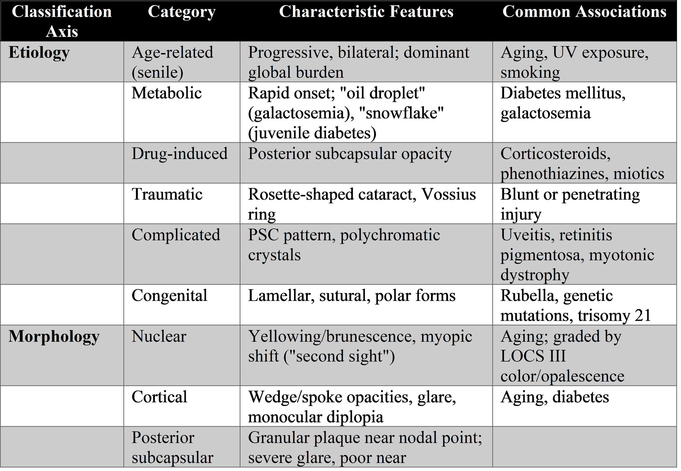
Morphological (anatomical) classification follows the layered anatomy of the lens and carries direct clinical meaning. Nuclear cataract, the most common age-related form, reflects the hardening, yellowing, and opacification of the central nucleus; patients typically develop a myopic shift — the elderly patient who delightedly discards reading glasses, the famous "second sight" — with distance blur, reduced contrast, and progressive brunescence, graded from faint yellow to near-black "cataracta nigra." Cortical cataract consists of wedge-shaped, spoke-like opacities of swollen, liquefied cortex beginning in the periphery; it is often asymptomatic until opacities reach the visual axis, when glare and light scatter, particularly oncoming headlights at night, become disabling. Posterior subcapsular cataract (PSC) forms a granular plaque just anterior to the posterior capsule, lies near the optical nodal point of the eye, and thus causes disproportionate disability for its size: severe glare, poor vision in bright light (when the pupil constricts around the opacity), and early loss of near vision. PSC is the hallmark of steroid use, diabetes, uveitis, and radiation, and characteristically affects younger patients. Less common morphological forms include anterior subcapsular, lamellar (zonular) cataract — the commonest congenital morphology, affecting a specific fetal layer — sutural, polar, and coronary cataracts.
Classification by maturity describes the natural history of an untreated cortical cataract: the incipient stage of peripheral opacities with clear central vision; the immature stage with partial opacification; the mature stage, in which the entire cortex is opaque and the fundus cannot be viewed; the hypermature stage, in which liquefied cortex leaks through an intact capsule, shrinking the lens and allowing the dense nucleus to sink inferiorly — the Morgagnian cataract. Hypermaturity is not a curiosity but a hazard: leaked lens proteins can obstruct the trabecular meshwork (phacolytic glaucoma), provoke granulomatous inflammation (phacoantigenic uveitis), or the capsule may rupture; hypermature lenses also carry increased surgical risk, including zonular dialysis and nucleus dislocation.
Severity grading for research and increasingly for clinical documentation is dominated by the Lens Opacities Classification System III (LOCS III), developed by Chylack and colleagues, which grades nuclear opalescence and nuclear color on scales of 0.1–6.9 and cortical and posterior subcapsular opacity on scales of 0.1–5.9 against standardized slit-lamp and retroillumination photographs. LOCS III remains the gold standard for cataract grading in research, though its subjectivity limits clinical use; objective imaging with Scheimpflug densitometry and anterior-segment OCT offers reproducible quantitative alternatives, and artificial-intelligence systems trained on LOCS III have achieved diagnostic accuracies above 90%, with the strongest performance for cortical and posterior subcapsular grading — a development with obvious implications for screening in underserved regions where slit-lamp expertise is scarce.
3.3 Other lens pathology: dislocation, subluxation, and developmental anomalies
Lens pathology is not confined to opacification. Ectopia lentis — displacement of the lens from its normal position — results from zonular weakness or rupture. Trauma is the commonest cause; systemic causes include Marfan syndrome (superotemporal subluxation, in perhaps 60–80% of affected patients), homocystinuria (inferonasal subluxation, a distinction beloved of examiners), Weill-Marchesani syndrome (microspherophakia with anterior dislocation and secondary glaucoma), aniridia, and pseudoexfoliation syndrome, the latter of enormous surgical importance as a cause of late intraocular lens-bag dislocation years after otherwise successful cataract surgery. A completely dislocated lens may fall into the anterior chamber — an ocular emergency risking corneal decompensation and pupillary block glaucoma — or sink into the vitreous, where it may be tolerated or may provoke inflammation and glaucoma. Developmental anomalies complete the picture: microphakia and spherophakia, lenticonus and lentiglobus (the anterior lenticonus of Alport syndrome, accompanied by the oil-droplet reflex), coloboma of the lens, and the persistent fetal vasculature spectrum with its characteristic retrolental fibrovascular plaque.
3.4 Clinical presentation and diagnostic evaluation
The symptoms of cataract are deceptively simple — painless, progressive visual loss — but the texture of the complaint points to the morphology: the myopic shift and dimmed color perception of nuclear sclerosis; the monocular diplopia and glare of cortical change; the sunshine blindness and lost reading vision of posterior subcapsular opacity. Patients describe halos around lights, fading and yellowing of colors, frequent changes of spectacle prescription, and the paradox of seeing better in dim light in PSC. Examination begins with visual acuity and refraction, proceeds to slit-lamp biomicroscopy through a dilated pupil — the definitive assessment, in which the opacity is localized, graded, and its density judged — and includes assessment of the red reflex (diminished, mottled, or absent), pupillary responses (the absence of a relative afferent defect is a reassuring sign that the visual loss is lenticular rather than retinal or neural), and measurement of intraocular pressure. When the fundus cannot be seen, B-scan ultrasonography excludes retinal detachment and tumor before surgery. The preoperative assessment for surgery adds biometry — keratometry and axial length measurement by optical or immersion methods — to calculate intraocular lens power, endothelial cell count where cornea guttata is present, and screening for risk factors: pseudoexfoliation, small pupil, posterior synechiae, dense brunescent nuclei, alpha-blocker use (tamsulosin famously causing intraoperative floppy iris syndrome), anticoagulation, and, in unilateral or atypical cases, investigation for an underlying cause. In children, the evaluation widens to a search for metabolic and genetic etiologies, and the clock is unforgiving: amblyopia makes timing the dominant variable.
3.5 Methods of treatment
Non-surgical management. No drug has been proven to prevent, slow, or reverse human cataract, and the clinician should say so honestly: antioxidant vitamin supplementation has failed in large trials; aspirin, aldose reductase inhibitors, and various "anticataract" drops have no established efficacy. What non-surgical management can offer is real but modest — updated spectacles, stronger lighting, magnification, and glare control — together with risk-factor modification: smoking cessation, ultraviolet protection, meticulous glycemic control, and avoidance of unnecessary corticosteroids. For an early, asymptomatic cataract in a patient whose visual needs are met, watchful waiting is not neglect but good medicine; surgery is indicated not by the appearance of the lens but by its effect on the patient's life.
The indications and principles of surgery. The indication for cataract surgery is functional: vision that interferes with the patient's daily activities, occupational needs, or safety — driving standards being a frequent practical benchmark — after informed discussion. Secondary indications include lens-induced glaucoma or uveitis, a cataract obscuring management of retinal disease, and refractive aims in the era of refractive lens exchange. The operation has three constants regardless of technique: remove the opaque lens, preserve the capsular bag, and replace the optical power, usually with an intraocular lens.
Phacoemulsification, the global standard of care, exemplifies how far the operation has come. Through a clear corneal incision of roughly 2.2–2.8 millimeters, the surgeon creates a continuous curvilinear capsulorhexis, mobilizes the lens by hydrodissection, fragments and aspirates the nucleus with ultrasonic energy, removes the cortex, and implants a foldable IOL into the capsular bag — typically under topical anesthesia, as a day case, without sutures, with visual recovery within days. Modern phaco platforms modulate power and fluidics to minimize energy and endothelial trauma, and torsional and transversal ultrasound modes have improved efficiency in dense nuclei. Phacoemulsification is the fruit of Kelman's insight that the lens could be emulsified rather than extracted whole, and it has been refined for half a century into one of the highest-volume, highest-success procedures in medicine.
Femtosecond laser–assisted cataract surgery (FLACS) automates the critical manual steps: it constructs the corneal incisions, performs a geometrically precise capsulotomy, fragments the nucleus, and can address astigmatism with arcuate incisions. The theoretical advantages — capsulotomy reproducibility, reduced phaco energy, improved IOL centration — are real but modest, and the evidence base has matured into an honest appraisal. A 2025 systematic review and meta-analysis of 41 randomized trials encompassing 9,310 eyes found FLACS superior for early and some long-term visual outcomes, capsulotomy accuracy, and effective lens position, but with no difference in visual outcomes at intermediate follow-up and no safety advantage overall; both techniques are effective and safe (Song et al., 2025). FLACS shows its clearest value in challenging scenarios — dense and white cataracts, shallow anterior chambers, posterior polar cataracts, and eyes with low endothelial reserve — while its cost-effectiveness for routine surgery remains debated. The sensible synthesis is that FLACS is a tool of precision for selected eyes rather than a universal replacement for skilled phacoemulsification.
Extracapsular and manual techniques retain vital importance. Manual small-incision cataract surgery (MSICS), in which the nucleus is expressed whole through a self-sealing scleral tunnel, delivers outcomes approaching phacoemulsification at a fraction of the cost and equipment burden, and it remains the workhorse of high-volume programs in South Asia and sub-Saharan Africa — indeed, it is arguably superior for rock-hard cataracts in eyes with compromised endothelium. Classical extracapsular and intracapsular extraction survive chiefly as historical footnotes and fallbacks for special circumstances, the intracapsular operation now reserved largely for complete lens dislocation.
Intraocular lenses are where cataract surgery became refractive surgery. Modern foldable acrylic monofocal IOLs, placed in the capsular bag, provide excellent distance vision with outstanding biocompatibility; aspheric designs compensate corneal spherical aberration; toric IOLs correct pre-existing astigmatism and have made "glasses-free distance vision" a realistic goal for many. Presbyopia-correcting lenses are the contemporary frontier. Multifocal IOLs — diffractive trifocals offering distance, intermediate, and near foci — deliver high rates of spectacle independence at the price of photic phenomena (halos, glare) and reduced contrast sensitivity, making careful patient selection paramount; they are generally avoided in eyes with macular disease, glaucoma, or irregular corneas. Extended depth-of-focus (EDOF) lenses stretch a single focal point into a range, trading some near acuity for markedly fewer dysphotopsias, and "enhanced monofocal" designs occupy the middle ground. Adjustable IOLs — whose power can be fine-tuned postoperatively by ultraviolet light (the light-adjustable lens) — and accommodative designs continue to evolve, and 2024–2025 has seen incremental refinement across all classes rather than revolution. Monovision, with one eye set for distance and one for near, remains a time-honored, low-technology alternative. The unifying principle is that the choice of IOL is now a conversation about the patient's visual life — reading, driving, screens, occupation, tolerance of optical side effects — not merely a calculation.
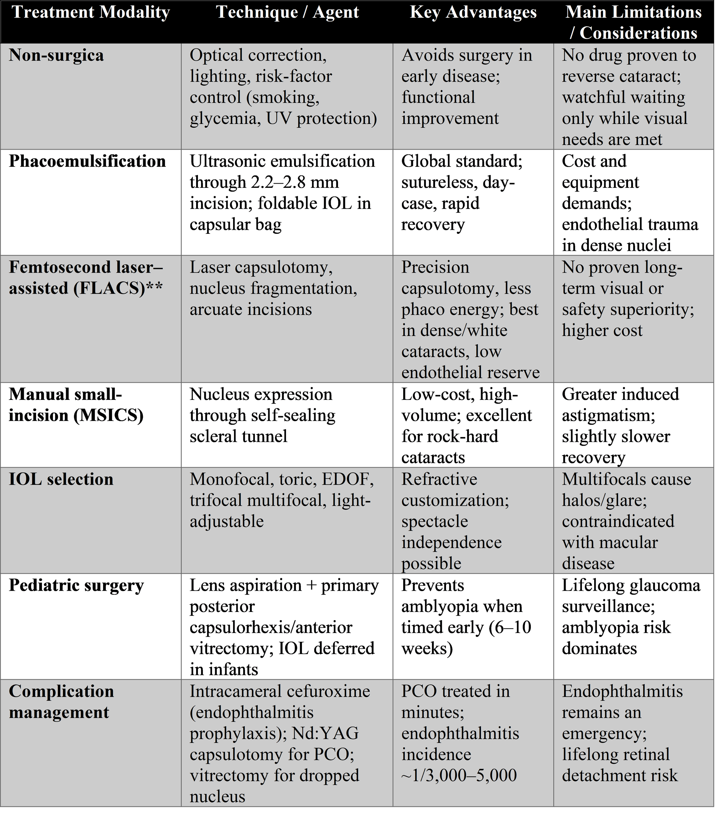
Pediatric cataract surgery is a different discipline. Timing is dictated by the amblyogenic window: dense unilateral congenital cataracts are operated within the first six to eight weeks of life, bilateral by about eight to ten weeks. The surgery itself is modified for the soft pediatric lens, elastic capsule, and intense inflammatory response — with anterior vitrectomy and primary posterior capsulorhexis to address the near-certain visual axis opacification of young children — and the question of IOL implantation in infancy was addressed by the Infant Aphakia Treatment Study, which showed no visual advantage but more adverse events and reoperations with IOLs in infants under seven months, tilting practice toward aphakic contact lens correction in the youngest and IOL implantation in older children. Lifelong surveillance for aphakic glaucoma, which may develop years or decades later, is mandatory. The refractive endpoint is moving target: the eye grows, so the implanted power is chosen to leave the child hyperopic, with emmetropization expected over time.
Complications and their management deserve honest enumeration. Intraoperative complications — posterior capsule rupture with vitreous loss, zonular dialysis, dropped nuclear fragments into the vitreous (requiring secondary vitrectomy), suprachoroidal hemorrhage, and the rare expulsive hemorrhage — have all become less common with modern techniques but remain the surgeon's constant preoccupation. Early postoperative complications include corneal edema, intraocular pressure spikes, toxic anterior segment syndrome, and the most feared, acute endophthalmitis, whose incidence of roughly 1 in 3,000–5,000 cases has been driven lower by intracameral cefuroxime; it presents with pain, hypopyon, and vitritis and demands emergent intravitreal antibiotics, with the Endophthalmitis Vitrectomy Study guiding the role of vitrectomy. Late complications include cystoid macular edema (the Irvine–Gass syndrome, treated with topical NSAIDs and steroids), retinal detachment (a modestly but persistently increased lifelong risk, greatest in young myopic males), and — most common of all — posterior capsule opacification, the "after-cataract," which clouds the bag months to years later in a substantial minority of patients and is treated definitively in minutes by Nd:YAG laser capsulotomy, with square-edged IOL designs having substantially reduced its incidence.
Emerging and frontier therapies complete the picture. Pharmacological reversal of cataract remains investigational: the chaperone-based lanosterol and 25-hydroxycholesterol approaches, which showed striking crystallin-solubilizing effects in animal lenses in 2015 and 2022 respectively, have yet to translate into validated human efficacy, and the older oxysterol and N-acetylcarnosine stories are cautionary tales of unmet promise. Artificial intelligence has already found a practical role in screening and grading. Robotic assistance, intraoperative OCT, and swept-source biometry with ever-refined IOL power formulas (Barrett, Kane, EVO, and AI-driven formulas outperforming legacy regression formulas especially in long and short eyes) define the current trajectory: toward a fully image-guided, personalized, refractively predictable procedure.
4. Discussion
Several unifying reflections emerge from this survey. The first is the coherence of lens pathology: nearly every clinical phenomenon — the myopic shift of nuclear sclerosis, the glare of posterior subcapsular opacity, the glaucoma of hypermaturity, the amblyopia of congenital cataract — follows directly from the anatomy and biochemistry of the lens. This makes lens disease one of the most satisfying areas of clinical medicine to reason through, and one of the least forgiving of rote learning: the examiner's "why" always has an answer rooted in the tissue.
The second reflection concerns the evolution of the therapeutic goal. For most of history, cataract surgery aimed at making the blind see. Modern surgery aims higher: at making the seeing see optimally — at a chosen distance, with corrected astigmatism, increasingly without spectacles. This reframing has elevated the importance of biometry, IOL selection, and the doctor–patient conversation, and it has introduced new failure modes: a technically perfect operation can still disappoint a patient whose expectations were for spectacle independence that a monofocal lens was never going to deliver. Managing expectations has become a clinical skill as real as wound construction.
The third is the equity paradox. Cataract is simultaneously the most curable and the most neglected of the great blinding diseases. The surgical backlog in low- and middle-income countries numbers in the tens of millions; the barriers are cost, workforce, and geography, not knowledge. High-volume, high-quality, low-cost models — MSICS-based surgical camps and institutional systems like the Aravind model — demonstrate that the constraint is organizational. Global initiatives targeting effective cataract surgical coverage, including the WHO's call for a 30-percentage-point increase by 2030, frame the next chapter of cataract care as one of delivery science. The most sophisticated FLACS platform in the world does nothing for the patient who never reaches a surgeon.
Finally, the limitations of this review should be acknowledged. It is a narrative synthesis rather than a systematic review; its coverage of a vast field necessarily compresses subspecialty detail; IOL technology and AI applications are moving quickly enough that specifics will date; and practice patterns vary legitimately between health systems. The student is encouraged to read the primary trials and consensus documents cited.
5. Conclusion
The pathology of the lens is, at its core, the story of a tissue that must remain transparent for a lifetime and of the predictable ways it fails — through oxidation, osmotic stress, trauma, drug toxicity, and the simple accumulation of years. Its classification, by cause, location, and maturity, converts that biology into a clinical grammar that guides recognition and management. And its treatment stands as one of medicine's clearest triumphs: from couching through intracapsular extraction to modern microincisional phacoemulsification with foldable refractive IOLs, each generation has restored sight more safely and more completely than the last. The contemporary challenges are equally clear: delivering this cure to the tens of millions who still await it, perfecting the refractive promises we now make, and — one day, perhaps — preventing or reversing the opacity itself. Until then, the cloudy lens remains what it has been for two centuries: the most gratifying diagnosis in ophthalmology, because it is the one we can unfailingly fix.
References
Chylack LT Jr, Wolfe JK, Singer DM, et al. The Lens Opacities Classification System III. Arch Ophthalmol. 1993;111(6):831–836.
Gali HE, Sella R, Afshari NA. Cataract grading systems: a review of past and present. Curr Opin Ophthalmol. 2019;30(1):13–18.
Song X, et al. Comparing the efficacy and safety between femtosecond laser-assisted cataract surgery and conventional phacoemulsification cataract surgery: systematic review and meta-analysis. Can J Ophthalmol. 2025;60(1):e1–e10.
Tang G, et al. Artificial intelligence in cataract grading system: a LOCS III-based hybrid model achieving high-precision classification. Front Med (PMC12454382). 2025.
Lee CM, Afshari NA. The global state of cataract blindness. Curr Opin Ophthalmol. 2017;28(1):98–103.
Liu YC, Wilkins M, Kim T, et al. Cataracts. Lancet. 2017;390(10094):600–612.
Kelman CD. Phaco-emulsification and aspiration: a new technique of cataract removal. Am J Ophthalmol. 1967;64(1):23–35.
Infant Aphakia Treatment Study Group. A randomized clinical trial comparing contact lens with intraocular lens correction of monocular aphakia during infancy. Arch Ophthalmol. 2010;128(7):810–818.
The Endophthalmitis Vitrectomy Study Group. Results of the Endophthalmitis Vitrectomy Study. Arch Ophthalmol. 1995;113(12):1479–1496.
Makley LN, McMenimen KA, DeVree BT, et al. Pharmacological chaperone for α-crystallin partially restores transparency in cataract models. Science. 2015;350(6261):674–677.
Riaz Y, Mehta JS, Wormald R, et al. Surgical interventions for age-related cataract. Cochrane Database Syst Rev. 2006;(4):CD001323.
de Silva SR, Evans JR, Kirthi V, et al. Multifocal versus monofocal intraocular lenses after cataract extraction. Cochrane Database Syst Rev. 2016;(12):CD003169.
Hashemi H, Pakzad R, Yekta A, et al. Global and regional prevalence of age-related cataract: a comprehensive systematic review and meta-analysis. Eye (Lond). 2020;34(8):1357–1370.
Pleyer U, et al. Intracameral cefuroxime and endophthalmitis prophylaxis (ESCRS study). J Cataract Refract Surg. 2013;39(3):378–385 (summary of ESCRS Endophthalmitis Study, 2007).
Michael R, Bron AJ. The ageing lens and cataract: a model of normal and pathological ageing. Phil Trans R Soc B. 2011;366(1568):1278–1292.
Sheeladevi S, Lawrenson JG, Fielder AR, et al. Global prevalence of childhood cataract: a systematic review. Eye (Lond). 2016;30(9):1160–1169.
Vasavada AR, Vasavada V. Current and future directions in IOL technology and pediatric cataract surgery. Indian J Ophthalmol. 2023;71(4) (and related reviews of premium IOLs).
World Health Organization. World Report on Vision. Geneva: WHO; 2019; and WHO SPECS 2030 initiative documents, 2021–2024.