
Symptomatology and General Principles of Treatment of Chronic Hepatitis and Chronic Pancreatitis
1. Kebekova Aizat Ashimzhanovna
2. Masheera Mumtaz
3. Kumari Pooja
(1. Lecturer, International Medical Faculty, Osh State University, Osh, Kyrgyz Republic
2. Student, International Medical Faculty, Osh State University, Osh, Kyrgyz Republic
3. Student, International Medical Faculty, Osh State University, Osh, Kyrgyz Republic)
Abstract
Chronic hepatitis and chronic pancreatitis are two common chronic inflammatory diseases of the digestive system. Both develop slowly, often with a long period of mild or nonspecific symptoms, and both may lead to irreversible organ damage if they are not recognised and treated in time. Chronic hepatitis is defined as inflammation of the liver lasting more than six months, and may progress to cirrhosis, liver failure and hepatocellular carcinoma. Chronic pancreatitis is a progressive fibro-inflammatory disease of the pancreas that causes permanent loss of exocrine and endocrine function and chronic pain. This article reviews the aetiology, clinical features (symptomatology), diagnostic approach and general principles of treatment of both conditions, with emphasis on current international guidelines.
Keywords: chronic hepatitis, chronic pancreatitis, symptomatology, antiviral therapy, pancreatic enzyme replacement therapy, treatment principles
1. Introduction
Diseases of the liver and pancreas make up a significant part of the workload of gastroenterologists and general physicians. The liver and the pancreas are closely related anatomically and functionally: both take part in digestion and metabolism, both are connected to the duodenum through the biliary and pancreatic ducts, and both are commonly damaged by alcohol. Knowledge of the symptoms and basic management of chronic hepatitis and chronic pancreatitis is therefore essential for every medical graduate.
The aim of this article is to describe the main clinical syndromes of chronic hepatitis and chronic pancreatitis and to summarise the general principles of their treatment.
2. Chronic Hepatitis
2.1 Definition and aetiology
Chronic hepatitis is a diffuse inflammatory-dystrophic disease of the liver lasting for at least 6 months, characterised histologically by inflammatory infiltration of the portal tracts and parenchyma, hepatocyte necrosis and varying degrees of fibrosis, but without the formation of regenerative nodules typical of cirrhosis.
The main causes are:
• Viral: hepatitis B virus (HBV), hepatitis C virus (HCV), hepatitis D virus (HDV, only together with HBV). Hepatitis A does not become chronic; hepatitis E is rarely chronic, mainly in immunosuppressed patients.
• Autoimmune hepatitis.
• Alcohol-related liver disease.
• Metabolic dysfunction-associated steatotic liver disease (MASLD, formerly NAFLD) and steatohepatitis.
• Drug-induced (e.g. methyldopa, isoniazid, nitrofurantoin, amiodarone, methotrexate).
• Hereditary and metabolic diseases: Wilson disease, alpha-1 antitrypsin deficiency, haemochromatosis.
• Cholestatic diseases (primary biliary cholangitis, primary sclerosing cholangitis), which overlap with chronic hepatitis in clinical practice.
Worldwide, chronic HBV and HCV infection are the leading causes of chronic hepatitis, cirrhosis and liver cancer.
2.2 Pathogenesis in brief
In viral hepatitis, hepatocyte injury is caused mainly by the immune response of the host to infected cells rather than by direct cytopathic action of the virus. Persistent inflammation leads to repeated cycles of necrosis and regeneration and to activation of hepatic stellate cells, which produce collagen and cause progressive fibrosis. In autoimmune hepatitis, loss of tolerance to hepatocyte antigens results in T-cell-mediated injury. In alcoholic and metabolic hepatitis, oxidative stress, lipotoxicity and inflammatory mediators play the leading role.
2.3 Symptomatology
Many patients, especially with chronic hepatitis B and C, are asymptomatic or have only minimal complaints for years, and the disease is discovered incidentally by raised transaminases. When symptoms are present, they can be grouped into clinical syndromes.
1. Asthenovegetative syndrome. This is the most common complaint: fatigue, weakness, reduced work capacity, sleep disturbance, irritability and difficulty concentrating.
2. Pain syndrome. Dull, aching pain or a sense of heaviness in the right upper quadrant, usually mild, aggravated by errors in diet, alcohol and physical exertion. It is caused by stretching of the liver capsule.
3. Dyspeptic syndrome. Nausea, bitter taste in the mouth, loss of appetite, intolerance of fatty food, bloating, unstable stools.
4. Cholestatic syndrome. Jaundice of the skin and sclera, pruritus, dark urine, pale stools. It is more typical for active disease or for cholestatic variants.
5. Cytolytic syndrome. Biochemical signs of hepatocyte damage: raised alanine aminotransferase (ALT) and aspartate aminotransferase (AST), with ALT usually higher in viral hepatitis; in alcoholic hepatitis AST is typically higher than ALT (ratio about 2:1).
6. Mesenchymal-inflammatory syndrome. Reflects activation of the immune system: raised gamma-globulins and thymol turbidity, positive autoantibodies in autoimmune hepatitis, fever, arthralgia.
7. Hepatocellular (synthetic) insufficiency. In advanced disease: reduced albumin, prolonged prothrombin time (INR increase), bleeding tendency, hypocholesterolaemia.
8. Portal hypertension. Present when fibrosis progresses to cirrhosis: splenomegaly, ascites, oesophageal varices, collateral veins.
9. Extrahepatic manifestations. Spider naevi, palmar erythema, gynaecomastia, hormonal disturbances, arthralgia, glomerulonephritis, cryoglobulinaemic vasculitis (HCV), thyroiditis, skin rashes.
Physical examination may show an enlarged, firm, slightly tender liver with a smooth edge, and in some cases a moderately enlarged spleen. Signs of chronic liver disease (spider naevi, palmar erythema) suggest more advanced damage.
2.4 Diagnosis
The diagnostic work-up has three aims: to establish the aetiology, to assess the activity of inflammation and to stage fibrosis.
• Biochemistry: ALT, AST, gamma-glutamyl transferase (GGT), alkaline phosphatase, bilirubin, albumin, INR.
• Aetiological tests: HBsAg, anti-HBc, HBeAg/anti-HBe, HBV DNA; anti-HCV and HCV RNA; anti-HDV; autoantibodies (ANA, SMA, anti-LKM1), immunoglobulin G; ceruloplasmin and urinary copper; ferritin and transferrin saturation; alpha-1 antitrypsin.
• Imaging: ultrasound of the abdomen, Doppler, elastography.
• Non-invasive fibrosis assessment: transient elastography (FibroScan), FIB-4 index, APRI score.
• Liver biopsy: remains the reference standard for grading of activity and staging of fibrosis, and is especially important in autoimmune hepatitis and unclear cases.
2.5 General principles of treatment
Treatment of chronic hepatitis is based on four principles: (1) elimination or control of the cause, (2) suppression of inflammation and prevention of fibrosis progression, (3) general measures and avoidance of liver-damaging factors, and (4) prevention and early detection of complications.
A. General measures (basic regimen)
• Complete abstinence from alcohol.
• Avoidance of hepatotoxic drugs, herbal supplements and occupational toxins; careful dosing of paracetamol (not more than 2 g per day in advanced disease).
• Avoidance of heavy physical exertion and psychological stress during exacerbation; moderate regular activity during remission.
• Diet (Pevzner table No. 5): regular meals 4–5 times per day; adequate protein (1–1.5 g/kg/day unless hepatic encephalopathy is present); limitation of animal fats, fried and smoked foods and spices; sufficient vegetables, fruit and fibre. In MASLD, weight reduction of 7–10% improves steatohepatitis and fibrosis.
• Vaccination against hepatitis A and B in non-immune patients.
B. Aetiological treatment
Chronic hepatitis B. The aim is long-term suppression of HBV DNA, normalisation of ALT and prevention of cirrhosis and hepatocellular carcinoma. Treatment is indicated in patients with elevated HBV DNA and ALT, significant fibrosis or cirrhosis. The preferred drugs are nucleos(t)ide analogues with a high barrier to resistance, entecavir or tenofovir (TDF or TAF), usually given long-term. Pegylated interferon alfa for a defined period of 48 weeks is an alternative in selected patients. Functional cure (HBsAg loss) is uncommon (EASL, 2017; AASLD, 2018).
Chronic hepatitis C. Treatment has been transformed by direct-acting antivirals (DAAs), for example sofosbuvir/velpatasvir or glecaprevir/pibrentasvir, given orally for 8–12 weeks. Sustained virological response (SVR, undetectable HCV RNA 12 weeks after therapy) is achieved in more than 95% of patients and is equivalent to cure. Treatment is recommended for all patients with chronic HCV infection (EASL, 2020).
Chronic hepatitis D. Peginterferon alfa has been used; bulevirtide, an entry inhibitor, is approved in Europe for compensated disease.
Autoimmune hepatitis. Immunosuppression with prednisolone, followed by addition of azathioprine as a steroid-sparing agent, induces remission in most patients; treatment is often prolonged and relapse is common after withdrawal.
Alcoholic hepatitis. Abstinence is the essential treatment; nutritional support is important.
MASLD/steatohepatitis. Lifestyle modification, control of diabetes, dyslipidaemia and hypertension; resmetirom is a newer option for non-cirrhotic steatohepatitis with moderate-to-advanced fibrosis in some countries.
Wilson disease. Lifelong copper chelation (D-penicillamine or trientine) or zinc.
Drug-induced hepatitis. Withdrawal of the causative agent.
C. Pathogenetic and symptomatic therapy
• Ursodeoxycholic acid is effective in primary biliary cholangitis and may reduce cholestasis in other conditions.
• "Hepatoprotectors" (silymarin, essential phospholipids, ademetionine and others) are widely used, but the evidence for their effect on outcomes is limited; they should be considered supportive and never a replacement for aetiological therapy.
• Cholestatic pruritus: cholestyramine, rifampicin.
• Treatment of complications: diuretics for ascites, non-selective beta-blockers and endoscopic band ligation for varices, lactulose for encephalopathy.
D. Monitoring and follow-up
Patients require regular clinical and laboratory monitoring (ALT, viral load, INR, albumin) and assessment of fibrosis. Patients with cirrhosis need ultrasound every 6 months, with or without alpha-fetoprotein, to screen for hepatocellular carcinoma, and endoscopic screening for varices. Liver transplantation is considered in decompensated cirrhosis and in selected cases of hepatocellular carcinoma.
3. Chronic Pancreatitis
3.1 Definition and aetiology
Chronic pancreatitis is a long-lasting, progressive fibro-inflammatory disease of the pancreas in individuals with genetic, environmental and other risk factors, leading to irreversible destruction of the parenchyma, fibrosis, ductal changes, pain, and finally loss of exocrine and endocrine function (Whitcomb et al., 2016).
Risk factors are summarised by the TIGAR-O classification:
• T – Toxic-metabolic: alcohol (the most frequent cause in adults), smoking, hypercalcaemia, hypertriglyceridaemia, chronic renal failure.
• I – Idiopathic.
• G – Genetic: mutations in PRSS1, SPINK1, CFTR, CTRC genes.
• A – Autoimmune: type 1 (IgG4-related) and type 2.
• R – Recurrent and severe acute pancreatitis.
• O – Obstructive: pancreatic duct stones or strictures, tumours, pancreas divisum, sphincter of Oddi dysfunction.
Smoking is an independent risk factor and acts synergistically with alcohol.
3.2 Pathogenesis in brief
Repeated injury to the pancreas, from toxins, ductal obstruction or premature activation of trypsinogen, causes inflammation. Persistent inflammation activates pancreatic stellate cells, which produce extracellular matrix, leading to progressive fibrosis, atrophy of acini, ductal strictures, formation of calculi and loss of islets. The result is exocrine and endocrine insufficiency, with chronic pain explained by ductal hypertension, parenchymal inflammation and changes in pancreatic nerves.
3.3 Symptomatology
1. Pain syndrome. Abdominal pain is the main symptom in most patients. It is usually located in the epigastrium, radiates to the back, may be girdle-like, and is often aggravated by meals or alcohol. It may be relieved by leaning forward. The pain may be episodic with attacks of acute pancreatitis, or constant. In late "burnt-out" stages the pain may decrease as parenchyma is destroyed.
2. Exocrine pancreatic insufficiency. Appears when more than 90% of lipase secretion is lost. Manifestations: steatorrhoea (bulky, pale, greasy, foul-smelling stools that are difficult to flush), diarrhoea, bloating, flatulence, weight loss, malnutrition. Deficiency of fat-soluble vitamins (A, D, E, K) may lead to osteoporosis, night blindness and coagulation disorders; vitamin B12 deficiency is also possible.
3. Endocrine insufficiency. Pancreatogenic diabetes mellitus (type 3c), usually late in the course. It is characterised by loss of both insulin and glucagon, so patients are prone to hypoglycaemia and brittle glucose control.
4. Dyspeptic syndrome. Nausea, vomiting, early satiety, loss of appetite and food aversion due to fear of pain.
5. Complications and their symptoms.
• Pseudocysts: palpable mass, pain, early satiety, infection or bleeding.
• Common bile duct stricture: jaundice and cholangitis.
• Duodenal stenosis: vomiting after meals.
• Splenic vein thrombosis: left-sided (sinistral) portal hypertension with gastric varices.
• Pancreatic ascites and pleural effusion due to ductal leaks.
• Pancreatic cancer: chronic pancreatitis increases the risk, and new weight loss, change in the pain pattern or jaundice should prompt investigation.
Physical findings are often poor: epigastric tenderness, signs of weight loss and nutritional deficiency, and sometimes a palpable pseudocyst.
3.4 Diagnosis
• Imaging: Abdominal ultrasound (screening); computed tomography (calcifications, ductal dilatation, atrophy, pseudocysts); magnetic resonance cholangiopancreatography (MRCP) for duct anatomy; endoscopic ultrasound (EUS) is the most sensitive for early changes.
• Pancreatic function tests: faecal elastase-1 (a value below 200 µg/g suggests exocrine insufficiency; values below 100 µg/g indicate severe insufficiency); the secretin test is the most sensitive but is available in few centres; 72-hour faecal fat is rarely used.
• Laboratory: serum amylase and lipase are often normal in chronic disease; fasting glucose and HbA1c to detect diabetes; vitamins A, D, E, K and B12, calcium, magnesium, zinc and albumin for nutritional status.
• Aetiological work-up: history of alcohol and smoking, serum triglycerides and calcium, IgG4, genetic testing in young patients or with family history.
3.5 General principles of treatment
The aims are to relieve pain, correct exocrine and endocrine insufficiency, treat complications, slow the progression of disease and improve nutrition and quality of life. Management should be multidisciplinary, involving a gastroenterologist, pancreatic surgeon, endoscopist, dietitian, diabetologist and, when needed, a pain specialist and addiction specialist (Löhr et al., 2017; Gardner et al., 2020).
A. Lifestyle and dietary measures
• Complete and permanent abstinence from alcohol and smoking. This is the most important measure and reduces pain and slows progression.
• Small, frequent meals (5–6 per day) with adequate energy and protein intake. Strict low-fat diets are not recommended because they worsen malnutrition; fat should be tolerated with adequate enzyme therapy.
• Oral nutritional supplements; enteral tube feeding if oral intake is inadequate; parenteral nutrition rarely.
B. Management of pain
• Analgesia follows a stepwise approach: paracetamol and NSAIDs (with caution), then weak opioids such as tramadol, and strong opioids only when necessary and for a limited period because of the risk of dependence.
• Neuropathic pain modulators (pregabalin, gabapentin, tricyclic antidepressants) are helpful in selected patients.
• Pancreatic enzymes may be tried for pain, although evidence is limited.
• Endoscopic therapy (ERCP with sphincterotomy, stone extraction, stent placement for strictures; extracorporeal shock-wave lithotripsy for large stones) is used for obstructive causes of pain.
• Surgery is considered in patients with intractable pain and ductal obstruction, or when complications exist. Options include lateral pancreaticojejunostomy (Puestow), duodenum-preserving head resection (Beger, Frey procedures), and in selected cases pancreatoduodenectomy or total pancreatectomy with islet autotransplantation. Evidence suggests that early surgery may give better long-term pain control than prolonged endoscopic treatment in suitable patients with a dilated duct.
• Coeliac plexus block has limited and short-lived benefit.
C. Pancreatic enzyme replacement therapy (PERT)
PERT is the cornerstone of treatment of exocrine insufficiency. Indications include steatorrhoea, weight loss and malnutrition, with or without low faecal elastase.
• Enteric-coated microsphere preparations (pancrelipase) are preferred.
• Usual starting dose: about 40,000–50,000 lipase units with main meals and 20,000–25,000 units with snacks; the dose can be increased (up to 10,000 units/kg/day lipase is the usual upper limit) according to symptoms and nutritional response.
• Capsules must be taken during the meal (divided at the start, middle and end), not swallowed before or long after.
• When response is inadequate, acid suppression with a proton-pump inhibitor may improve enzyme efficacy; bacterial overgrowth should also be excluded.
• Fat-soluble vitamins (A, D, E, K) and other deficiencies should be supplemented; bone density should be checked because of the risk of osteoporosis.
D. Treatment of endocrine insufficiency
Pancreatogenic diabetes is managed with diet, and insulin when necessary. Metformin may be used in mild cases. Because of the risk of hypoglycaemia (reduced glucagon), glycaemic targets should be individualised and avoid overly strict control.
E. Treatment of complications
• Symptomatic or enlarging pseudocysts: endoscopic transmural drainage (preferred) or surgical drainage.
• Biliary stricture: endoscopic stenting or surgical bypass.
• Duodenal obstruction: endoscopic dilatation or surgical bypass.
• Autoimmune pancreatitis (type 1): corticosteroids (prednisolone), with immunomodulators for relapses.
• Suspected malignancy: urgent evaluation and surgical referral.
F. Follow-up
Regular review (at least annually) should monitor symptoms, nutrition, bone health, glucose control, and alcohol and smoking cessation. Patients with hereditary pancreatitis have an especially high risk of pancreatic cancer and may require surveillance.
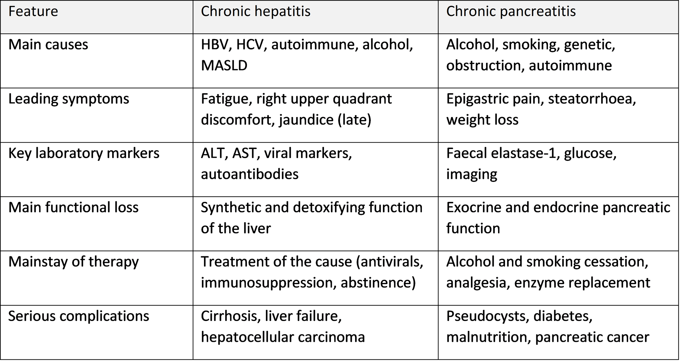
4. Comparison of the Two Diseases
5. Conclusion
Chronic hepatitis and chronic pancreatitis are chronic, progressive diseases with a long latent course and a wide variety of symptoms. In chronic hepatitis the dominant syndromes are asthenovegetative, dyspeptic, cytolytic, cholestatic and mesenchymal-inflammatory, and later hepatocellular failure and portal hypertension. In chronic pancreatitis the leading features are pain, exocrine insufficiency with steatorrhoea and malnutrition, and diabetes. Treatment in both cases rests on elimination of the cause (antiviral or immunosuppressive therapy in hepatitis; abstinence from alcohol and smoking in pancreatitis), a rational diet and regimen, correction of functional deficiencies, prevention and treatment of complications and long-term follow-up. Modern antiviral drugs, especially direct-acting antivirals for hepatitis C, have made cure of an important cause of chronic hepatitis possible, while a multidisciplinary approach to chronic pancreatitis improves pain control and nutrition. Early diagnosis and patient education remain the most important measures to prevent cirrhosis, cancer and pancreatic failure.
References
1. European Association for the Study of the Liver. EASL 2017 Clinical Practice Guidelines on the management of hepatitis B virus infection. Journal of Hepatology. 2017;67(2):370–398.
2. Terrault NA, Lok ASF, McMahon BJ, et al. Update on prevention, diagnosis, and treatment of chronic hepatitis B: AASLD 2018 hepatitis B guidance. Hepatology. 2018;67(4):1560–1599.
3. European Association for the Study of the Liver. EASL recommendations on treatment of hepatitis C: Final update of the series. Journal of Hepatology. 2020;73(5):1170–1218.
4. Mack CL, Adams D, Assis DN, et al. Diagnosis and management of autoimmune hepatitis in adults and children: 2019 practice guidance and guidelines from the AASLD. Hepatology. 2020;72(2):671–722.
5. Whitcomb DC, Frulloni L, Garg P, et al. Chronic pancreatitis: an international draft consensus proposal for a new mechanistic definition. Pancreatology. 2016;16(2):218–224.
6. Löhr JM, Dominguez-Munoz E, Rosendahl J, et al. United European Gastroenterology evidence-based guidelines for the diagnosis and therapy of chronic pancreatitis (HaPanEU). United European Gastroenterology Journal. 2017;5(2):153–199.
7. Gardner TB, Adler DG, Forsmark CE, et al. ACG clinical guideline: chronic pancreatitis. American Journal of Gastroenterology. 2020;115(3):322–339.
8. Kasper DL, Fauci AS, Hauser SL, et al. Harrison's Principles of Internal Medicine. 21st ed. McGraw-Hill; 2022.