
Symptomatology and General Principles of Treatment of Chronic Hepatitis and Liver Cirrhosis
1. Kebekova Aizat Ashimzhanovna
2. Khan Tanaz
3. Sable Aditya
(1. Lecturer, International Medical Faculty, Osh State University, Osh, Kyrgyz Republic
2. Student, International Medical Faculty, Osh State University, Osh, Kyrgyz Republic
3. Student, International Medical Faculty, Osh State University, Osh, Kyrgyz Republic)
Abstract
Background: Chronic hepatitis and liver cirrhosis are important causes of chronic liver disease worldwide. Persistent hepatic inflammation may lead to progressive fibrosis, distortion of normal liver architecture, portal hypertension, and hepatic insufficiency.
Objective: To review the clinical manifestations, diagnostic approaches, disease progression, and general principles of treatment of chronic hepatitis and liver cirrhosis.
Methods: This narrative review summarizes established medical literature and guidance from the World Health Organization (WHO), the American Association for the Study of Liver Diseases (AASLD), and standard medical textbooks. It is not a systematic review or original clinical study.
Results: Chronic hepatitis may be asymptomatic or present with fatigue, weakness, anorexia, hepatomegaly, and jaundice. Advanced chronic liver disease may progress to cirrhosis, with portal hypertension and impaired hepatic function. Management is cause-specific and includes antiviral or immunosuppressive therapy when indicated, lifestyle and nutritional measures, prevention and treatment of complications, surveillance for hepatocellular carcinoma, and transplantation assessment in selected patients.
Conclusion: Early diagnosis, treatment of the underlying cause, and structured monitoring are central to reducing progression and complications.
Keywords: chronic hepatitis; liver cirrhosis; hepatic fibrosis; portal hypertension; ascites; hepatic encephalopathy; antiviral therapy.
1. Introduction
The liver is essential for carbohydrate, protein, and lipid metabolism; synthesis of albumin and coagulation factors; bile production; storage of vitamins and minerals; and metabolism of drugs and toxins. Chronic liver disease can progressively impair these functions.
Chronic hepatitis describes persistent inflammation of the liver, commonly lasting longer than six months. It may be caused by viral infection, autoimmune disease, metabolic dysfunction, alcohol-related injury, or medications and toxins.
Cirrhosis is an advanced stage of chronic liver disease characterized histologically by diffuse fibrosis and regenerative nodules with distortion of the normal hepatic architecture. It may remain compensated for years before complications develop.
2. Etiology of Chronic Hepatitis
The cellular and molecular mechanisms underlying chronic liver injury converge on a final common pathway of microvascular disruption, persistent immune activation, and progressive fibrogenesis, regardless of the initial toxic, viral, or metabolic insult.
Chronic viral hepatitis—driven by persistent infection with HBV, HCV, or HDV—instigates parenchymal damage primarily through immune-mediated destruction of infected hepatocytes rather than direct viral cytopathic effects. Cytotoxic T-lymphocytes recognize viral antigens presented on human leukocyte antigen class I molecules, initiating cell apoptosis and continuous necroinflammation. In HDV co-infection or superinfection, this immunopathogenesis is intensified, resulting in rapid lobular architecture breakdown and accelerated extracellular matrix accumulation. Over years of continuous tissue injury and regeneration, persistent lymphocyte infiltration sustains a pro-inflammatory microenvironment that continuously activates hepatic stellate cells.
Alcohol-related liver disease develops through direct cellular toxicity and metabolic stress. Ethanol metabolism via alcohol dehydrogenase and cytochrome P450 2E1 generates acetaldehyde, a highly reactive compound that forms protein adducts, impairs intracellular lipid processing, and triggers structural membrane degradation. This metabolic strain induces hepatocellular steatosis, while parallel mitochondrial injury and reactive oxygen species generation provoke steatohepatitis. Dysregulated lipid peroxidation, combined with endotoxin translocation through a compromised gut mucosal barrier, amplifies Kupffer cell activation, promoting diffuse pericellular and perivenular fibrosis that steadily evolves toward cirrhosis.
Autoimmune hepatitis is characterized by a breakdown in self-tolerance toward hepatocyte autoantigens, driving an uninhibited T-cell-mediated autoimmune cascade. Activated helper T-lymphocytes recruitment triggers plasma cell proliferation, resulting in characteristic elevations of serum immunoglobulin G and distinct autoantibody profiles. The continuous immune attack at the interface between the portal tracts and the lobular parenchyma—known as interface hepatitis—causes piecemeal hepatocyte necrosis. Without targeted immunosuppression, this persistent stromal inflammation leads to severe structural remodeling and rapid bridging fibrosis.
Metabolic dysfunction-associated steatotic liver disease (MASLD) and its active inflammatory form, metabolic dysfunction-associated steatohepatitis (MASH), arise from peripheral insulin resistance, systemic lipotoxicity, and metabolic dysfunction commonly tied to obesity and type 2 diabetes. Excess free fatty acids overflow into hepatocytes, driving intrahepatic triglyceride accumulation. When intracellular lipid storage mechanisms are overwhelmed, toxic lipid intermediates induce endoplasmic reticulum stress, mitochondrial dysfunction, and cellular ballooning. This lipotoxic state triggers inflammatory pathways and activates resident immune cells, turning simple hepatic steatosis into progressive necroinflammation and matrix deposition.
Drug- and toxin-induced liver injury operates through both intrinsic (predictable, dose-dependent) and idiosyncratic (unpredictable, immune-mediated or metabolic) mechanisms. Prolonged exposure to specific therapeutic agents, environmental toxins, or botanical supplements causes direct mitochondrial dysfunction, bile canalicular transport inhibition, or reactive metabolite generation. These disruptions trigger localized hepatocyte apoptosis or necrosis, which, if unresolved, sustains a chronic inflammatory reaction capable of simulating primary viral or autoimmune liver diseases.
Inherited and metabolic disorders cause progressive organ damage through the toxic intracellular accumulation of endogenous metabolites. In Wilson disease, mutations in the ATP7B gene impair biliary copper excretion and prevent ceruloplasmin binding, resulting in toxic free copper accumulation within hepatocytes that triggers oxidative stress, cellular necrosis, and secondary tissue fibrosis. Hereditary hemochromatosis is driven by dysregulated hepcidin expression, causing uncontrolled intestinal iron absorption and progressive parenchymal iron overload; the resulting Fenton reaction generates free hydroxyl radicals that damage cellular lipids and DNA. Alpha-1 antitrypsin deficiency involves the synthesis of a misfolded mutant glycoprotein that cannot be secreted; its accumulation within the endoplasmic reticulum of hepatocytes induces marked conformational stress, autophagic degradation pathways, and secondary chronic necroinflammation.
3. Pathogenesis of Chronic Hepatitis and Fibrosis
Persistent hepatocyte injury activates inflammatory pathways and hepatic stellate cells. Activated stellate cells produce extracellular matrix proteins, including collagen. Excessive matrix deposition leads to fibrosis. With ongoing injury, fibrous septa and regenerative nodules may develop, progressively disrupting hepatic blood flow and function.
A simplified sequence is: persistent liver injury → inflammation → hepatic fibrosis → architectural distortion and regenerative nodules → cirrhosis → portal hypertension and/or hepatic insufficiency. The rate of progression varies according to cause, duration of injury, comorbidities, and individual factors.
4. Symptomatology of Chronic Hepatitis
Chronic hepatitis may be clinically silent for a prolonged period. When symptoms occur, they are often nonspecific and may not correlate closely with the degree of fibrosis.
4.1 General symptoms
Constitutional and non-specific symptoms are frequently the earliest indicators of underlying chronic liver pathology, reflecting systemic metabolic derangement and sustained immune activation.
Profound fatigue, generalized physical weakness, and reduced exercise tolerance dominate the initial symptomatic presentation across diverse liver etiologies. These symptoms stem directly from impaired hepatic glycogen storage, altered gluconeogenesis, and secondary systemic sarcopenia. Additionally, failure of the injured liver to clear circulating pro-inflammatory cytokines, alongside alterations in central neurotransmitter signaling within the hypothalamic-pituitary-adrenal axis, contributes significantly to chronic physical and mental exhaustion.
Gastrointestinal symptoms manifest as persistent anorexia, nausea, and occasional postprandial vomiting. These features arise from delayed gastric emptying, dysregulated gastrointestinal peptide secretion, and altered splanchnic hemodynamics. The loss of appetite is exacerbated by altered bile acid composition reaching the proximal intestine, which disrupts normal luminal lipid digestion and triggers neuro-visceral signals of early satiety and digestive intolerance.
Unintentional weight loss and generalized malaise highlight progressive systemic metabolic deterioration. Malnutrition in this setting is multifactorial, driven by reduced caloric intake due to sitophobia and nausea, concurrent intestinal malabsorption, and an elevated basal metabolic rate secondary to chronic low-grade inflammation. Accelerated breakdown of host adipose and skeletal muscle tissue occurs to meet baseline energy demands, culminating in overt physical depletion.
Sustained low-grade fever occurs selectively in active inflammatory, autoimmune, or necroinflammatory hepatic states. This pyrexial response is mediated by the ongoing release of endogenous pyrogens, including interleukin-1, interleukin-6, and tumor necrosis factor-alpha, from activated intrahepatic Kupffer cells and infiltrating lymphocytes during active lobular parenchymal destruction.
4.2 Gastrointestinal and hepatic symptoms
Localized right upper quadrant symptoms and overt physical signs of biliary impairment reflect structural enlargement of the hepatic parenchyma and altered bilirubin handling pathways.
A sensation of dull discomfort or continuous heaviness localized to the right upper quadrant arises from mechanical distension of Glisson's capsule. Because the visceral parenchyma lacks pain innervation, nociception is triggered exclusively when acute inflammation, cellular swelling, or lipid accumulation rapidly expands the liver volume, stretching the somatic pain fibers embedded within the surrounding fibrous capsule.
Physical examination reveals hepatomegaly, characterized by an inferior extension of the liver margin below the right costal margin. On palpation, the liver edge presents as firm and smooth, often demonstrating mild tenderness on deep inspiration due to active parenchymal congestion and capsular tension.
Disturbances in bilirubin conjugation and excretion pathways produce the classic triad of jaundice, dark urine, and pale stools. When intrahepatic cholestasis or significant hepatocellular necrosis occurs, impaired canalicular excretion causes conjugated bilirubin to back-leak into the systemic circulation. Hydrophilic conjugated hyperbilirubinemia undergoes renal filtration, resulting in dark, tea-colored urine (bilirubinuria). Concurrently, the lack of stercobilinogen reaching the intestinal tract turns the stools pale or clay-colored. Systemic deposition of excess bilirubin pigment into cutaneous tissues and scleral elastin yields overt cutaneous and scleral icterus.
Pruritus is a characteristic symptom of cholestatic liver disorders, driven by the systemic retention of substances normally excreted in bile. Impaired canalicular flow leads to the systemic accumulation of bile salts, endogenous opioids, and pruritogenic lysophosphatidic acid generated by the circulating enzyme autotaxin. These neuroactive compounds stimulate peripheral C-fiber unmyelinated nerve endings within the skin, producing intense, refractory itching that classically worsens at night and predominantly affects the palms of the hands and soles of the feet.
4.3 Extrahepatic manifestations
Depending on the cause, extrahepatic features may include arthralgia, skin manifestations, renal involvement, vasculitis, and mixed cryoglobulinemia (particularly associated with HCV). Autoimmune hepatitis may coexist with other autoimmune disorders.
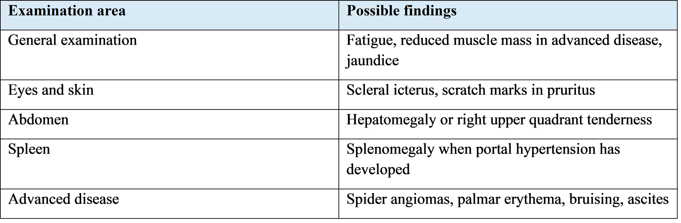
5. Clinical Examination in Chronic Hepatitis
6. Diagnosis of Chronic Hepatitis
Laboratory investigations and targeted etiological panels provide a comprehensive biochemical and serological framework for evaluating chronic liver disease, allowing clinicians to distinguish between hepatocellular and cholestatic injury, quantify true synthetic impairment, and pinpoint the specific underlying cause.
Standard Biochemical Evaluation
Serum aminotransferases—specifically alanine aminotransferase (ALT) and aspartate aminotransferase (AST)—serve as sensitive markers of active hepatocellular injury and cell membrane leakage. ALT is localized predominantly within the hepatocyte cytoplasm, rendering it more liver-specific, whereas AST is present in both cytoplasmic and mitochondrial compartments of hepatic, cardiac, skeletal muscle, and renal tissues. Elevated levels indicate ongoing necroinflammation; however, aminotransferase concentrations reflect the acute velocity of cell damage rather than functional reserve, often declining in end-stage cirrhosis due to severe loss of viable parenchymal mass.
Cholestatic liver enzymes quantify biliary epithelial injury and impaired bile excretion. Alkaline phosphatase (ALP) is localized to the microvillus membrane of bile canaliculi, rising in response to intrahepatic or extrahepatic biliary obstruction and localized ductal injury. Gamma-glutamyl transferase (GGT) is a microsomal membrane enzyme sensitive to biliary inflammation and enzyme induction; measuring GGT concurrently confirms the hepatic origin of an isolated ALP elevation, excluding bone or physiological growth-related sources.
Bilirubin fractioning clarifies excretory capacity and differentiates systemic hemolytic states from hepatobiliary disorders. Total serum bilirubin measures the sum of conjugated and unconjugated fractions. Elevated direct (conjugated) bilirubin confirms hepatic uptake and intracellular conjugation remain functional but highlights impaired canalicular excretion or biliary flow obstruction.
True hepatic synthetic function is evaluated through serum albumin concentrations and the prothrombin time or International Normalized Ratio (PT/INR). Because albumin is synthesized exclusively by hepatocytes with a long biological half-life of approximately twenty days, falling serum levels reflect chronic, long-standing reduction in functional mass or severe baseline nutritional impairment. Conversely, vitamin K-dependent coagulation factors (II, VII, IX, and X) synthesized by the liver have rapid turnover rates; a prolonged PT or elevated INR serves as an acute and sensitive measure of declining synthetic capability.
General hematological and metabolic panels complement specific liver biochemistry. A complete blood count identifies cytopenias resulting from hypersplenism or chronic blood loss. Serum electrolytes and renal function markers assess secondary circulatory volume changes and watch for hepatorenal syndrome, while blood glucose monitoring detects impaired hepatic glycogen storage or coexisting metabolic dysfunction.
Etiological Testing Protocols
Diagnostic confirmation of hepatitis B virus (HBV) relies on a structured panel of serological markers and viral load quantification tailored to the clinical context. Hepatitis B surface antigen (HBsAg) confirms active infection when present beyond six months, while total antibody to hepatitis B core antigen (anti-HBc) confirms past or ongoing exposure. Characterization of viral replication relies on hepatitis B e-antigen (HBeAg) and anti-HBe status, supplemented by quantitative real-time PCR for HBV DNA to measure active viral replication, determine phase of infection, and establish baseline thresholds for antiviral therapy.
Assessment of hepatitis C virus (HCV) follows a sequential two-step diagnostic algorithm. Initial screening uses a third-generation enzyme immunoassay for anti-HCV antibodies to detect past exposure. A positive antibody result must be followed by quantitative HCV RNA testing via PCR; detection of circulating viral RNA confirms active, ongoing infection and dictates the need for direct-acting antiviral therapy, whereas undetectable HCV RNA signifies resolved prior infection.
Hepatitis D virus (HDV) evaluation is indicated in HBsAg-positive individuals, particularly those with severe disease, rapid progression, or relevant epidemiological risk factors. Initial screening measures serum anti-HDV antibodies (total or IgG/IgM). Positive serology requires confirmation of active viral replication using quantitative HDV RNA PCR to guide combined antiviral management.
Suspected autoimmune hepatitis (AIH) requires quantitative serum immunoglobulin G (IgG) measurement paired with targeted autoantibody panels. Diagnostic markers include antinuclear antibodies (ANA) and smooth muscle antibodies (SMA) for type 1 AIH, and anti-liver kidney microsome type 1 (anti-LKM-1) or anti-liver cytosol type 1 (anti-LC1) antibodies for type 2 AIH. Elevated IgG combined with characteristic autoantibody titers provides strong diagnostic evidence, often supporting the clinical decision to perform a confirmative liver biopsy.
Metabolic and inherited liver disease screening relies on specialized biochemical panels when common viral and toxic etiologies are excluded. Hereditary hemochromatosis is evaluated via fasting transferrin saturation and serum ferritin levels; a transferrin saturation exceeding forty-five percent warrants genetic testing for HFE gene mutations. Wilson disease screening involves measuring total serum ceruloplasmin levels, serum free copper, and twenty-four-hour urinary copper excretion, alongside slit-lamp examination for Kayser-Fleischer rings. Alpha-1 antitrypsin (AAT) deficiency is investigated by measuring quantitative serum AAT levels, followed by Pi-typing (phenotyping) or genetic allele testing when serum concentrations are significantly depressed.
6.3 Imaging and fibrosis assessment
Abdominal ultrasonography evaluates liver morphology, biliary dilation, spleen size, ascites, and focal lesions. Doppler ultrasonography assesses portal and hepatic vascular flow. Transient elastography estimates liver stiffness and can help assess fibrosis. Liver biopsy is reserved for selected cases in which histology is needed to establish diagnosis, grade inflammation, or clarify uncertain findings.
7. General Principles of Treatment of Chronic Hepatitis
Treatment is determined by the cause of hepatitis, fibrosis stage, comorbidities, and the patient's overall clinical condition. A hepatology or gastroenterology assessment is appropriate when disease is persistent, advanced, or diagnostically uncertain.
7.1 General measures
Preventative interventions, supportive care, and systematic longitudinal monitoring form the cornerstone of disease modification in chronic liver disease, serving to arrest fibrotic progression, prevent secondary infectious insults, and catch clinical decompensation early.
Comprehensive Supportive Care and Risk Mitigation
Abstinence from alcohol is a universal, non-negotiable requirement across all etiologies of chronic liver disease. Ethanol exposure accelerates necroinflammation and fibrosis by compounding oxidative stress, disrupting intestinal barrier integrity, and increasing hepatic stellate cell activation. Complete cessation halts synergistic parenchymal destruction, slows the progression to cirrhosis, and significantly lowers the baseline risk of malignant transformation into hepatocellular carcinoma.
Nutritional stabilization targets both metabolic overload and sarcopenia. In metabolic liver diseases such as MASLD and MASH, structured lifestyle modifications aimed at achieving a 7% to 10% reduction in total body weight reduce intrahepatic lipid accumulation and resolve inflammatory ballooning. Conversely, in advanced liver disease where hypermetabolism and impaired glycogen storage drive rapid muscle breakdown, nutritional management emphasizes preserving lean body mass. Patients require a high-protein diet (1.2 to 1.5 grams per kilogram of body weight daily) with frequent small meals and a carbohydrate-rich late-evening snack to minimize overnight gluconeogenesis and muscle catabolism.
Pharmacological vigilance requires continuous auditing of prescribed medications, over-the-counter products, and botanical supplements. Compromised hepatic microcirculation and reduced cytochrome P450 enzyme activity alter drug clearance, predisposing patients to drug-induced liver injury. Essential therapeutic regimens must not be discontinued abruptly without specialist review, but non-essential hepatotoxic drugs and high-risk herbal products should be eliminated. Acetaminophen, while preferred over nonsteroidal anti-inflammatory drugs due to the risk of renal failure or variceal bleeding, must be capped at a maximum of two grams daily to avoid glutathione depletion.
Preventative immunization safeguards against superadded viral hepatitis, which can trigger acute-on-chronic liver failure. All non-immune patients with chronic liver disease must undergo serological screening for hepatitis A virus (HAV) and hepatitis B virus (HBV) susceptibility. Indicated vaccinations against HAV and HBV should be administered early in the disease course, as humoral immune responses decline significantly as hepatic cirrhosis advances.
Infection control and harm reduction protocols prevent de novo acquisition or transmission of bloodborne pathogens. Standard precautions—including single-use sterile injection equipment, safe needle disposal, rigorous screening of blood products, and targeted barrier protection—effectively mitigate horizontal transmission risks for hepatitis B, C, and D in healthcare and community settings.
7.2 Cause-specific treatment
Chronic hepatitis B: antiviral treatment is recommended for patients who meet guideline-based criteria. High-barrier nucleos(t)ide analogues such as tenofovir or entecavir are commonly used. Treatment suppresses HBV replication and reduces the risk of progression, but it does not usually eradicate covalently closed circular DNA; long-term monitoring is therefore important.
Chronic hepatitis C: direct-acting antiviral (DAA) regimens can cure most infections. Regimen selection depends on factors such as prior treatment, cirrhosis status, renal function, drug interactions, and local guidance.
Autoimmune hepatitis: immunosuppressive treatment commonly uses corticosteroids, with azathioprine in suitable patients. Diagnosis and treatment require specialist assessment and monitoring for adverse effects.
MASLD/MASH: management focuses on sustained lifestyle measures, weight management where appropriate, physical activity, and treatment of diabetes, dyslipidemia, hypertension, and other metabolic risks. Selected patients may qualify for disease-specific pharmacotherapy under current local guidance.
Drug-induced liver injury: the suspected causative agent should be reviewed and stopped when clinically appropriate. Severe cases require urgent assessment and supportive or cause-specific treatment.
8. Liver Cirrhosis
8.1 Definition
Liver cirrhosis is an advanced chronic liver disease characterized by diffuse fibrosis and regenerative nodules that alter normal hepatic architecture. These changes increase intrahepatic vascular resistance and impair hepatic synthetic, metabolic, and detoxification functions.
8.2 Major causes
● Chronic hepatitis B, C, and, in selected patients, D.
● Alcohol-related liver disease.
● MASH and other metabolic liver disorders.
● Autoimmune hepatitis.
● Cholestatic disorders, including primary biliary cholangitis and primary sclerosing cholangitis.
● Inherited disorders such as Wilson disease, hemochromatosis, and alpha-1 antitrypsin deficiency.
9. Pathophysiology and Clinical Stages of Cirrhosis
Fibrous septa and regenerative nodules distort the hepatic vascular architecture. Increased resistance to portal blood flow causes portal hypertension. Splanchnic vasodilation and neurohormonal activation contribute to sodium and water retention, ascites, and reduced effective circulating volume. Loss of functioning hepatocyte mass contributes to jaundice, coagulopathy, hypoalbuminemia, and impaired detoxification.
Compensated cirrhosis refers to cirrhosis without major clinical decompensating events. Decompensated cirrhosis is characterized by complications such as ascites, variceal hemorrhage, hepatic encephalopathy, or jaundice attributable to liver failure.
10. Symptomatology of Liver Cirrhosis
In the early stages, symptoms are often subtle and nonspecific. Patients typically experience persistent fatigue, weakness, a reduced appetite, nausea, or feeling full prematurely. Over time, these issues often lead to noticeable weight loss, muscle wasting, general abdominal discomfort, and a decreased tolerance for exercise.
As the condition progresses, cutaneous and systemic signs become more evident. These manifest as jaundice and itchy skin, along with vascular markers like spider angiomas and palmar erythema. Complex hemostatic changes frequently cause easy bruising or bleeding, while ongoing nutritional deficiencies contribute to worsening peripheral muscle wasting.
Advanced progression leads to gastrointestinal and portal hypertensive manifestations. Increased portal pressure often causes ascites and abdominal distension, while hypersplenism leads to splenomegaly and low platelet counts. Additionally, esophageal or gastric varices can develop and pose a significant bleeding risk, and visible collateral veins may form across the abdominal wall.
10.4 Endocrine manifestations
Possible findings include gynecomastia, testicular atrophy, reduced libido, menstrual irregularities, and altered body-hair distribution. These findings are variable and are not specific to cirrhosis.
10.5 Hepatic encephalopathy
Hepatic encephalopathy is a spectrum of neuropsychiatric dysfunction associated with liver insufficiency and/or portosystemic shunting. Manifestations range from altered sleep pattern and impaired attention to confusion, disorientation, and reduced consciousness. Asterixis may be present. Precipitating factors include infection, gastrointestinal bleeding, constipation, dehydration, electrolyte disturbance, and sedative medicines.
11. Diagnosis and Severity Assessment of Cirrhosis
11.1 Imaging and endoscopy
Ultrasonography may show a nodular liver contour, altered echotexture, splenomegaly, ascites, or collateral vessels. Doppler studies evaluate portal venous flow. Elastography estimates liver stiffness. Upper gastrointestinal endoscopy is used to assess varices when indicated by the patient's risk profile and current guidance. CT or MRI may be required to characterize focal liver lesions or vascular complications.
11.2 Severity scores
Child–Pugh classification uses bilirubin, albumin, PT/INR, ascites, and encephalopathy to classify severity as class A, B, or C. The MELD-based score, including MELD-Na where used, estimates short-term mortality risk and assists transplant prioritization. Scores support, but do not replace, clinical judgment.
12. General Principles of Treatment of Liver Cirrhosis
Management aims to treat the underlying cause, prevent first decompensation where possible, identify complications early, maintain nutrition and function, and assess transplant suitability when appropriate.
12.1 General management
Management begins with strict lifestyle and disease-specific measures, primarily complete abstinence from alcohol alongside targeted treatment for the underlying liver condition. From a nutritional standpoint, patients require adequate energy and protein intake, and routine protein restriction must be avoided, even when managing hepatic encephalopathy. Comprehensive care also includes routine screening for sarcopenia and malnutrition, which can be addressed through individualized dietetic support and strategies like a late-evening snack to preserve lean mass.
Medication safety and preventive clinical measures are equally crucial. Healthcare providers should regularly review all prescriptions and strictly avoid non-steroidal anti-inflammatory drugs in individuals with ascites or elevated renal risk unless a specialist explicitly advises otherwise. Additionally, patients should receive appropriate vaccinations and preventive treatments, paired with ongoing monitoring of renal function, serum electrolytes, nutritional status, and clinical signs of liver decompensation.
12.2 Ascites
New-onset ascites generally requires diagnostic paracentesis to determine its cause and exclude spontaneous bacterial peritonitis. Management commonly includes dietary sodium restriction and diuretics such as spironolactone, with furosemide added when appropriate. Renal function, serum sodium, and potassium require monitoring. Large-volume paracentesis is used for tense or refractory ascites; albumin is administered after large-volume removal according to guideline recommendations. Selected patients may be assessed for transjugular intrahepatic portosystemic shunt (TIPS).
12.3 Portal hypertension and varices
Prevention of variceal hemorrhage is based on individual risk assessment. Non-selective beta-blockers, including carvedilol in suitable patients, or endoscopic variceal ligation may be used according to current guidance and clinical circumstances. Acute variceal bleeding is a medical emergency requiring hospital treatment, vasoactive medication, antibiotic prophylaxis, and urgent endoscopic management.
12.4 Hepatic encephalopathy
Management requires identifying and correcting precipitating factors. Lactulose is commonly used for overt hepatic encephalopathy and is titrated to achieve regular soft bowel movements. Rifaximin may be added for recurrent episodes in appropriate patients. Nutrition should be maintained, and routine protein restriction should be avoided.
12.5 Spontaneous bacterial peritonitis
Spontaneous bacterial peritonitis should be suspected in a patient with cirrhosis and ascites who develops fever, abdominal pain, encephalopathy, renal deterioration, or unexplained clinical decline. Diagnosis is based on ascitic fluid analysis, including polymorphonuclear leukocyte count. Prompt antibiotics and albumin in selected patients are important; prophylaxis is reserved for defined high-risk situations.
12.6 Hepatocellular carcinoma surveillance
Patients with cirrhosis generally require hepatocellular carcinoma surveillance at six-month intervals, commonly with liver ultrasonography with or without alpha-fetoprotein (AFP), according to applicable guidelines and patient suitability.
12.7 Liver transplantation
Liver transplantation should be considered for selected patients with decompensated cirrhosis, significant complications, or high predicted mortality. Referral should not be delayed until terminal illness. Eligibility and timing are determined by a specialist transplant team.
13. Complications of Liver Cirrhosis
Liver disease can lead to several severe vascular and fluid-related complications. Increased resistance in the portal venous system causes portal hypertension, which promotes the formation of portosystemic collateral vessels. This elevated pressure frequently leads to ascites and abdominal distension, creating a environment where spontaneous bacterial peritonitis can develop. Furthermore, the high pressure can cause fragile esophageal and gastric varices to expand, posing a severe risk of variceal hemorrhage.
Systemic organ systems and neurological functions are also heavily impacted as liver function deteriorates. The accumulation of neurotoxins can result in hepatic encephalopathy, altering mental status and cognitive capacity. Renal function is similarly vulnerable, with patients facing acute kidney injury and hepatorenal syndrome. Pulmonary involvement may also occur in select cases, manifesting as hepatopulmonary syndrome or portopulmonary hypertension.
Hematologic and nutritional decline represent additional major challenges in advanced liver disease. Impaired synthetic function and hypersplenism contribute to complex coagulopathy, anemia, and thrombocytopenia. Simultaneously, metabolic dysfunction often triggers progressive malnutrition, sarcopenia, and physical frailty. Finally, long-standing chronic liver damage significantly elevates the risk of developing hepatocellular carcinoma.
14. Prevention and Prognosis
Prevention includes hepatitis B vaccination, testing and treatment of viral hepatitis, safe injection practices, blood safety, avoidance of harmful alcohol use, and management of metabolic risk factors. Patients with established chronic liver disease benefit from regular follow-up and vaccination against hepatitis A when susceptible and indicated.
Prognosis depends on the underlying etiology, fibrosis stage, hepatic functional reserve, comorbidities, and occurrence of decompensation. Patients with compensated cirrhosis generally have a better prognosis than those with decompensated disease. Effective treatment of the cause can reduce progression and, in some cases, improve fibrosis; established cirrhosis still requires ongoing surveillance even after etiological treatment.
15. Discussion
Chronic hepatitis and cirrhosis are related but distinct clinical entities. Chronic hepatitis describes persistent inflammation, while cirrhosis is defined by advanced architectural distortion and fibrosis. Patients may have few symptoms despite significant fibrosis, so risk-based testing and non-invasive fibrosis assessment are important.
The treatment landscape has changed substantially. Antiviral therapy can suppress HBV and cure most HCV infections. Cause-directed treatment, alcohol abstinence, metabolic risk reduction, and immunosuppression for appropriately diagnosed autoimmune hepatitis can reduce ongoing injury. In cirrhosis, prevention and early management of portal hypertensive and hepatic complications are central to care.
A multidisciplinary approach involving primary care clinicians, hepatologists, dietitians, radiologists, endoscopists, and transplant teams can improve continuity and outcomes. Management should follow current national and specialty guidelines because recommendations evolve.
16. Conclusion
Chronic hepatitis and liver cirrhosis are important causes of long-term illness and premature mortality. Clinical manifestations range from nonspecific fatigue and anorexia to portal hypertension, ascites, variceal hemorrhage, hepatic encephalopathy, and liver failure. Accurate etiological diagnosis, cause-specific therapy, nutritional care, complication prevention, and structured surveillance are essential. Timely specialist referral and liver transplantation assessment are important for selected patients with advanced disease.
17. References
1. World Health Organization. Guidelines for the prevention, diagnosis, care and treatment for people with chronic hepatitis B infection. Geneva: WHO; 2024.
2. World Health Organization. Hepatitis B. Fact sheet. Geneva: WHO. Available at: https://www.who.int/news-room/fact-sheets/detail/hepatitis-b
3. Kaplan DE, Ripoll C, Thiele M, et al. AASLD Practice Guidance on risk stratification and management of portal hypertension and varices in cirrhosis. Hepatology. 2024;79(5):1180–1211. doi:10.1097/HEP.0000000000000647.
4. Biggins SW, Angeli P, Garcia-Tsao G, et al. Diagnosis, evaluation, and management of ascites, spontaneous bacterial peritonitis and hepatorenal syndrome. Hepatology. 2021;74(2):1014–1048.
5. European Association for the Study of the Liver. EASL Clinical Practice Guidelines on the management of liver diseases. Consult the current disease-specific guideline at: https://easl.eu/publications/clinical-practice-guidelines/
6. American Association for the Study of Liver Diseases. Practice guidance and clinical resources. Available at: https://www.aasld.org/practice-guidelines
7. Jameson JL, Fauci AS, Kasper DL, Hauser SL, Longo DL, Loscalzo J, editors. Harrison's Principles of Internal Medicine. 21st ed. New York: McGraw Hill.
8. Kumar V, Abbas AK, Aster JC. Robbins & Cotran Pathologic Basis of Disease. 10th ed. Philadelphia: Elsevier.