
Anatomical Variations of the Brachial Plexus and Their Clinical Significance
1. Tanabaeva Aiperi
2. Fauzia Perveen
3. Muskan
4. Mirza Nasir Khan
5. Selim Rahman
6. MD Shahnawaz
(1. Professor, International Medical Faculty, Osh State University, Osh, Kyrgyz Republic
2. Student, International Medical Faculty, Osh State University, Osh, Kyrgyz Republic
3. Student, International Medical Faculty, Osh State University, Osh, Kyrgyz Republic
4. Student, International Medical Faculty, Osh State University, Osh, Kyrgyz Republic
5. Student, International Medical Faculty, Osh State University, Osh, Kyrgyz Republic
6. Student, International Medical Faculty, Osh State University, Osh, Kyrgyz Republic)
Abstract:
The brachial plexus represents a complex somatic nerve network formed by the ventral rami of the lower four cervical nerves (C5–C8) and the first thoracic nerve (T1), supplying motor and sensory innervation to the upper extremity. While textbooks outline a standardized architecture of roots, trunks, divisions, cords, and terminal branches, cadaveric dissections and high-resolution imaging consistently demonstrate that deviations from this classical pattern occur in over half of the general population. These variations arise early in embryonic development as mesenchymal limb buds interact with migrating spinal axons, where subtle shifts in neurotrophic signaling or vascular patterning alter neural trajectories.
Clinically, unrecognized anatomical variants carry significant consequences across multiple specialties. In regional anesthesia, prefixed or postfixed configurations, alongside aberrant fascicular pathways through the scalene musculature, frequently account for incomplete nerve blocks or inadvertent phrenic nerve paresis during interscalene and supraclavicular approaches. For orthopedic, trauma, and reconstructive surgeons operating in the posterior cervical triangle, infraclavicular fossa, or axilla, unexpected neural communications—such as the absence of the musculocutaneous nerve, duplicated median nerve roots, or anomalous branching patterns around the axillary artery—substantially elevate the risk of iatrogenic traction or transection injuries. Furthermore, atypical neurovascular relationships complicate diagnostic electrophysiology and obscure the clinical presentation of thoracic outlet syndrome and compressive neuropathies.
This review synthesizes the embryological mechanisms underlying brachial plexus morphogenesis, categorizes high-yield structural variations from the cervical roots to the terminal branches, and evaluates their practical implications in surgical exploration and ultrasound-guided regional analgesia. Integrating detailed anatomical awareness with preoperative sonographic mapping remains essential to minimizing perioperative morbidity and improving patient outcomes.
Keywords: Brachial plexus; Anatomical variations; Peripheral nerves; Regional anesthesia; Axillary artery; Cadaveric dissection; Iatrogenic nerve injury; Embryology.
1. Introduction:
In standard anatomical pedagogy, the brachial plexus is presented as a predictable, almost geometric network of peripheral nerves originating from the anterior primary rami of the fifth through eighth cervical spinal nerves (C5–C8) and the first thoracic spinal nerve (T1). As these ventral rami emerge from the intervertebral foramina and traverse the interscalene groove between the anterior and middle scalene muscles, they unite to form three distinct trunks—superior (C5–C6), middle (C7), and inferior (C8–T1). Passing deep to the clavicle into the cervicoaxillary canal, each trunk bifurcates into anterior and posterior divisions, which subsequently reorganize around the second part of the axillary artery to establish the lateral, posterior, and medial cords. From these cords arise the major terminal branches responsible for the motor control and cutaneous sensation of the entire upper limb, notably the musculocutaneous, axillary, radial, median, and ulnar nerves.
While this classical organizational blueprint serves as an indispensable framework for medical students and clinicians, extensive cadaveric investigations and intraoperative findings demonstrate that strict adherence to the textbook model is the exception rather than the rule. Across diverse global populations, structural deviations at one or more levels of the plexus have been documented in roughly 45% to 55% of dissected upper extremities. These anomalies range from subtle variations in spinal root contributions to complete absence of specific cords, unusual neural communications between terminal branches, and atypical spatial relationships with adjacent vascular structures such as the subclavian and axillary arteries.
Understanding these variations extends far beyond academic curiosity. Modern clinical practice relies heavily on precise neuroanatomical localization—whether an anesthesiologist is depositing local anesthetic around nerve trunks under real-time ultrasound guidance, a trauma surgeon is exploring a lacerated axilla, or a neurologist is interpreting discordant nerve conduction studies. Unrecognized plexus anomalies can lead to incomplete surgical anesthesia, unexpected motor deficits following routine orthopedic procedures, or diagnostic misattribution in patients presenting with upper-limb radiculopathy or thoracic outlet compression.
2. Embryological Basis of Brachial Plexus Variations:
To appreciate why the brachial plexus exhibits such remarkable structural plasticity, one must examine the chronology of upper limb morphogenesis during the fourth and fifth weeks of human embryonic development. The upper limb bud appears around day 24 as a localized outgrowth of somatopleuric lateral plate mesoderm covered by an ectodermal cap, situated opposite the lower cervical and first thoracic somites. Almost immediately, motor axons sprout from the ventral horn neuroblasts of the developing spinal cord, while sensory axons extend peripherally from neural crest-derived dorsal root ganglia.
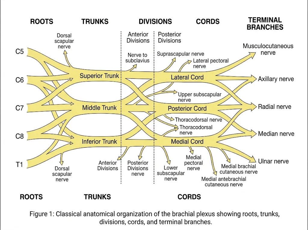
Rather than growing along rigid, pre-drawn anatomical tracks, advancing axonal growth cones navigate the mesenchymal limb bud through a dynamic interplay of chemoattractant and chemorepellent molecular cues. Guidance families—including ephrins, netrins, semaphorins, and Slit proteins—interact with specific surface receptors on the axonal tips to steer them toward target myotomes and dermatomes. At the base of the limb bud, these migrating axons converge into a dense, undifferentiated neural plate before splitting into dorsal (extensor) and ventral (flexor) premuscular masses.
Crucially, neurogenesis in the embryonic limb occurs simultaneously with vasculogenesis. Initially, the developing upper extremity is supplied by a diffuse capillary plexus that progressively remodels into a dominant axial artery—the precursor to the subclavian, axillary, and brachial arteries. Under normal developmental conditions, the axillary artery establishes its definitive course between the lateral and medial cords of the brachial plexus. However, if an alternate capillary channel persists and enlarges instead of the primary axial trunk, the resulting A vascular loop can physically split, displace, or reroute developing nerve fascicles. Consequently, an anomalous axillary artery branching pattern frequently coexists with altered cord formation or atypical median nerve roots. Furthermore, minor cranio-caudal shifts in the segmental origin of the limb bud relative to the neural tube during somite differentiation directly explain why adjacent spinal segments (such as C4 or T2) are occasionally recruited into the plexus architecture.
3. Root and Trunk Level Variations:
Variations at the proximal origin of the brachial plexus primarily involve cranio-caudal shifts in spinal segment recruitment, alterations in trunk union patterns, and aberrant spatial trajectories through the scalene musculature of the posterior cervical triangle. Because these proximal structures lie tightly bundled within a narrow fascial compartment alongside major subclavian vessels, even minor morphological deviations carry immediate implications for regional nerve blockade and thoracic outlet pathology.
3.1 Pre-Fixed and Post-Fixed Configurations:
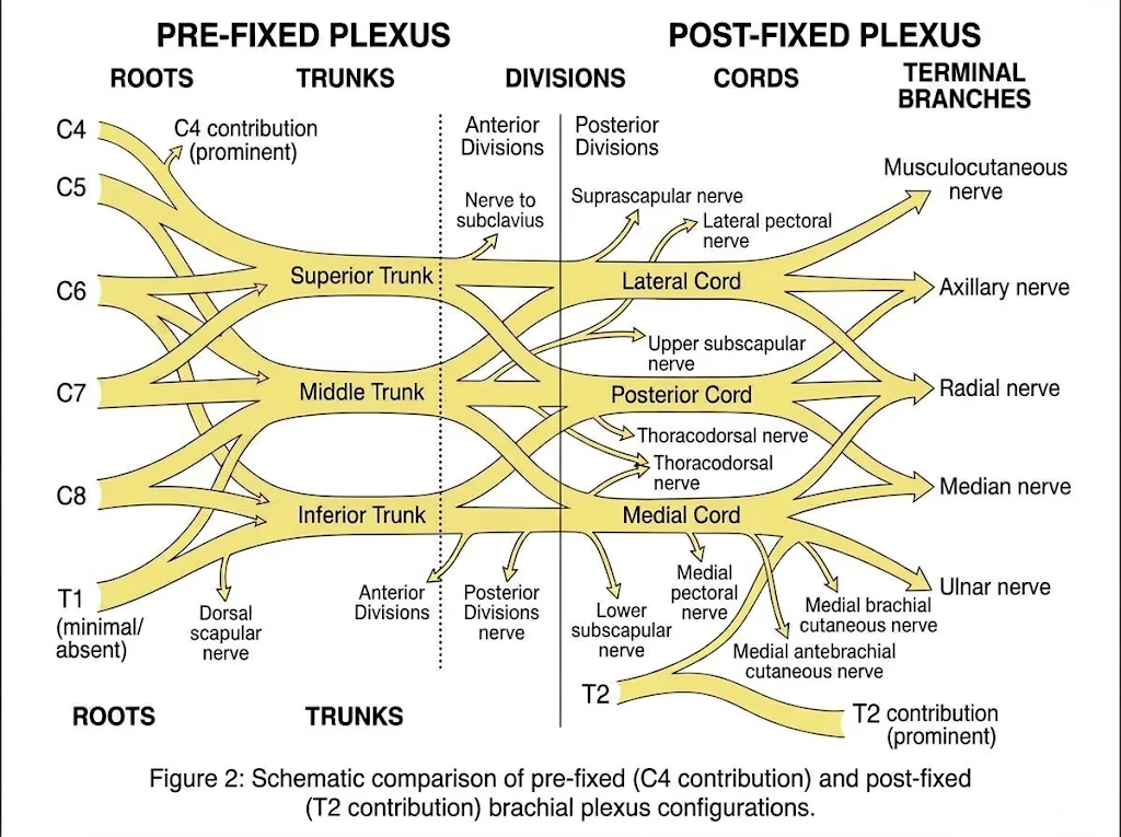
In the classical anatomical arrangement, the brachial plexus draws its neural fibers exclusively from the C5 through T1 ventral rami. However, slight cranial or caudal shifts in the embryonic positioning of the upper limb bud relative to the neuraxis frequently alter this segmental blueprint (Figure 2):
● Pre-Fixed Brachial Plexus: When the plexus shifts cranially, the fourth cervical ventral ramus (C4) contributes a substantial communicating branch to C5, while the contribution from T1 is markedly diminished and T2 is entirely absent. Reported in approximately 15% to 25% of cadaveric specimens, a pre-fixed configuration elevates the overall functional center of the plexus. Notably, C4 fibers entering the superior trunk often travel alongside—or share fascicular origins with—the phrenic nerve (C3–C5), increasing susceptibility to diaphragmatic hemiparesis during proximal cervical procedures.
● Post-Fixed Brachial Plexus: Conversely, a caudal shift produces a post-fixed configuration, observed in roughly 1% to 5% of individuals. Here, the C5 root is slender or lacks a direct contribution from C4, whereas the second thoracic ventral ramus (T2) sends a prominent ascending fascicle through the first intercostal space to join T1 in forming the inferior trunk. Because the T1–T2 union must ascend sharply over the neck of the first rib to reach the cervicoaxillary canal, post-fixed plexuses are predisposed to mechanical traction and vascular-neural compression against the first rib or a cervical rib.
3.2 Trunk Formation Anomalies:
Beyond segmental shifts, the fusion of ventral rami into three distinct trunks exhibits considerable variability:
● Absence or Non-Union of the Superior Trunk: In roughly 2% to 4% of upper limbs, the C5 and C6 ventral rami fail to unite into a common superior trunk. Instead, they descend independently through the interscalene space and split directly into their respective anterior and posterior divisions before reaching the lateral cord.
● Absent Middle Trunk or Bifurcated C7: While C7 typically continues as the middle trunk, it occasionally fuses prematurely with C6 to form a combined upper-middle trunk or splits early within the interscalene groove into separate anterior and posterior branches.
● Direct Entry of T1 into the Posterior Cord: Rarely, the C8 and T1 roots remain separate rather than forming an inferior trunk, with C8 joining C7 while T1 continues independently into the medial cord without contributing to the posterior divisions.
3.3 Relationships with the Scalene Musculature:
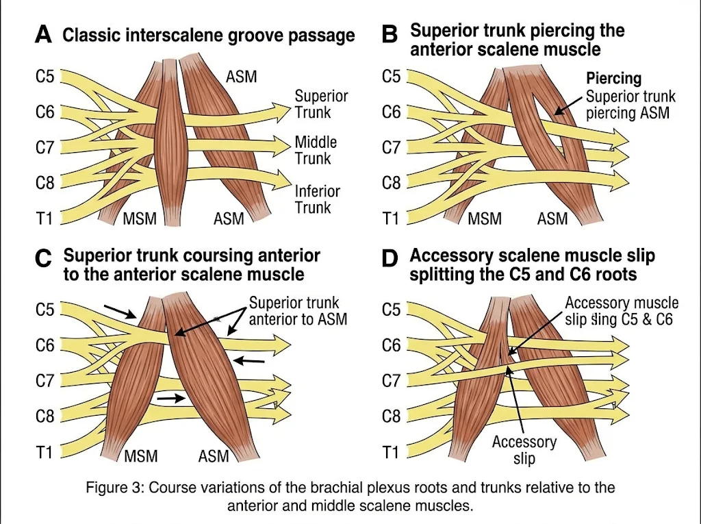
Classically, all five ventral rami emerge between the anterior scalene muscle (ASM) and the middle scalene muscle (MSM) within the interscalene groove, positioned posterosuperior to the subclavian artery. Anatomical dissections and high-frequency ultrasonography reveal that only about 60% to 70% of individuals conform to this textbook alignment (Figure 3).
In the most common variant (occurring in 15% to 30% of specimens), the C5 root—or the entire superior trunk—pierces directly through the belly of the anterior scalene muscle rather than passing behind it. Less frequently, C5 and C6 course entirely anterior to the anterior scalene muscle, lying superficially beneath the prevertebral fascia right next to the phrenic nerve. Furthermore, the presence of an accessory muscular slip—known as the scalenus minimus (present in up to 30%–40% of individuals)—frequently passes between the C7 and C8 roots or separates the inferior trunk from the subclavian artery. When muscular hypertrophy, fibrous bands, or repetitive overhead motion occurs in patients with intramuscular or split-trunk trajectories, the entrapped fascicles experience dynamic compression, manifesting clinically as neurogenic thoracic outlet syndrome (nTOS).
4. Division and Cord Level Anomalies:
As the three primary trunks pass beneath the clavicle and enter the cervicoaxillary canal, each splits into an anterior and a posterior division. While textbooks present this bifurcation as an orderly, predictable sorting of flexor and extensor compartments, dissection records consistently demonstrate that the infraclavicular plexus is a site of substantial structural plasticity. Variations at this level alter both the internal fascicular mixing of the nerves and their spatial orientation around the second part of the axillary artery.
4.1 Atypical Divisional Reunions and Accessory Slips:
In the standard anatomical arrangement, the anterior divisions of the superior and middle trunks merge to form the lateral cord, the anterior division of the inferior trunk continues independently as the medial cord, and all three posterior divisions converge to create the posterior cord. However, anomalous divisional splitting frequently disrupts this symmetry. A common finding is the bifurcation of the anterior division of the middle trunk into two distinct slips: one joining the superior trunk’s anterior division as expected, and an aberrant slip descending obliquely across the axillary artery to fuse with the medial cord. Conversely, the posterior division of the inferior trunk is occasionally absent or hypoplastic; in such specimens, the posterior cord is formed exclusively by the posterior divisions of the superior and middle trunks, leaving the C8 and T1 posterior fibers to travel via variant communicating rami.
4.2 Cord Fusion, Absence, and Duplication:
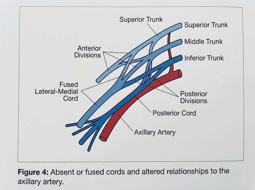
The cords themselves are named strictly for their classical relationship to the second part of the axillary artery—situated lateral, medial, and posterior to the vessel. When divisional mergers fail to segregate normally, entire cords may be absent, duplicated, or fused into a single composite trunk:
● Absence of the Posterior Cord: Rather than forming a unified posterior cord, the posterior divisions may bypass consolidation altogether, giving rise directly to the radial, axillary, thoracodorsal, and subscapular nerves from independent divisional stems.
● Fusion of the Lateral and Medial Cords: In a notable subset of upper limbs, the anterior divisions of all three trunks converge into a single, large anterior common cord positioned anterolateral to the axillary artery. From this solitary trunk, the musculocutaneous, median, and ulnar nerves arise sequentially without ever forming separate lateral and medial cords.
● Duplication of the Lateral Cord: A split superior trunk or an early bifurcation of its anterior division can yield two parallel lateral cords—one positioned superiorly and one inferolaterally—that reunite distally just prior to giving off the lateral root of the median nerve.
4.3 Altered Relationships to the Axillary Artery:
The spatial architecture of the infraclavicular fossa depends heavily on the embryological timing of angiogenesis relative to axonal outgrowth. Normally, the axillary artery develops from the seventh cervical intersegmental artery, threading neatly between the precursors of the lateral and medial cords while leaving the posterior cord dorsal to it. When an anomalous segmental vessel—such as the sixth or eighth intersegmental artery—persists as the dominant axillary trunk, the entire cord arrangement shifts.
In these vascular-driven variants, all three cords may lie entirely lateral or anterolateral to the axillary artery, leaving the medial and posterior aspects of the vessel devoid of major neural structures. In rarer instances, the axillary artery pierces directly through the substance of a fused cord or becomes entrapped between a duplicated lateral cord, creating an anatomical bottleneck susceptible to dynamic vascular compression during shoulder abduction. Furthermore, a high-origin profunda brachii or subscapular artery occasionally passes between the posterior and medial cords, splaying them apart and distorting the palpable landmarks relied upon during infraclavicular regional anesthesia and surgical exploration of the axilla.
5. Terminal Branch Variations and Neural Communications:
Distal to the pectoralis minor, the cords terminate into the principal peripheral nerves of the upper extremity. Even when proximal roots, trunks, and cords follow a textbook layout, terminal branching and inter-nervous anastomoses in the axilla and brachium frequently diverge from classical descriptions. Because these nerves supply the primary flexors, extensors, pronators, and cutaneous territories of the upper limb, variations here carry direct consequences for motor mapping, sensory deficits, and operative exposure.
5.1 Musculocutaneous Nerve Variations:
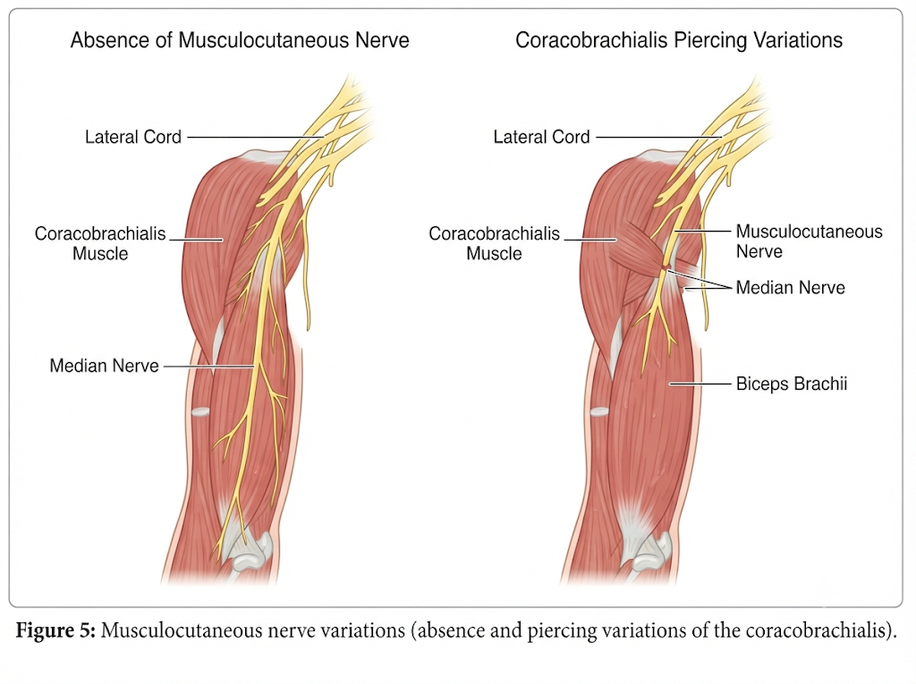
Classically, the musculocutaneous nerve arises from the lateral cord (carrying C5–C7 fibers), pierces the coracobrachialis muscle roughly 5 to 8 cm distal to the coracoid process, courses between the biceps brachii and brachialis, and continues distally as the lateral antebrachial cutaneous nerve. Anatomical surveys reveal several recurring deviations from this route:
● Non-Piercing Course: In a recognized proportion of limbs, the musculocutaneous nerve passes medial or posterior to coracobrachialis without piercing its belly, traveling directly into the interval between biceps brachii and brachialis before giving off motor branches to the flexors.
● High or Multiple Piercing Points: Rather than entering via a single belly perforation, the nerve may pierce coracobrachialis closer to its proximal attachment or split into two slips that perforate the muscle separately, increasing vulnerability during coracoid osteotomy or anterior shoulder procedures.
● Apparent Absence of the Nerve: In rarer cases, a distinct musculocutaneous trunk is absent or fails to separate early from the median nerve; motor branches to coracobrachialis, biceps brachii, and brachialis emerge directly from the lateral root or main trunk of the median nerve, while the sensory branch leaves later in the arm to become the lateral antebrachial cutaneous nerve.
5.2 Median Nerve Formation Anomalies:
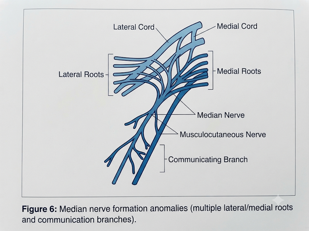
Normally, the median nerve is formed anterior or anterolateral to the third part of the axillary artery by the union of a single lateral root from the lateral cord (C5–C7) and a single medial root from the medial cord (C8–T1). Variations in root number, fusion site, and adjacent communications occur with notable regularity:
● Supernumerary Roots: The median nerve may receive two or three lateral roots from the lateral cord or two medial roots from the medial cord. These extra roots sometimes join at staggered levels—producing an elongated Y- or H-shaped junction or a small neural ring encircling the axillary artery or one of its branches.
● Distal or Proximal Fusion: Instead of uniting just anterior to the axillary artery, the lateral and medial roots may merge more proximally near the axilla's apex or more distally in the upper third of the arm, altering the length and mobility of the roots during humeral or axillary dissection.
● Musculocutaneous–Median Communicating Branches: Fine or substantial connecting rami frequently bridge the musculocutaneous and median nerves in the middle third of the arm. Depending on direction, fibers may pass from the musculocutaneous nerve into the median nerve (often carrying C6/C7 fibers destined for forearm flexors or hand intrinsics) or from the median nerve into the musculocutaneous nerve.
5.3 Posterior Cord Terminal Branch Variations and Neural Communications:
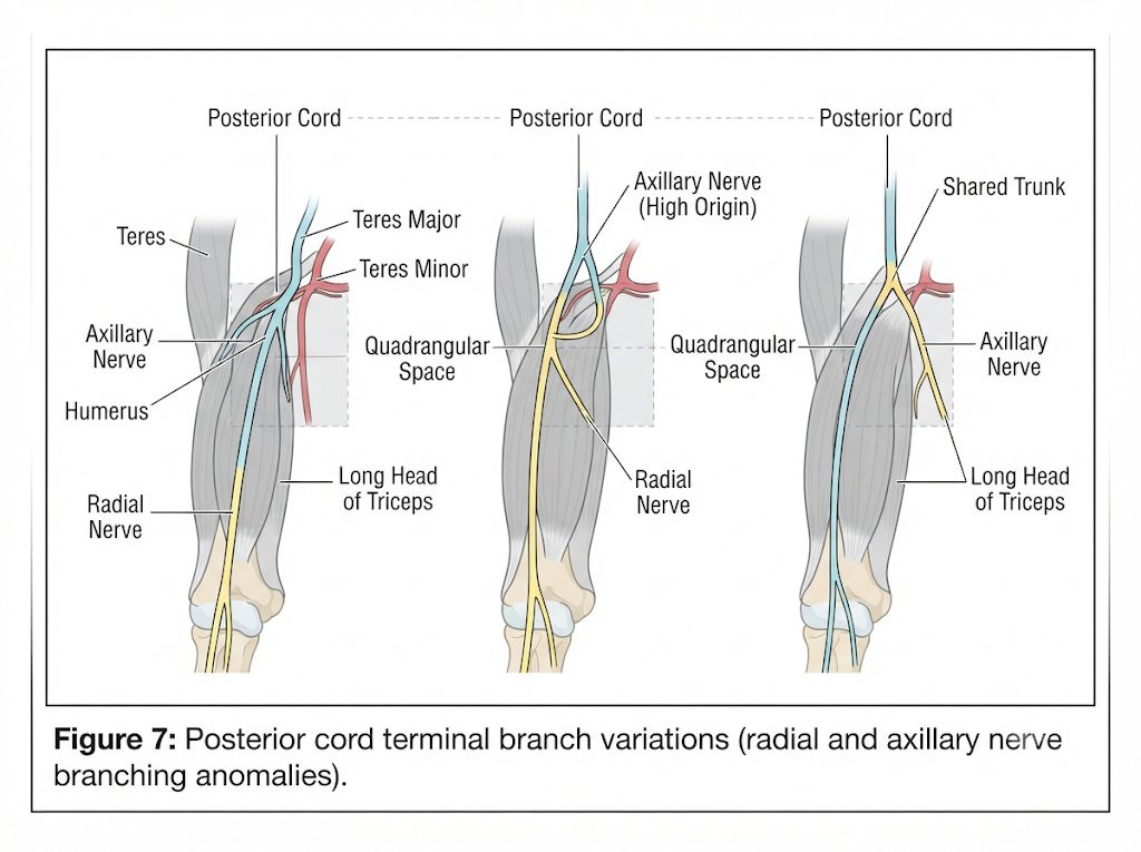
The posterior cord typically terminates by dividing into the axillary nerve and the radial nerve (C5–T1), after giving off the upper subscapular, thoracodorsal, and lower subscapular nerves. Variations in this region often involve shared origins, early bifurcations, and inter-branch communications:
● Aberrant Axillary and Radial Origins: The axillary and radial nerves may arise from a common short stem before separating near the inferior border of subscapularis, or one nerve may originate higher than usual while the other branches more distally. Occasionally, the posterior cord splits into three terminal branches rather than two—for instance, giving off a separate trunk to the long or medial head of triceps alongside the axillary and radial nerves.
● Early Triceps Branching: Branches to the long and medial heads of the triceps brachii sometimes arise in the axilla or very proximal arm rather than within the radial groove, meaning a proximal radial nerve injury in the axilla does not always spare elbow extension uniformly.
● Inter-Nerve Communications in the Axilla and Arm: Communicating twigs are occasionally documented between the radial nerve and the posterior cutaneous nerve of the arm or forearm, between the axillary and radial nerves near the quadrangular space, or between the medial cord/ulnar nerve and the medial brachial cutaneous/intercostobrachial nerves. Such interconnections help explain why focal nerve lesions sometimes produce smaller-than-expected sensory or motor loss.
6. Clinical, Surgical, and Anesthetic Significance:
Anatomical variations of the brachial plexus are far more than academic curiosities found in the dissection hall; they directly dictate clinical outcomes across operating rooms, trauma bays, and outpatient clinics. When neural elements deviate from their textbook pathways, routine procedures carry heightened risks of iatrogenic injury, diagnostic misinterpretation, and incomplete analgesia. Understanding these patterns allows clinicians to anticipate anomalies before a scalpel or block needle ever touches the skin.
6.1 Regional Anesthesia and Ultrasound-Guided Nerve Blocks:
The widespread adoption of high-frequency ultrasound has transformed upper-extremity regional anesthesia, yet anatomical variants remain a leading cause of block failure and inadvertent intraneural injection. During an interscalene brachial plexus block, clinicians typically look for the classic "stoplight" sign—three hypoechoic nodules stacked vertically between the anterior and middle scalene muscles. However, when the C5 or C6 ventral rami pierce the belly of the anterior scalene muscle or course entirely anterior to it, standard injection within the interscalene groove leaves these aberrant roots unanesthetized, resulting in spared dermatomes over the lateral shoulder and proximal arm.
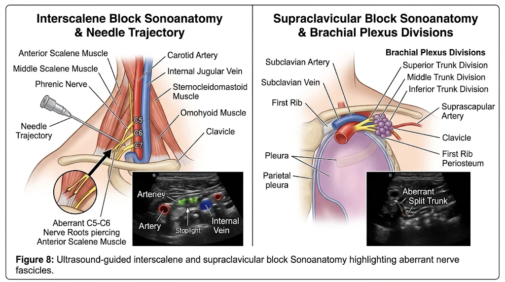
Similarly, variations in trunk formation alter the sonoanatomy at the supraclavicular level. A split superior trunk or an early-branching suprascapular nerve may sit outside the main perineural sheath, evading local anesthetic deposited in the traditional "corner pocket" between the subclavian artery and the first rib. Furthermore, in patients with a pre-fixed plexus, significant C4 contributions may require supplemental superficial cervical plexus coverage to achieve surgical anesthesia for clavicle or shoulder arthroplasty. Real-time dynamic scanning—tracing nerve roots proximally from the transverse processes down into the supraclavicular fossa—remains the most reliable safeguard against both missed blocks and accidental needle trauma to displaced fascicles.
6.2 Orthopedic, Trauma, and Reconstructive Surgery:
Surgeons operating around the clavicle, shoulder girdle, and proximal humerus rely heavily on consistent anatomical landmarks to protect neural structures. Open reduction and internal fixation of midshaft clavicle fractures, for instance, places aberrant supraclavicular nerve branches and high-riding cords at risk during subperiosteal dissection and drill advancement. In anterior shoulder procedures—such as the Latarjet coracoid transfer or arthroscopic Bankart repair—an altered relationship between the musculocutaneous nerve and the coracobrachialis muscle creates a well-documented hazard. When the musculocutaneous nerve enters the muscle abnormally high (within 2 to 3 centimeters of the coracoid process) or fails to pierce it altogether, routine medial retraction can produce severe traction neuropraxia.
In the axilla, variations in cord fusion and vascular relationships complicate axillary lymph node dissections, radical mastectomies, and vascular repairs of the axillary artery. When the lateral and medial cords fuse distally or form a single common cord anterior to the axillary artery, circumferential vascular control becomes treacherous. Similarly, a duplicated axillary artery passing between the roots of the median nerve increases the likelihood of simultaneous neurovascular injury during penetrating trauma or tumor resection.
6.3 Thoracic Outlet Syndrome and Compression Neuropathies:
Structural deviations at the root and trunk levels are primary anatomical drivers of neurogenic thoracic outlet syndrome (nTOS). In a standard configuration, the roots and trunks pass cleanly through the interscalene triangle bounded by the anterior scalene, middle scalene, and first rib. When the inferior trunk (C8–T1) or a post-fixed T2 contribution elevates over a cervical rib or a fibrous band extending from an elongated C7 transverse process, chronic mechanical irritation ensues.
Aberrant intramuscular pathways compound this vulnerability. Roots or trunks that traverse through the substance of the anterior scalene muscle or become entrapped by a slip of the scalenus minimus muscle experience dynamic compression every time the patient elevates the arm or inhales deeply. Clinically, these patients present with diffuse medial forearm paresthesia, intrinsic hand muscle wasting (Gilliatt-Sumner hand), and vascular symptoms that often mimic cervical radiculopathy or distal ulnar entrapment. Recognizing these variants on high-resolution MR neurography helps surgeons plan targeted scalenectomy and neurolysis rather than performing incomplete first-rib resections alone.
6.4 Electrodiagnostic Pitfalls and Neurosurgical Nerve Transfers:
Variant neural communications in the arm and forearm frequently confound electrodiagnostic testing and clinical neurological exams. Anastomoses between the musculocutaneous and median nerves—or distal Martin-Gruber and Riche-Cannieu connections—allow motor axons to bypass their expected peripheral pathways. A patient with a complete proximal median nerve laceration, for example, may retain unexpected thenar muscle function if fibers traveled via the musculocutaneous nerve before rejoining the median trunk in the mid-arm. Without recognizing this dual innervation, clinicians can underestimate injury severity or mislocalize lesions during nerve conduction studies.
Conversely, these exact variants offer strategic advantages during reconstructive neurosurgery. In brachial plexus avulsion injuries, surgeons routinely perform nerve transfers—such as the Oberlin procedure, which transfers redundant flexor carpi ulnaris fascicles from the ulnar nerve to the biceps branch of the musculocutaneous nerve. Preoperative identification of an absent musculocutaneous nerve, where biceps and brachialis motor branches arise directly from the median nerve, fundamentally changes the surgical blueprint, allowing the reconstructive team to select alternative donor fascicles and avoid damaging the sole motor supply to the anterior compartment of the arm.
7. Conclusion and Future Perspectives:
7.1 Synthesis of Anatomical Variability:
Although classical medical textbooks present the brachial plexus as an orderly sequence of five roots, three trunks, six divisions, three cords, and five terminal branches, decades of cadaveric dissections, fetal studies, and high-resolution imaging demonstrate that deviation from this archetype is the rule rather than the exception. Over half of all human upper-limb plexuses exhibit at least one structural anomaly, ranging from proximal shifts in spinal segment contribution (pre-fixed and post-fixed configurations) to distal cord fusions, absent terminal nerves, and complex neural anastomoses in the arm. These variations stem directly from subtle fluctuations in embryological axon guidance, mesenchymal signaling gradients, and early subclavian-axillary vascular remodeling during the fourth and fifth weeks of gestation.
7.2 Imperatives for Clinical Practice:
For the practicing clinician, recognizing that the "textbook" brachial plexus exists in only a subset of patients is essential for patient safety. In regional anesthesia, reliance on surface landmarks or static ultrasound views risks incomplete blockade or inadvertent nerve trauma when roots pierce the anterior scalene muscle or trunks split prematurely. In orthopedic, vascular, and neurosurgical procedures around the neck, clavicle, and axilla, awareness of altered cord-artery relationships and high-origin terminal branches prevents catastrophic iatrogenic injuries and guides successful nerve transfer reconstructions. Similarly, neurologists and electrophysiologists must account for peripheral communicating branches—such as musculocutaneous-median anastomoses—to avoid misinterpreting atypical motor or sensory deficits following trauma.
7.3 Future Directions in Anatomical Mapping:
As imaging technologies continue to advance, preoperative mapping of brachial plexus architecture is moving from cadaveric extrapolation to individualized, patient-specific visualization. Emerging modalities—including high-frequency micro-ultrasound, three-dimensional magnetic resonance neurography, and diffusion tensor tractography—increasingly allow clinicians to trace variant fascicles non-invasively before invasive interventions. Integrating these imaging insights with foundational anatomical knowledge will remain vital for minimizing procedural morbidity and advancing precision care in upper-extremity medicine.
References:
Aggarwal, A., Puri, N., Aggarwal, A. K., Harjeet, K., & Sahni, D. (2010). Anatomical variation in formation of brachial plexus and its branching. Surgical and Radiologic Anatomy, 32(9), 891–894. https://doi.org/10.1007/s00276-010-0683-8 Cited by: 54
Bozhikova, E., Novakov, S., Petleshkova, T., Harizanova, Z., & Uzunov, N. (2025). Variations in the Terminal Branches of the Brachial Plexus in the Axillary Fossa. BioMed Research International, 2025. https://doi.org/10.1155/bmri/5547451 Cited by: 7
Butz, J. (2014). Bilateral variations of brachial plexus involving the median nerve and lateral cord: An anatomical case study with clinical implications. Australasian Medical Journal, 7(5), 227–231. https://doi.org/10.4066/amj.2014.2070 Cited by: 27
Chrysikos, D., Athanasopoulos, A., Georgakopoulos, P., Antonopoulos, I., Samolis, A., & Troupis, T. (2020). Anatomical Variation of a Communicating Branch between the Musculocutaneous and the Median Nerve: A Case Report. Acta Medica Academica, 49(1), 71. https://doi.org/10.5644/ama2006-124.286 Cited by: 17
Emamhadi, M. (n.d.). Anatomical Variations of Brachial Plexus in Adult Cadavers. Cited by: 121
Fumo, C. (n.d.). first case report, anatomical study, and comprehensive review of. Cited by: 6
Guerri‐Guttenberg, R. A., & Ingolotti, M. (2009). Classifying musculocutaneous nerve variations. Clinical Anatomy, 22(6), 671–683. https://doi.org/10.1002/ca.20828 Cited by: 183
Guo, C., Ma, J., Ma, X., et al. (2017). Supraclavicular block versus interscalene brachial plexus block for shoulder surgery: A meta-analysis of clinical control trials. International Journal of Surgery, 45, 85–91. https://doi.org/10.1016/j.ijsu.2017.07.098 Cited by: 64
Kirik, A., Mut, S. E., Daneyemez, M. K., & Secer, H. I. (2017). Anatomical variations of brachial plexus in fetal cadavers. Turkish Neurosurgery. https://doi.org/10.5137/1019-5149.jtn.21339-17.2 Cited by: 36
Kubwimana, O., Ndata, A., Ivang, A., et al. (2022). Musculocutaneous and median nerve branching: anatomical variations. Case Series from UR clinical anatomy and literature review. African Health Sciences, 22, 263–268. https://doi.org/10.4314/ahs.v22i1.33 Cited by: 13
Leijnse, J. N., de Bakker, B. S., & D'Herde, K. (2019). The brachial plexus – explaining its morphology and variability by a generic developmental model. Journal of Anatomy, 236, 862–882. https://doi.org/10.1111/joa.13123 Cited by: 59
Nichols, A. M., Patel, D. B., Geske, N. L., & McMillan, W. (2023). A Case Report on Brachial Plexus Anomaly, Embryological Basis, and Clinical Implications. Cureus. https://doi.org/10.7759/cureus.49504 Cited by: 6
Pandey, S., & Shukla, V. (2006). Anatomical variations of the cords of brachial plexus and the median nerve. Clinical Anatomy, 20, 150–156. https://doi.org/10.1002/ca.20365 Cited by: 154
Patel, N. T., & Smith, H. F. (2023). Clinically Relevant Anatomical Variations in the Brachial Plexus. Diagnostics, 13, 830. https://doi.org/10.3390/diagnostics13050830 Cited by: 35
Porter, S. B., Garner, H. W., Schoch, B. S., et al. (2022). Anomalous Course of the Brachial Plexus Identified During Ultrasound-Guided Brachial Plexus Nerve Block. Turkish Journal of Anaesthesiology and Reanimation, 50, 312–314. https://doi.org/10.5152/tjar.2022.21064 Cited by: 8
Uysal, İ. İ., Şeker, M., Karabulut, A. K., Büyükmumcu, M., & Ziylan, T. (2003). Brachial Plexus Variations in Human Fetuses. Neurosurgery, 53, 676–684. https://doi.org/10.1227/01.neu.0000079485.24016.70 Cited by: 267
Woźniak, J., Kędzia, A., & Dudek, K. (2012). Brachial plexus variations during the fetal period. Anatomical Science International, 87, 223–233. https://doi.org/10.1007/s12565-012-0150-1 Cited by: 30
Yadav, N., Saini, N., & Ayub, A. (2014). Anatomical variations of interscalene brachial plexus block: Do they really matter? Saudi Journal of Anaesthesia, 8, 142. https://doi.org/10.4103/1658-354x.125981 Cited by: 13