
Topographic Anatomy and Cranio-Cerebral Projections of the Cerebral Region of the Head: Boundaries, Stratification, Neurovascular Architecture, and the Krönlein-Brusov Scheme
1. Tanabaeva Aiperi
2. Yashfeen Fatima
3. Misbah Ashraf
4. Najmu Sehar
5. Sohaib Akhtar
6. Shaikh Arslan Imran
(1. Professor, International Medical Faculty, Osh State University, Osh, Kyrgyz Republic
2. Student, International Medical Faculty, Osh State University, Osh, Kyrgyz Republic
3. Student, International Medical Faculty, Osh State University, Osh, Kyrgyz Republic
4. Student, International Medical Faculty, Osh State University, Osh, Kyrgyz Republic
5. Student, International Medical Faculty, Osh State University, Osh, Kyrgyz Republic
6. Student, International Medical Faculty, Osh State University, Osh, Kyrgyz Republic)
Abstract:
Background: The cerebral region of the head (regio cerebralis capitis) constitutes a structurally intricate anatomical territory where superficial soft tissues, osteological landmarks, and critical intracranial structures converge within confined spatial planes. In acute neurosurgical practice and clinical morphology, rapid and accurate localization of underlying cortical sulci, gyri, and meningeal vessels relative to external surface landmarks remains essential for safe surgical access. Despite rapid advancements in intraoperative neuronavigation and stereotactic imaging, classical craniometric projection methods continue to serve as indispensable diagnostic and operative tools, particularly in emergency trauma settings and foundational medical education.
Objective: This review systematically synthesizes the regional topography of the cerebral section of the head, detailing its anatomical boundaries, layered soft-tissue and osseous stratification, cellular spaces, arterial and venous networks, sensory-motor innervations, and the geometric principles governing the Krönlein-Brusov cranio-cerebral topography scheme.
Methods: A structured morphological and clinical synthesis was conducted using established anatomical nomenclature (Terminologia Anatomica), classical operative surgery frameworks, and contemporary neurosurgical literature. Key anatomical relationships were organized sequentially from superficial to deep planes, correlating external craniometric reference points with intracranial vascular and cortical projections.
Results: The cerebral region is demarcated superiorly and laterally by the supraorbital margins, zygomatic arches, mastoid processes, and superior nuchal lines, subdividing functionally into the frontoparietooccipital (regio frontoparietooccipitalis) and temporal (regio temporalis) regions. Each subdivision exhibits distinct fascial and cellular compartmentation—most notably the subaponeurotic and subperiosteal spaces—which dictate the spread of hematomas and purulent infections. Vascularization is characterized by extensive extracranial anastomoses among branches of the external and internal carotid arteries, paired with valveless emissary and diploic venous channels that create direct pathways between superficial tissues and dural venous sinuses. Furthermore, construction of the Krönlein-Brusov grid—utilizing two horizontal baselines (the German horizontal and supraorbital lines) intersected by three vertical lines (anterior, middle, and posterior)—reliably projects the central sulcus (Rolandic fissure), lateral sulcus (Sylvian fissure), and the bifurcation trunks of the middle meningeal artery onto the external cranial surface.
Conclusion: A rigorous command of layered cranial topography, neurovascular distribution, and the Krönlein-Brusov projection system bridges theoretical anatomical knowledge with bedside surgical precision. Integrating these spatial principles minimizes iatrogenic neurovascular injury during trepanation, decompressive craniectomy, and soft-tissue surgical interventions, reinforcing the enduring clinical value of classical topographic anatomy.
Keywords: Topographic anatomy; Cerebral region of the head; Cranio-cerebral topography; Krönlein-Brusov scheme; Frontoparietooccipital region; Temporal region; Middle meningeal artery; Craniometric landmarks; Scalp stratification; Operative neurosurgery.
INTRODUCTION:
Understanding the surgical anatomy of the cerebral section of the head (regio cerebralis capitis) forms the bedrock of safe neurosurgical practice and clinical diagnosis. Unlike other regions of the human body where soft tissues offer generous mobility and wide operative margins, the cranial vault houses vital neural and vascular structures within a rigid, unyielding osseous container. Every millimeter of tissue—from the dense, hair-bearing epidermis down to the periosteum, calvarial diploë, and meningeal envelopes—carries distinct anatomical behaviors that directly govern how trauma unfolds, how infections spread, and how surgical incisions must be planned.
In everyday clinical emergencies, physicians frequently encounter patients with acute epidural hematomas, depressed skull fractures, or penetrating cranial trauma where minutes dictate neurological survival. While modern neurosurgical centers rely heavily on frameless stereotactic navigation and high-resolution computed tomography, these technologies are not universally available at the bedside, in rural trauma units, or during rapid damage-control procedures. Consequently, surgeons must still master the classical art of surface-to-deep anatomical projection—translating external bony palpation into an exact mental map of the underlying cerebral cortex and meningeal vasculature.
Historically, anatomists and surgeons sought reliable geometric frameworks to standardize this mapping across varying skull shapes and sizes. Developed by Swiss surgeon Rudolf Ulrich Krönlein and later refined by Russian anatomist N.N. Brusov to include deeper vascular landmarks, the Krönlein-Brusov scheme remains the gold standard of cranio-cerebral topography. By intersecting simple horizontal and vertical reference lines anchored to constant osteological landmarks—the infraorbital margin, the external acoustic meatus, and the zygomatic arch—clinicians can accurately pinpoint the central sulcus (Rolandic fissure), the lateral sulcus (Sylvian fissure), and the anterior and posterior branches of the middle meningeal artery directly onto the intact scalp.
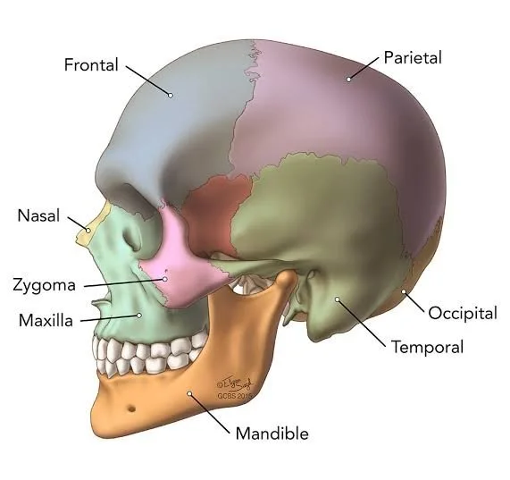
Figure 1: Osteological divisions of the cranial vault. Source: Goodman Campbell Brain and Spine
This article presents a structured, clinically oriented analysis of the cerebral region of the head designed for medical students, surgical trainees, and academic researchers. To build a complete spatial picture from the surface inward, the discussion is organized into four core sections:
● Regional Boundaries and Superficial Soft-Tissue Layers: Examining the anatomical borders of the cerebral head and the five-layered architecture of the frontoparietooccipital region.
● Topographical Stratification of the Temporal Region and Cellular Spaces: Analyzing the multi-layered musculo-fascial anatomy of the temporal fossa and the clinical significance of its closed cellular compartments.
● Neurovascular Architecture: Detailing the dense arterial anastomoses, valveless emissary venous drainage pathways, and trigeminal-cervical sensory innervation patterns.
● Cranio-Cerebral Projections and the Krönlein-Brusov Scheme: Providing a step-by-step geometric construction of the reference grid alongside its direct applications in trepanation and emergency vascular ligation.
1. Regional Boundaries and Superficial Soft-Tissue Layers of the Cerebral Region:
1.1 External Demarcation and Topographical Subdivisions:
In classical operative surgery and clinical morphology, the head is separated into two primary anatomical territories—the cerebral section (regio cerebralis) and the facial section (regio facialis)—by a continuous osteological boundary line. Starting anteriorly at the glabella, this demarcation traces laterally along the supraorbital margins of the frontal bone to the frontozygomatic sutures, follows the superior border of the zygomatic arch backward toward the external acoustic meatus, curves inferiorly around the apex of the mastoid process, and runs medially along the superior nuchal line to terminate at the external occipital protuberance (inion). Every structure situated superior and posterior to this line belongs to the cerebral region of the head, enveloping the neurocranium and the cerebral hemispheres.
To facilitate localized surgical access and systematic anatomical study, the cerebral region is further subdivided into three paired and unpaired topographic zones:
● The Frontoparietooccipital Region (Regio frontoparietooccipitalis): An extensive, unpaired vaulted territory bounded anteriorly by the supraorbital margins and glabella, posteriorly by the superior nuchal lines and external occipital protuberance, and laterally on each side by the superior temporal lines (lineae temporales superiores).
● The Temporal Region (Regio temporalis): Paired lateral zones bounded superiorly and posteriorly by the superior temporal line, anteriorly by the frontal process of the zygomatic bone, and inferiorly by the zygomatic arch, which separates the superficial temporal region from the deeper infratemporal fossa.
● The Mastoid Region (Regio mastoidea): Paired posteroinferior zones corresponding externally to the mastoid process of the temporal bone, bounded anteriorly by the posterior wall of the external acoustic meatus, superiorly by the temporal line extension (supramastoid crest), and inferiorly by the mastoid tip.
1.2 Stratification of the Frontoparietooccipital Region (SCALP Layers):
The soft tissues covering the cranial vault in the frontoparietooccipital region exhibit a remarkably uniform, five-layered arrangement classically remembered by the mnemonic SCALP, progressing sequentially from the external surface down to the calvarial bone. Unlike the loose, mobile integument of the neck or limbs, the outer three layers here are bound together into a single functional biomechanical unit.
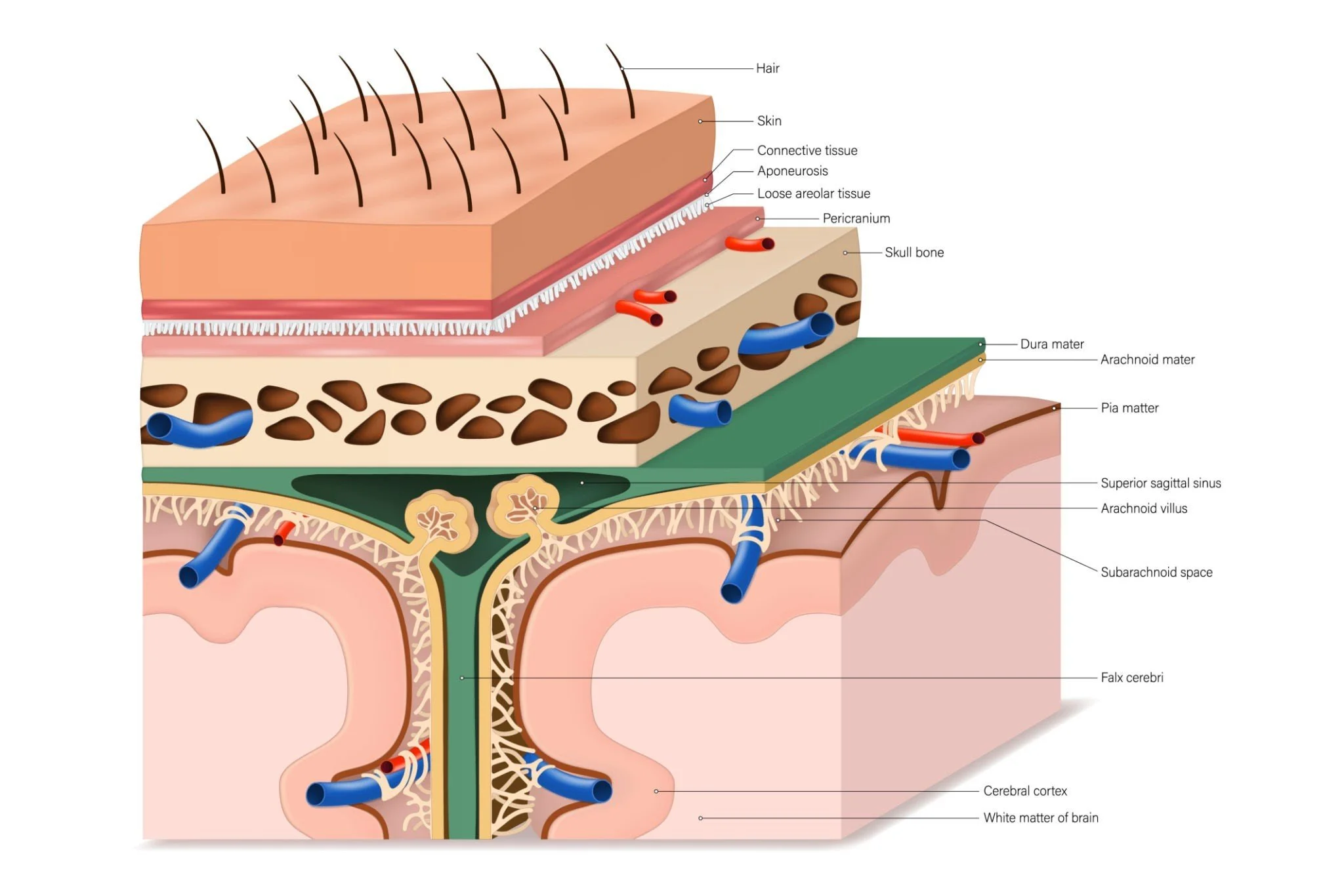
Figure 2: Stratification of the scalp, calvaria, and cranial meninges.
Skin (Cutis): The cutaneous layer of the cranial vault is among the thickest in the human body, measuring between 3 mm in the frontal zone and up to 8 mm over the occipital protuberance. It contains a dense concentration of hair follicles, sebaceous glands, and sweat glands. Clinically, this high glandular density predisposes the scalp to sebaceous (pilar) cysts and localized folliculitis. Because the dermis is tightly anchored to the underlying aponeurosis, skin incisions here cannot easily be undermined without releasing deeper fibrous septa.
Subcutaneous Connective Tissue (Tela subcutanea): Directly beneath the dermis lies a dense fibro-fatty layer divided into small lobules by tough, vertically oriented fibrous retinacula (retinacula cutis) that bind the skin firmly to the epicranial aponeurosis. Running directly within these rigid fibrous compartments are the primary trunks of the subcutaneous arteries, veins, and sensory nerves. When a scalp laceration severs an artery, these unyielding fibrous septa mechanically prevent the vessel walls from retracting or constricting, explaining why even minor scalp wounds bleed profusely until direct external pressure or galeal suturing is applied.
Epicranial Aponeurosis (Galea aponeurotica) and Occipitofrontalis Muscle: Forming the musculoaponeurotic foundation of the vault, the galea aponeurotica is a dense, inelastic tendinous sheet stretching across the vertex. Anteriorly, it anchors into the paired frontal bellies (venter frontalis) of the occipitofrontalis muscle, which insert into the dermis of the eyebrows and glabella without bony attachment; posteriorly, it attaches to the paired occipital bellies (venter occipitalis) arising from the supreme nuchal lines. Laterally, the galea thins out as it passes over the superior temporal line to continue into the temporoparietal fascia. From a surgical standpoint, the direction of scalp lacerations dictates wound behavior: transverse incisions that sever the galea parallel to the coronal suture gape widely due to opposing traction from the frontal and occipital bellies, whereas sagittal wounds remain closely approximated.
Loose Areolar Connective Tissue (Textus cellulosus subaponeuroticus): Situated between the galea aponeurotica and the underlying pericranium lies a delicate, avascular, sponge-like layer of loose connective tissue. This stratum serves as the natural gliding plane that allows the upper three layers of the scalp to move freely as a single unit over the cranium—a biomechanical arrangement known surgically as the scalping layer, as traumatic avulsion injuries typically shear along this exact plane. Because this subaponeurotic space lacks internal fibrous partitions across the vault, fluid collections—such as subgaleal hematomas or purulent effusions—can diffuse rapidly across the entire cranial dome, halting only at the bony attachments of the occipitofrontalis muscle posteriorly, the zygomatic arches laterally, and descending anteriorly into the loose tissues of the upper eyelids to produce delayed periorbital ecchymosis ("raccoon eyes") without an underlying orbital fracture.
Pericranium (Pericranium): The deepest soft-tissue layer is the external periosteum of the calvarial bones. While it adheres loosely across the smooth convexity of the frontal and parietal bones—allowing surgeons to elevate it effortlessly with a periosteal elevator—it becomes densely fused along the cranial sutures (coronal, sagittal, and lambdoid), where Sharpey's fibers plunge directly into the sutural connective tissue. Consequently, subperiosteal hematomas (such as neonatal cephalhematomas) remain strictly confined within the boundaries of a single cranial bone and never cross suture lines.
1.3 Osseous Architecture of the Calvaria:
Beneath the pericranium lies the bony vault of the skull (calvaria), engineered as a lightweight yet impact-resistant trilaminar structure:
● External Table (Lamina externa): A thick, resilient layer of compact cortical bone capable of absorbing and distributing blunt mechanical forces across the cranial curvature.
● Diploë: A central spongy cancellous layer housing red bone marrow and an intricate network of thin-walled, valveless diploic veins (venae diploicae—frontal, anterior temporal, posterior temporal, and occipital). Notably, the diploë is absent in newborns and develops gradually during early childhood as the cranial bones thicken.
● Internal Table (Lamina interna): A thinner, more brittle layer of compact bone lined internally by the periosteal layer of the dura mater. Because of its lower elasticity, blunt cranial trauma can fracture the internal table (lamina vitrea) independently while leaving the outer table intact, placing the underlying middle meningeal vessels at risk of laceration.
2. Topographical Stratification of the Temporal Region and Cellular Spaces:
2.1 Anatomical Complexity of the Temporal Fossa:
While the frontoparietooccipital vault follows a uniform five-layered architecture, the lateral wall of the cerebral region—specifically the temporal region (regio temporalis)—possesses a distinctly more intricate, multi-tiered musculo-fascial and cellular stratification. Bounded superiorly and posteriorly by the superior temporal line, anteriorly by the frontal process of the zygomatic bone, and inferiorly by the zygomatic arch, this territory houses the powerful fan-shaped temporalis muscle alongside critical neurovascular structures such as the superficial temporal vessels, the auriculotemporal nerve, and the deep temporal arteries. In neurosurgical approaches—particularly pterional, frontotemporal, and subtemporal craniotomies—navigating these layered planes without injuring the frontal branch of the facial nerve (ramus frontalis nervi facialis) or triggering postoperative masticatory fibrosis requires exact knowledge of each fascial division and its intervening cellular spaces.
2.2 Layer-by-Layer Stratification from Superficial to Deep:
Dissecting inward from the lateral cranial surface toward the squamous part of the temporal bone, nine distinct anatomical strata and cellular planes are encountered:
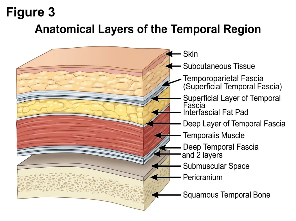
Skin (Cutis): Noticeably thinner, more pliable, and less densely populated with hair follicles than the skin of the vertex, particularly near the anterior hairline and preauricular margin.
Subcutaneous Connective Tissue (Tela subcutanea): A relatively loose, poorly lobulated adipose layer compared to the dense retinacular compartment of the vault. Within this plane ascend the main trunk and parietal/frontal branches of the superficial temporal artery (arteria temporalis superficialis), the accompanying superficial temporal vein, and the auriculotemporal nerve (nervus auriculotemporalis).
Superficial Temporal Fascia / Temporoparietal Fascia (Fascia temporoparietalis): Representing the lateral continuation of the galea aponeurotica over the superior temporal line, this thin, vascularized fibromuscular sheet connects inferiorly with the superficial musculoaponeurotic system (SMAS) of the face. Crucially, the temporal (frontal) branch of the facial nerve courses directly along the deep surface or within the substance of this fascial layer as it crosses the zygomatic arch roughly 1.5 to 2.0 cm anterior to the tragus.
Loose Subaponeurotic Areolar Plane (Innominate Fascia): Situated immediately beneath the temporoparietal fascia, this avascular gliding plane allows free mobility of the overlying scalp and superficial fascia over the dense deep temporal fascia below.
Deep Temporal Fascia (Fascia temporalis): A glistening, remarkably tough aponeurotic membrane anchored superiorly along the superior temporal line. Inferiorly, roughly 2 to 3 cm above the zygomatic arch, this single dense sheet bifurcates into two distinct laminae:
○ Superficial Lamina (Lamina superficialis): Attaches to the lateral margin of the superior border of the zygomatic arch.
○ Deep Lamina (Lamina profunda): Attaches to the medial margin of the superior border of the zygomatic arch, continuing deep to the arch to blend with the parotidomasseteric fascia.
Interfascial Adipose Space (Spatium interaponeuroticum): Enclosed between the diverging superficial and deep laminae of the deep temporal fascia just above the zygomatic arch, this triangular compartment contains loose adipose tissue, the middle temporal artery (arteria temporalis media), and branches of the zygomaticotemporal nerve. In modern neurosurgery, performing an interfascial dissection through this fat pad protects the frontal branch of the facial nerve when mobilizing a frontotemporal myocutaneous flap.
Subaponeurotic Temporal Space (Spatium subaponeuroticum temporale): Located between the deep surface of the deep temporal fascia and the superficial belly of the temporalis muscle, this closed cellular compartment contains a cushion of loose fat that facilitates smooth sliding of the muscle fibers during mastication.
Temporalis Muscle (Musculus temporalis): A broad, bipennate masticatory muscle originating from the entire bony floor of the temporal fossa and the deep surface of the temporal fascia, converging inferiorly into a thick tendon that passes medial to the zygomatic arch to insert onto the coronoid process and anterior ramus of the mandible.
Deep Temporal Cellular Space (Spatium temporale profundum) and Pericranium: Beneath the muscular belly lies a deep connective tissue plane traversed by the anterior and posterior deep temporal arteries and nerves (branches of the maxillary and mandibular divisions, respectively), which enter the muscle directly from its deep osteal aspect. Below this plane lies the thin periosteum tightly adherent to the squamous temporal bone, parietal bone, and greater wing of the sphenoid (pterion).
2.3 Clinical Significance of the Closed Cellular Spaces:
The architectural arrangement of the three temporal cellular compartments—interfascial, subaponeurotic, and deep temporal—carries profound clinical implications for both trauma and deep head-and-neck infections:
● Self-Limiting vs. Descending Spread: Because the deep temporal fascia seals firmly along the superior temporal line and the zygomatic arch, hematomas or suppurative collections within the interfascial and subaponeurotic spaces remain strictly bounded above the zygomatic arch, producing localized convex swelling of the temple without tracking into the cheek.
● Pathways to the Deep Face: Conversely, the deep temporal cellular space beneath the temporalis muscle communicates directly inferiorly beneath the zygomatic arch with the infratemporal fossa, the pterygopalatine fossa, and the buccal fat pad (Bichat's fat pad). Consequently, odontogenic abscesses originating from upper molar infections or deep infratemporal hematomas can ascend upward beneath the zygomatic arch into the deep temporal space, presenting clinically as painful temporal swelling accompanied by severe trismus due to irritation of the temporalis muscle fibers.
3. Neurovascular Architecture: Blood Supply, Venous Drainage, and Innervation:
3.1 Arterial Network and Anastomotic Rings
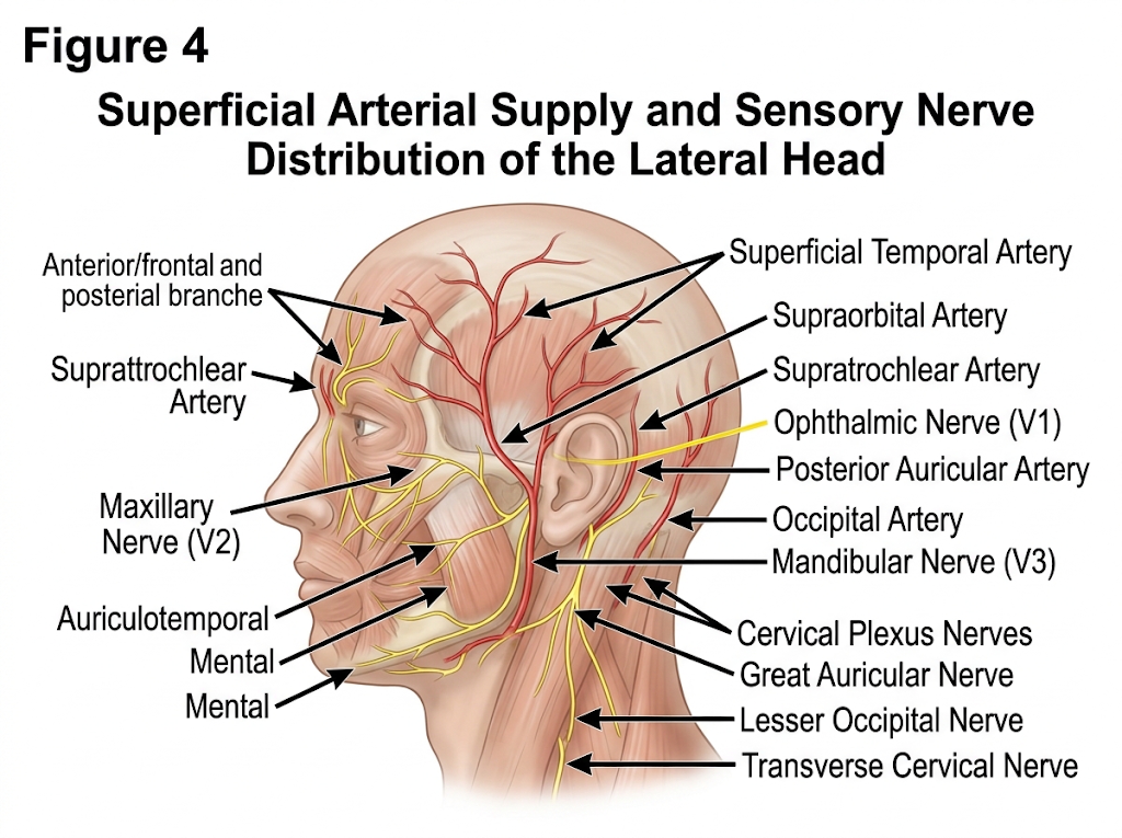
The soft tissues of the cerebral region receive a rich, centripetally directed arterial supply derived from both the internal carotid and external carotid systems, creating a dense subdermal anastomotic network across the cranial vault.
● Anterior (Internal Carotid) Territory: Branches of the ophthalmic artery—specifically the supratrochlear artery (a. supratrochlearis) and supraorbital artery (a. supraorbitalis)—exit their respective orbital notches to ascend over the forehead, supplying the frontal scalp and pericranium.
● Lateral and Posterior (External Carotid) Territory: Three main trunks supply the remaining vault: the superficial temporal artery (a. temporalis superficialis), ascending anterior to the tragus and dividing into frontal and parietal branches; the posterior auricular artery (a. auricularis posterior), ascending behind the auricle; and the occipital artery (a. occipitalis), piercing the trapezius attachment to vascularize the posterior scalp up to the vertex. Deep to the temporalis muscle, the anterior and posterior deep temporal arteries (from the maxillary artery) supply the muscular belly and underlying squamous bone.
Because these five paired vessels form free anastomoses across the midline and between anterior and posterior territories, large scalp flaps survive reliably on narrow pedicles, though arterial lacerations produce brisk bidirectional hemorrhage.
3.2 Venous Drainage and Intracranial Connections:
Superficial scalp veins accompany their corresponding arteries—forming the supratrochlear, supraorbital, superficial temporal, posterior auricular, and occipital venous trunks—before draining into the facial, retromandibular, and external jugular systems. Clinically, these extracranial veins communicate directly with the diploic veins within the calvarial cancellous bone and with the intracranial dural venous sinuses via valveless emissary veins (venae emissariae—notably the parietal, mastoid, and occipital emissary veins). Because these channels lack valves, bidirectional blood flow permits superficial scalp or temporal infections to propagate inward, risking septic dural sinus thrombosis or meningitis.
3.3 Sensory and Motor Innervation:
Sensory innervation of the cerebral head divides sharply along a coronal plane passing through the vertex and external acoustic meatus:
● Anterior to the Interauricular Line: Supplied by all three divisions of the trigeminal nerve (CN V)—the supratrochlear and supraorbital nerves anteriorly; the zygomaticotemporal nerve over the anterior temple; and the auriculotemporal nerve ascending with the superficial temporal vessels.
● Posterior to the Interauricular Line: Supplied by dorsal and ventral rami of the upper cervical spinal nerves specifically the lesser occipital nerve, the greater occipital nerve, and the third occipital nerve.
● Motor Innervation: The frontal and occipital bellies of the occipitofrontalis muscle are innervated by the temporal and posterior auricular branches of the facial nerve (CN VII), respectively, while the temporalis muscle receives motor fibers from the deep temporal nerves of the mandibular division.
4. Cranio-Cerebral Projections and the Krönlein-Brusov Scheme:
4.1 Principles of Cranio-Cerebral Topography:
Establishing a reliable geometric correlation between external cranial landmarks and deep intracranial neurovascular structures is fundamental in neurosurgery, particularly in emergency trauma scenarios where stereotactic neuronavigation is unavailable. The Krönlein-Brusov scheme provides a standardized, reproducible geometric grid that projects the central sulcus (Rolandic fissure), lateral sulcus (Sylvian fissure), and primary branches of the middle meningeal artery directly onto the intact scalp and skull.
4.2 Construction of the Krönlein-Brusov Grid:
To construct the classical Krönlein-Brusov topographic coordinate system on the lateral aspect of the head, a specific sequence of craniometric lines is drawn:
Baseline (Line I / Frankfort Horizontal): A horizontal baseline is drawn connecting the inferior orbital margin (margo infraorbitalis) anteriorly to the upper margin of the external acoustic meatus (porus acusticus externus) posteriorly.
Base Parallel (Line II): A second parallel horizontal line is drawn tangent to the superior margin of the zygomatic arch and the supraorbital margin of the frontal bone.
Vertical Lines (Lines III, IV, and V): Three perpendicular vertical lines are erected from the baseline:
○ Anterior Vertical Line (Line III): Drawn vertically upward from the midpoint of the zygomatic arch.
○ Middle Vertical Line (Line IV): Drawn vertically upward from the posterior border of the articular eminence of the temporal bone (coinciding with the preauricular point).
○ Posterior Vertical Line (Line V): Drawn vertically upward from the posterior margin of the mastoid process.
4.3 Projection of Intracranial Structures:
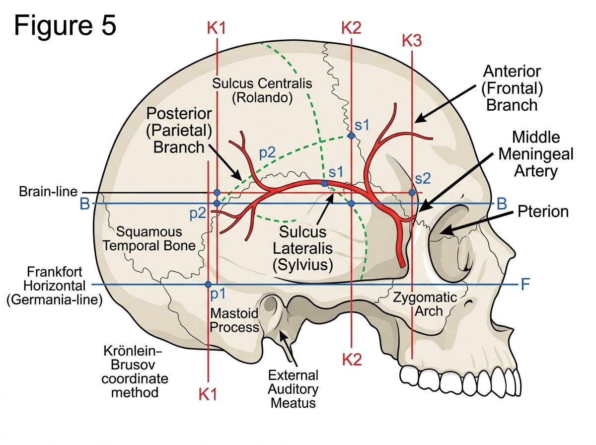
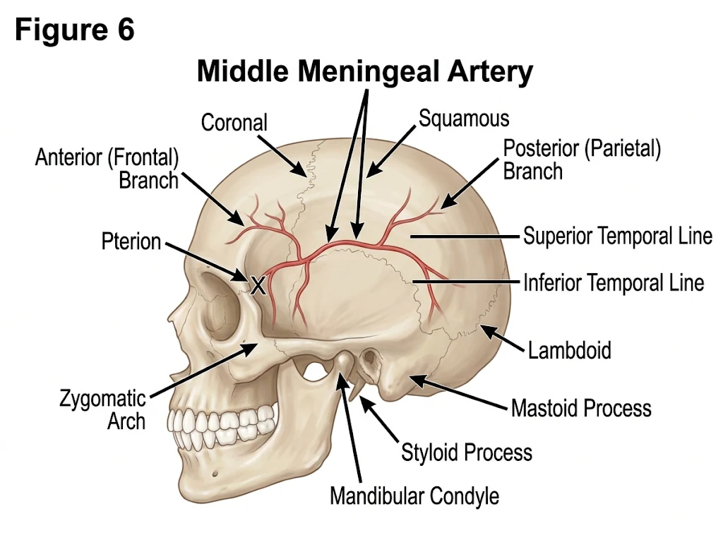
Using this geometric grid, the key cortical fissures and meningeal vessels are projected as follows:
● Middle Meningeal Artery (A. meningea media): The main trunk and its anterior branch are mapped using two intersection points: the lower point situated 1.5 cm above the zygomatic arch on the anterior vertical line, and the upper point located at the junction of the middle and lower thirds of the distance between the orbital margin and external acoustic meatus.
● Central Sulcus (Rolandic Fissure): Projected by connecting the point of intersection between the posterior vertical line (Line V) and the skull vertex with the point where the anterior vertical line (Line III) intersects the oblique Rolandic line (drawn from the root of the nose to the inion at an angle of roughly 67 degrees).
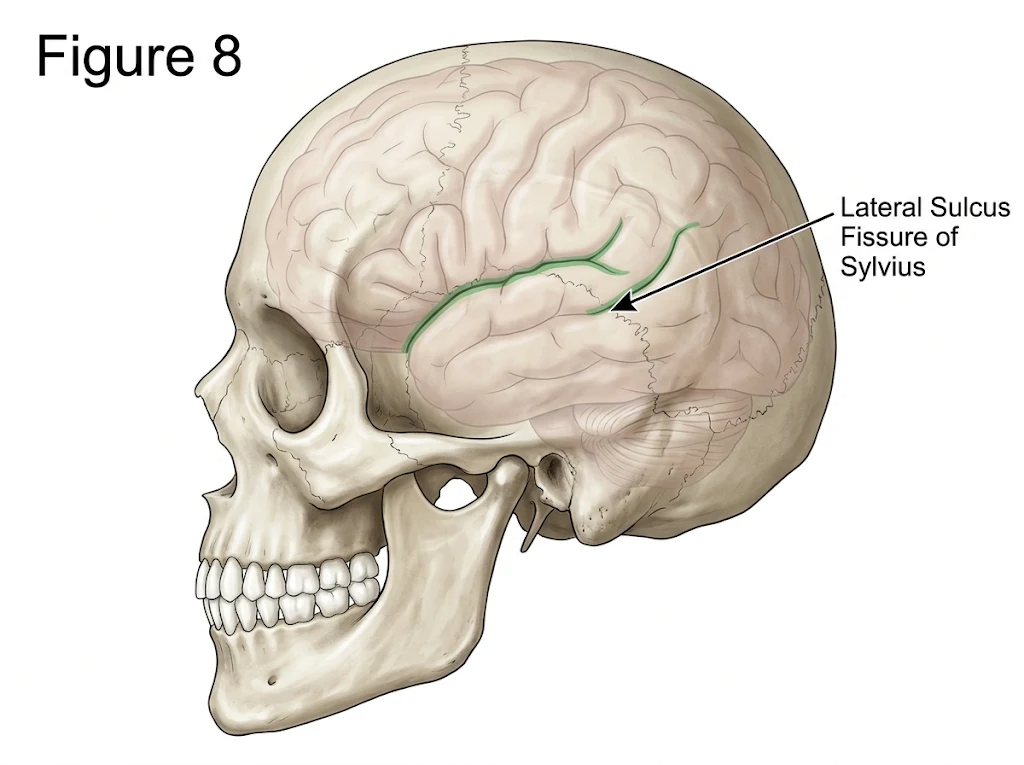
Conclusion:
A comprehensive command of the topographic anatomy of the cerebral region of the head—spanning from the dense, retinacular layers of the frontoparietooccipital scalp and the multi-tiered cellular compartments of the temporal fossa to the underlying neurovascular networks—remains indispensable in clinical medicine and operative neurosurgery. The distinct biomechanical properties of the galea aponeurotica, the closed fascial spaces of the temple, and the valveless connections linking superficial scalp veins with intracranial dural sinuses directly dictate how cranial trauma, subgaleal hematomas, and deep infections evolve clinically. Furthermore, the Krönlein-Brusov scheme bridges external osteological landmarks with deep cerebral sulci and meningeal vasculature, offering a rapid, reproducible geometric method for localizing the central sulcus, lateral sulcus, and middle meningeal artery during emergency trepanation and trauma care. Integrating these foundational anatomical principles with contemporary neurosurgical techniques minimizes iatrogenic injury and strengthens diagnostic precision at the bedside.
References:
Standring S, ed. Gray's Anatomy: The Anatomical Basis of Clinical Practice. 42nd ed. Elsevier; 2021.
Moore KL, Dalley AF, Agur AMR. Clinically Oriented Anatomy. 9th ed. Wolters Kluwer; 2023.
Kovanov VV, ed. Operative Surgery and Topographic Anatomy. Mir Publishers; 1985.
Ostroverkhov GE, Bomash YM, Lubotsky DN. Operative Surgery and Topographic Anatomy. MIA; 2013.
Rhoton AL Jr. The cerebrum. Neurosurgery. 2002;51(4 Suppl):S1-S51.
Yasargil MG, Reichman MV, Kubik S. Preservation of the frontotemporal branch of the facial nerve using the interfascial temporalis flap for pterional craniotomy. J Neurosurg. 1987;67(3):463-466.
Seckel BR. Facial Danger Zones: Avoiding Nerve Injury in Facial Plastic Surgery. 2nd ed. Quality Medical Publishing; 2010.
Tubbs RS, Salter EG, Oakes WJ. Superficial surgical landmarks for the transverse sinus and torcular herophili. J Neurosurg. 2000;93(2):279-281.
Ribas GC, Yasuda A, Ribas EC, Nishikuni K, Rodrigues AJ Jr. Surgical anatomy of microneurosurgical sulcal key points. Neurosurgery. 2006;59(4 Suppl 2):ONS177-ONS210.
Gusmão S, Silveira RL, Arantes A. Landmarks to the one-burr hole pterional craniotomy. Arq Neuropsiquiatr. 2003;61(4):1013-1017.
Kendir S, Acar HI, Comert A, et al. Window anatomy for neurosurgical approaches: laboratory investigation. J Neurosurg. 2009;111(2):365-370.
Davidge KM, van Furth WR, Agur A, Cusimano M. Naming the soft tissue layers of the temporoparietal region: unifying anatomic terminology across surgical disciplines. Neurosurgery. 2010;67(3 Suppl Operative):ons120-ons129.
Poblete T, Jiang X, Komune N, Matsushima K, Rhoton AL Jr. Preservation of the nerves to the frontalis muscle during pterional craniotomy. J Neurosurg. 2015;122(6):1274-1282.
Abul-Kasim K, Overgaard A, Maly P, et al. Age-related changes in calvarial thickness and diploic width: a computed tomography study. Surg Radiol Anat. 2009;31(7):515-521.
Federative International Programme for Anatomical Terminology (FIPAT). Terminologia Anatomica. 2nd ed. FIPAT; 2019.
Greenberg MS. Greenberg's Handbook of Neurosurgery. 10th ed. Thieme; 2023.