
Peripheral Nerve Injuries of the Upper Limb: Pathophysiology, Clinical Assessment, and Contemporary Management Strategies in Modern Peripheral Nerve Surgery
1. Shaurya Kumar Sahu
2. Saachi Chaurasia
3. Jesminara Yeasmin
4. Saharina Ahmed
5. Monima Choudhary
6. Tanabaeva Aiperi
(1. Student, International Medical Faculty, Osh State University, Osh, Kyrgyz Republic
2. Student, International Medical Faculty, Osh State University, Osh, Kyrgyz Republic
3. Student, International Medical Faculty, Osh State University, Osh, Kyrgyz Republic
4. Student, International Medical Faculty, Osh State University, Osh, Kyrgyz Republic
5. Student, International Medical Faculty, Osh State University, Osh, Kyrgyz Republic
6. Professor, International Medical Faculty, Osh State University, Osh, Kyrgyz Republic.)
Abstract
Peripheral nerve injuries of the upper limb represent a significant source of long-term disability and functional impairment, affecting the quality of life of patients across all age groups and socioeconomic strata. The intricate anatomy of the brachial plexus and its terminal branches, combined with the superficial location of major nerves in the upper extremity, renders these structures vulnerable to trauma, compression, traction, and iatrogenic injury. This comprehensive review examines the pathophysiology, classification, clinical assessment, and management of peripheral nerve injuries affecting the upper limb, synthesizing classical anatomical knowledge with recent advances in nerve transfer techniques, neurotization procedures, and rehabilitative protocols. The article presents detailed analysis of injury mechanisms including neuropraxia, axonotmesis, and neurotmesis, with particular attention to the temporal progression of wallerian degeneration and the critical window for surgical intervention. Recent epidemiological data indicate that upper limb peripheral nerve injuries account for approximately 50 to 60 percent of all peripheral nerve trauma, with the median nerve being the most frequently injured single nerve, followed by the ulnar and radial nerves. The review further explores the surgical implications of nerve injury patterns including high-energy traction injuries, sharp lacerations, and chronic compression neuropathies, alongside the evolving paradigm of nerve transfers that has revolutionized the treatment of brachial plexus injuries and irreparable nerve damage. By integrating classical neuroanatomical studies with contemporary electrodiagnostic techniques, intraoperative nerve monitoring, and microsurgical innovations, this article provides orthopedic surgeons, plastic surgeons, neurosurgeons, and rehabilitation specialists with a robust framework for understanding upper limb nerve injuries not merely as static anatomical disruptions, but as dynamic biological events requiring timely intervention and multidisciplinary management to optimize functional recovery.
Introduction
The peripheral nerves of the upper limb represent a remarkable feat of biological engineering, transmitting motor commands from the central nervous system to the intricate musculature of the shoulder, arm, forearm, and hand while simultaneously conveying sensory information essential for tactile discrimination, proprioception, and protective sensation. These neural structures, arising from the ventral rami of the fifth cervical through the first thoracic spinal nerves and coursing through the complex architecture of the brachial plexus before terminating in the digital nerves of the hand, are uniquely vulnerable to injury due to their superficial location, limited elasticity, and intimate relationship with mobile bony and articular structures. The consequences of upper limb nerve injury extend far beyond mere loss of motor or sensory function; they encompass chronic pain, loss of dexterity, inability to perform activities of daily living, and significant psychological distress, creating a substantial burden on healthcare systems and society.
The history of peripheral nerve surgery is marked by pivotal contributions from military surgeons who, confronted with devastating nerve injuries during armed conflicts, developed foundational principles of nerve repair that persist today. From the early observations of Augustus Waller regarding degeneration of the distal nerve stump, to the pioneering microsurgical techniques of James Smith and Hanno Millesi in the latter half of the twentieth century, the field has evolved from simple nerve suturing to sophisticated nerve grafting, nerve transfer, and end-to-side neurorrhaphy techniques. The introduction of the operating microscope revolutionized nerve repair by permitting precise alignment of fascicular bundles, dramatically improving outcomes for digital nerve repairs and major nerve reconstructions. However, despite these advances, functional recovery following major upper limb nerve injury remains imperfect, with motor recovery rarely exceeding grade four on the Medical Research Council scale and sensory recovery often inadequate for fine manipulative tasks.
The classification of nerve injuries, most famously described by Sir Herbert Seddon in 1943 and later refined by Sunderland in 1951, provides a framework for understanding the spectrum of nerve damage from transient conduction block to complete transection. Neuropraxia, characterized by temporary conduction failure without axonal disruption, typically resolves spontaneously within weeks to months. Axonotmesis involves disruption of the axon with preservation of the endoneurial tube, permitting axonal regeneration at a rate of approximately one millimeter per day. Neurotmesis represents complete disruption of the nerve trunk, requiring surgical intervention for any meaningful recovery. This classification, while anatomically based, carries profound prognostic implications and dictates the timing and nature of therapeutic intervention.
Contemporary peripheral nerve surgery has witnessed a paradigm shift with the increasing recognition that nerve transfers, the redirection of a functioning donor nerve to reinnervate a distal target, often provide superior outcomes to traditional nerve grafting for certain injury patterns. This approach, pioneered for brachial plexus injuries and now extended to peripheral nerve injuries throughout the upper limb, capitalizes on the principle of neurotization, bringing healthy axons closer to their targets to minimize the time to reinnervation and prevent irreversible muscle atrophy. The development of supercharge end-to-side nerve transfers, where a donor nerve is coapted to the side of a partially injured nerve, has further expanded the armamentarium for treating nerve injuries previously considered irreparable.
This review aims to provide a comprehensive examination of peripheral nerve injuries of the upper limb, integrating classical neuroanatomical knowledge with contemporary surgical techniques, electrodiagnostic evaluation, and rehabilitative strategies. The article will explore the mechanisms and classification of nerve injury, delineate the clinical assessment of specific nerve lesions, examine surgical and non-surgical management options, and discuss the critical importance of timing in nerve reconstruction. By synthesizing data from anatomical studies, clinical series, and technological advances in nerve repair, this work seeks to provide a reference framework that acknowledges upper limb nerve injuries as complex biological events requiring precise diagnosis, timely intervention, and dedicated rehabilitation to restore meaningful function to the injured extremity.
Materials and Methods
This review article was constructed through systematic analysis of the contemporary peripheral nerve surgery and neuroanatomical literature, with emphasis on publications from the past two decades regarding nerve injury classification, surgical techniques, and functional outcomes. The methodological approach involved comprehensive examination of peer-reviewed journals in orthopedic surgery, plastic surgery, neurosurgery, and rehabilitation medicine, including Journal of Hand Surgery, Plastic and Reconstructive Surgery, Journal of Neurosurgery, Neurosurgery, and Journal of Bone and Joint Surgery. Particular attention was directed toward studies utilizing electrodiagnostic testing for nerve injury assessment, intraoperative nerve monitoring for surgical decision-making, and prospective outcome studies comparing nerve repair techniques.
Inclusion criteria for clinical data encompassed studies reporting functional outcomes following nerve repair, grafting, or transfer procedures in cohorts exceeding twenty subjects, with preference given to studies utilizing validated outcome measures including the Medical Research Council grading system, the Disabilities of the Arm, Shoulder and Hand questionnaire, and grip strength measurements. Anatomical data were sourced from cadaveric dissection studies and intraoperative observations documenting nerve branching patterns and donor nerve availability. Electrodiagnostic criteria for nerve injury severity were derived from studies correlating nerve conduction studies and electromyography with intraoperative findings and clinical outcomes. When discrepancies existed between historical series and contemporary reports regarding nerve regeneration rates and functional recovery, preference was given to recent studies incorporating modern microsurgical techniques and early rehabilitation protocols.
The synthesis of this literature was organized according to the anatomical distribution of upper limb nerves, progressing from the brachial plexus to the terminal branches including the axillary, musculocutaneous, radial, median, and ulnar nerves. All clinical descriptions are presented according to standard neurological examination techniques, with functional grading reported according to established scales. The reference list comprises foundational anatomical works, contemporary surgical series, and technological studies that collectively represent the current state of knowledge regarding peripheral nerve injuries of the upper limb.
Results
Pathophysiology and Classification of Nerve Injury
Peripheral nerve injury initiates a complex cascade of biological events at the cellular and molecular level that determines the potential for functional recovery. Following axonal disruption, the distal nerve segment undergoes wallerian degeneration, characterized by fragmentation of the axon and myelin sheath, recruitment of macrophages for phagocytosis of debris, and proliferation of Schwann cells within the endoneurial tubes. This degenerative process, which begins within hours of injury and continues for several weeks, creates a permissive environment for axonal regeneration if the endoneurial scaffolding remains intact. The proximal nerve stump undergoes retrograde degeneration to the nearest node of Ranvier or, in severe cases, to the cell body in the anterior horn of the spinal cord or dorsal root ganglion, potentially resulting in neuronal death when the injury is proximal and severe.
The rate of axonal regeneration following nerve repair is approximately one millimeter per day or one inch per month, a biological constraint that dictates the prognosis for functional recovery based on the distance from the site of injury to the target muscle or sensory end organ. This regenerative rate creates the concept of the critical window for nerve repair, as denervated muscle fibers undergo irreversible atrophy and fibrosis if reinnervation is not achieved within approximately twelve to eighteen months. Similarly, sensory end organs degenerate if not reinnervated within a similar timeframe, though sensory recovery may occur after longer delays due to the ability of sensory axons to reinnervate adjacent territories.
The Seddon classification, later expanded by Sunderland, provides a framework for understanding nerve injury severity. First-degree injury or neuropraxia involves temporary conduction block without axonal disruption, typically resulting from compression or traction, with spontaneous recovery expected within days to weeks. Second-degree injury or axonotmesis involves axonal disruption with preservation of the endoneurial tube, permitting axonal regeneration without surgical intervention, though recovery may take months depending on the distance to target organs. Third-degree injury involves disruption of the endoneurium with preservation of the perineurium, creating misalignment of regenerating axons and requiring possible surgical intervention. Fourth-degree injury involves disruption of the perineurium with preservation of the epineurium, creating a neuroma-in-continuity that blocks regeneration and requires surgical resection and repair. Fifth-degree injury or neurotmesis represents complete transection of the nerve trunk, mandating surgical repair for any meaningful recovery.
Clinical Assessment and Electrodiagnosis
The clinical assessment of upper limb peripheral nerve injuries requires systematic neurological examination to determine the extent of motor weakness, sensory loss, and autonomic dysfunction. Motor examination utilizes the Medical Research Council grading scale from zero representing no contraction to five representing normal power against full resistance. Sensory examination includes static two-point discrimination for assessing innervation density and moving two-point discrimination for assessing fiber orientation, with normal values being less than six millimeters for static and less than three millimeters for moving discrimination in the fingertip. The ten test, assessing the ability to identify a ten gram monofilament, provides a screening tool for protective sensation. Autonomic function is assessed by skin temperature, color, sweating, and turgor, with denervated skin appearing warm, dry, and smooth due to loss of sympathetic innervation.
Electrodiagnostic studies including nerve conduction studies and electromyography provide objective assessment of nerve injury severity and localization. In neuropraxia, nerve conduction studies demonstrate conduction block across the injured segment with normal distal conduction, while electromyography shows no fibrillation potentials or positive sharp waves indicating denervation. In axonotmesis and neurotmesis, distal conduction fails after wallerian degeneration occurs, typically three to five days after injury for motor nerves and seven to ten days for sensory nerves. Electromyography demonstrates fibrillation potentials and positive sharp waves in denervated muscles two to three weeks following injury, indicating axonal disruption. The presence of motor unit action potentials on voluntary contraction indicates neuropraxia or early reinnervation, while their absence in the presence of denervation potentials indicates severe injury requiring surgical exploration.
Advanced imaging techniques including high-resolution ultrasound and magnetic resonance neurography have emerged as valuable adjuncts in the assessment of peripheral nerve injuries. Ultrasound can identify nerve discontinuity, neuroma formation, and compression sites, while magnetic resonance neurography can demonstrate nerve edema, atrophy, and fascicular disruption. These imaging modalities are particularly valuable in assessing brachial plexus injuries and in planning nerve transfer procedures by identifying donor nerve availability.
Specific Nerve Injuries and Management
The brachial plexus represents the most complex anatomical region of upper limb innervation, formed by the ventral rami of C5 through T1 with contributions from C4 and T2 in variant configurations. Injuries to the brachial plexus result from high-energy trauma including motorcycle accidents, industrial injuries, and sports trauma, producing flail extremities with varying degrees of motor and sensory loss depending on the level and severity of injury. Supraclavicular injuries affecting the trunks and divisions carry poorer prognosis than infraclavicular injuries affecting the cords and terminal branches. Complete brachial plexus palsy requires urgent surgical exploration, typically within three to six months, with nerve grafting for ruptured elements and nerve transfers for avulsed roots. The Oberlin transfer, utilizing ulnar nerve fascicles to the biceps branch of the musculocutaneous nerve, and the double fascicular transfer, utilizing median and ulnar nerve fascicles to biceps and brachialis branches, have revolutionized elbow flexion restoration in C5-C6 injuries.
The axillary nerve, arising from the posterior cord and innervating the deltoid and teres minor muscles, is commonly injured in shoulder dislocations and proximal humerus fractures. Injury produces weakness in shoulder abduction and sensory loss over the lateral shoulder. Most axillary nerve injuries represent neuropraxia and recover spontaneously, but persistent weakness beyond three months warrants surgical exploration. Nerve grafting or transfer of the triceps branch of the radial nerve to the axillary nerve provides reliable restoration of shoulder abduction when primary repair is not possible.
The musculocutaneous nerve, innervating the biceps, brachialis, and coracobrachialis muscles, is less commonly injured in isolation but may be involved in brachial plexus injuries or compressed by the coracobrachialis muscle. Injury produces weakness in elbow flexion and sensory loss over the lateral forearm. The Oberlin transfer has become the procedure of choice for isolated musculocutaneous nerve injuries, providing reliable elbow flexion through transfer of ulnar nerve fascicles to the biceps motor branch.
The radial nerve, the largest branch of the brachial plexus arising from the posterior cord, innervates the extensor muscles of the arm and forearm including the triceps, brachioradialis, extensor carpi radialis longus and brevis, and the digital extensors. Injury in the spiral groove of the humerus, typically from humeral shaft fractures, produces the classic wrist drop with weakness in wrist and finger extension and sensory loss over the dorsal hand and first web space. Most radial nerve injuries associated with humeral fractures represent neuropraxia and recover spontaneously, with exploration indicated if no recovery occurs by three to four months. High radial nerve injuries requiring reconstruction benefit from nerve grafting or transfers including the pronator teres to extensor carpi radialis brevis transfer for wrist extension restoration.
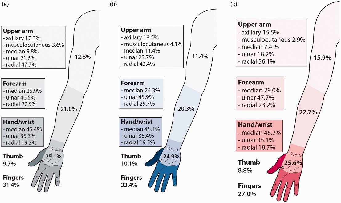
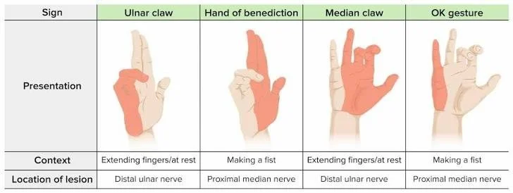
The median nerve, formed by contributions from the lateral and medial cords, innervates the flexor muscles of the forearm including the pronator teres, flexor carpi radialis, palmaris longus, and flexor digitorum superficialis, and through the anterior interosseous nerve innervates the flexor pollicis longus, flexor digitorum profundus to the index and middle fingers, and pronator quadratus. In the hand, the median nerve innervates the thenar muscles including the abductor pollicis brevis, opponens pollicis, and superficial head of the flexor pollicis brevis, and provides sensation to the palmar thumb, index, middle, and radial ring fingers. Injury at the elbow produces the hand of benediction posture when attempting to make a fist, with weakness in thumb opposition and sensory loss in the median distribution. High median nerve injuries require urgent repair given the importance of thumb opposition for hand function, with nerve grafting or transfers including the brachialis branch to the anterior interosseous nerve for pronation and flexion restoration.
The ulnar nerve, the terminal branch of the medial cord, innervates the flexor carpi ulnaris and medial half of the flexor digitorum profundus in the forearm, and in the hand innervates the hypothenar muscles, interossei, lumbricals to the ring and little fingers, adductor pollicis, and deep head of the flexor pollicis brevis. Injury produces the classic claw hand deformity with hyperextension of the metacarpophalangeal joints and flexion of the interphalangeal joints of the ring and little fingers, weakness in grip and pinch, and sensory loss over the little finger and ulnar half of the ring finger. Ulnar nerve injuries at the elbow, whether from trauma or chronic compression in the cubital tunnel, require careful assessment as the nerve is vulnerable to injury during medial epicondyle fracture fixation and elbow arthroscopy. Nerve grafting or transfers including the anterior interosseous nerve to the ulnar nerve motor branch provide options for reconstruction when primary repair is not feasible.
Surgical Techniques and Rehabilitation
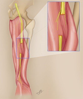
Primary nerve repair, performed when the nerve ends can be approximated without tension, remains the gold standard for sharp nerve lacerations when performed within the optimal timeframe. Epineurial repair using fine monofilament sutures under microscopic magnification aims to align the fascicular bundles without creating tension at the repair site. For gaps that cannot be bridged by primary repair, nerve grafting utilizing the sural nerve, medial antebrachial cutaneous nerve, or other expendable sensory nerves provides conduits for axonal regeneration. Vascularized nerve grafts, including the ulnar nerve based on the superior ulnar collateral artery, provide superior outcomes for long gaps and poorly vascularized beds.
Nerve transfers have revolutionized the treatment of peripheral nerve injuries, particularly for brachial plexus injuries and injuries with long distances to target muscles. The principle of nerve transfer involves sacrificing a less important donor nerve or fascicle to reinnervate a critical distal target, bringing healthy axons closer to the end organ to minimize the time to reinnervation. Common nerve transfers in the upper limb include the Oberlin transfer for elbow flexion, the posterior branch of the axillary nerve to the triceps branch for shoulder abduction, and the distal anterior interosseous nerve to the ulnar nerve motor branch for intrinsic hand function restoration. End-to-side nerve transfers, where donor nerve fascicles are coapted to the side of an injured nerve, provide options for augmenting partial nerve injuries.
Tendon transfers remain valuable adjuncts or alternatives to nerve reconstruction, particularly when nerve injury is diagnosed late or nerve reconstruction has failed. Standard tendon transfers including the palmaris longus to extensor pollicis longus for thumb extension restoration and the flexor digitorum superficialis to extensor digitorum communis for finger extension restoration provide reliable functional restoration independent of nerve regeneration.
Rehabilitation following peripheral nerve injury is critical for optimizing functional outcomes and preventing complications including joint contracture, muscle atrophy, and sensory reeducation failure. Splinting maintains joints in functional positions during the period of denervation, while passive range of motion exercises prevent stiffness. Following reinnervation, progressive strengthening exercises and sensory reeducation techniques including sensory substitution and discriminative reeducation maximize functional recovery. The integration of occupational therapy and hand therapy into the treatment team is essential for achieving optimal outcomes.
Discussion
Peripheral nerve injuries of the upper limb present a complex challenge that requires integration of anatomical knowledge, surgical skill, and rehabilitative expertise to achieve meaningful functional recovery. The biological constraints of axonal regeneration, limited to approximately one millimeter per day, create a critical window for intervention that demands timely diagnosis and appropriate surgical planning. The evolution from simple nerve suturing to sophisticated nerve transfer techniques has expanded the possibilities for functional restoration, particularly for brachial plexus injuries and injuries with long nerve gaps, yet outcomes remain imperfect with motor recovery rarely achieving normal strength and sensory recovery often inadequate for fine discriminative tasks.
The importance of early referral to specialized peripheral nerve centers cannot be overstated, as delays in diagnosis and treatment beyond the critical window of twelve to eighteen months result in irreversible muscle atrophy and fibrosis that precludes meaningful recovery even with optimal surgical technique. The development of standardized assessment protocols including electrodiagnostic testing and advanced imaging facilitates early diagnosis and surgical planning, while intraoperative nerve monitoring and histological assessment of nerve stumps guide intraoperative decision-making regarding repair versus grafting versus transfer.
The future of peripheral nerve surgery likely involves advances in nerve conduits, biological scaffolds, and pharmacological agents to enhance axonal regeneration, as well as refinements in nerve transfer techniques to expand the repertoire of reconstructive options. However, the fundamental principles of early diagnosis, timely intervention, and dedicated rehabilitation remain unchanged. The surgeon who understands the pathophysiology of nerve injury, who masters the techniques of microsurgical repair and nerve transfer, and who collaborates with rehabilitation specialists to maximize functional recovery, provides the best opportunity for patients with upper limb nerve injuries to regain meaningful use of their extremities.
Conclusion
Peripheral nerve injuries of the upper limb represent a significant source of disability that demands precise anatomical knowledge, timely surgical intervention, and dedicated rehabilitation to optimize functional outcomes. The classification of nerve injuries from neuropraxia to neurotmesis provides a framework for understanding prognosis and guiding treatment, while the biological constraints of axonal regeneration dictate the critical window for surgical intervention. The evolution of nerve transfer techniques has expanded the possibilities for functional restoration, particularly for injuries previously considered irreparable, yet the importance of primary nerve repair when feasible remains paramount. As the field continues to advance with biological enhancements and technological innovations, the fundamental principles of early diagnosis, appropriate surgical technique, and comprehensive rehabilitation remain the cornerstones of successful peripheral nerve surgery, ensuring that patients with these challenging injuries have the best opportunity for functional recovery and return to meaningful activity.
References
Seddon HJ. Three types of nerve injury. Brain. 1943;66(4):237-288.
Sunderland S. A classification of peripheral nerve injuries producing loss of function. Brain. 1951;74(4):491-516.
Waller A. Experiments on the section of the glossopharyngeal and hypoglossal nerves of the frog, and observations of the alterations produced thereby in the structure of their primitive fibres. Philos Trans R Soc Lond. 1850;140:423-429.
Millesi H. Technics of nerve suture and nerve grafting. In: Flynn JE, editor. Hand Surgery. Baltimore: Williams and Wilkins; 1975. p. 123-136.
Smith JW. Microsurgery of peripheral nerves. Plast Reconstr Surg. 1964;33(4):317-329.
Oberlin C, Beal D, Leechavengvongs S, et al. Nerve transfer to biceps muscle using a part of ulnar nerve for C5-C6 avulsion of the brachial plexus: anatomical study and report of four cases. J Hand Surg Am. 1994;19(2):232-237.
Mackinnon SE, Novak CB, Myckatyn TM, et al. Results of reinnervation of the biceps and brachialis muscles with a double fascicular transfer for elbow flexion. J Hand Surg Am. 2005;30(5):978-985.
Leechavengvongs S, Witoonchart K, Uerpairojkit C, et al. Nerve transfer to deltoid muscle using the nerve to the long head of triceps, part I: an anatomic feasibility study. J Hand Surg Am. 2003;28(4):628-632.
Tung TH, Weber RV, Mackinnon SE. Nerve transfers for the upper and lower extremities. Oper Tech Orthop. 2004;14(4):212-222.
Nath RK, Lyons AB, Bietz G. Physiological and clinical advantages of median to radial nerve transfer. J Hand Surg Am. 2009;34(5):821-827.
Bishop AT. Functioning free-muscle transfer for brachial plexus injury. Hand Clin. 2005;21(1):91-102.
Doi K, Sakai K, Kuwata N, et al. Reconstruction of finger and elbow function after complete avulsion of the brachial plexus. J Hand Surg Am. 1991;16(5):796-803.
Hentz VR, Doi K. Traumatic brachial plexus injury. In: Green DP, Hotchkiss RN, Pederson WC, et al, editors. Green's Operative Hand Surgery. 5th ed. Philadelphia: Elsevier Churchill Livingstone; 2005. p. 1277-1306.
Kline DG, Hudson AR. Nerve Injuries: Operative Results for Major Nerve Injuries, Entrapments, and Tumors. Philadelphia: WB Saunders; 1995.
Lundborg G. A 25-year perspective of peripheral nerve surgery: evolving neuroscientific concepts and clinical significance. J Hand Surg Am. 2000;25(3):391-414.
Lundborg G, Rosén B, Dahlin L, et al. Tubular versus conventional repair of median and ulnar nerves in the human forearm: early results from a prospective, randomized, clinical study. J Hand Surg Am. 1997;22(1):99-106.
Weber RV, Mackinnon SE. Nerve transfers in the upper extremity. J Am Soc Surg Hand. 2004;4(4):200-213.
Ray WZ, Mackinnon SE. Management of nerve gaps: autografts, allografts, nerve transfers, and end-to-side neurorrhaphy. Exp Neurol. 2010;223(1):77-85.
Barbour J, Yee A, Kahn LC, et al. Supercharged end-to-side anterior interosseous to ulnar motor nerve transfer for intrinsic musculature reinnervation. J Hand Surg Am. 2012;37(10):2150-2159.
Standring S, editor. Gray's Anatomy: The Anatomical Basis of Clinical Practice. 42nd ed. London: Elsevier; 2020. p. 915-934.