
Surgical Anatomy of the Thyroid Gland and its Clinical Importance during Thyroidectomy
1. Tanabaeva Aiperi
2. Adil Ali
3. Shahnawaz Ali
4. Aditya Bahir
(1. Professor, International Medical Faculty, Osh State University, Osh, Kyrgyz Republic
2. Student, International Medical Faculty, Osh State University, Osh, Kyrgyz Republic
3. Student, International Medical Faculty, Osh State University, Osh, Kyrgyz Republic
4. Student, International Medical Faculty, Osh State University, Osh, Kyrgyz Republic)
Abstract
The thyroid gland represents one of the most surgically accessible yet anatomically perilous endocrine organs, its intimate relationship with the recurrent laryngeal nerves, superior and inferior thyroid arteries, parathyroid glands, and tracheoesophageal complex demanding meticulous surgical technique to prevent devastating complications. This comprehensive review examines the surgical anatomy of the thyroid gland through the lens of contemporary endocrine surgical practice, synthesizing classical anatomical knowledge with recent advances in intraoperative nerve monitoring, high-resolution imaging, and minimally invasive surgical approaches. The article presents detailed analysis of the gland's embryological development, capsular architecture, vascular supply, and the critical neurovascular structures that define the operative field during thyroidectomy. Particular attention is directed toward the anatomical variations of the recurrent laryngeal nerve, including its relationship to the inferior thyroid artery and the ligament of Berry, non-recurrent laryngeal nerve configurations, and extralaryngeal branching patterns, which collectively influence surgical strategy and complication rates. Recent epidemiological data indicate that thyroid cancer incidence continues to rise globally, with concurrent increases in surgical volume necessitating renewed emphasis on anatomical education to mitigate risks of permanent hypoparathyroidism and recurrent laryngeal nerve injury, which persist despite technological advances. The review further explores the surgical implications of variant thyroid anatomy including pyramidal lobe persistence, agenesis of the isthmus, and ectopic thyroid tissue, alongside the critical distinction between true capsular dissection and extracapsular resection that defines nerve preservation. By integrating classical cadaveric studies with contemporary surgical series and intraoperative monitoring data, this article provides endocrine surgeons, surgical trainees, and anatomists with a robust framework for understanding the thyroid gland not merely as a target for resection, but as a complex anatomical region where millimeter-scale precision determines patient outcomes including voice quality, calcium homeostasis, and quality of life.
Introduction
The thyroid gland, first described in detail by the Roman anatomist Galen who named it "glandula thyreoides" for its shield-like appearance, remains one of the most frequently operated upon endocrine organs in modern surgical practice, with over 150,000 thyroidectomies performed annually in the United States alone and exponentially increasing volumes worldwide. Despite its superficial location and apparent surgical accessibility, the thyroid gland occupies a region of extraordinary anatomical complexity, where the surgeon must navigate the recurrent laryngeal nerves coursing through the tracheoesophageal groove, preserve the parathyroid glands and their delicate blood supply, and maintain the integrity of the external branch of the superior laryngeal nerve, all while achieving complete resection of pathological tissue. The consequences of anatomical ignorance in this region are severe and permanent: unilateral recurrent laryngeal nerve injury produces vocal cord paralysis and dysphonia that may necessitate medialization procedures, bilateral injury creates airway obstruction requiring emergency tracheostomy, and parathyroid devascularization results in hypocalcemia with its attendant neuromuscular symptoms and long-term metabolic consequences.
The evolution of thyroid surgery from the high-mortality procedures of the nineteenth century, when Theodor Billroth and Theodor Kocher first systematized the operation, to the modern era of nerve monitoring and minimally invasive techniques has been predicated upon increasingly sophisticated understanding of regional anatomy. Kocher's recognition that total thyroidectomy could be performed safely through meticulous capsular dissection, earning him the Nobel Prize in 1909, established the anatomical principles that remain foundational today: identification of the recurrent laryngeal nerve, preservation of the parathyroid glands with their blood supply, and precise ligation of the thyroid vessels close to the capsule. However, despite over a century of surgical experience, complications including recurrent laryngeal nerve palsy and permanent hypoparathyroidism persist at rates of 0.5 to 5 percent and 1 to 4 percent respectively in high-volume centers, with significantly higher rates in low-volume settings, underscoring the continued relevance of anatomical education and surgical precision.
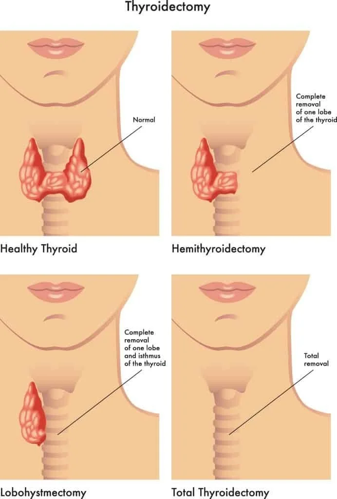
Contemporary endocrine surgery has witnessed transformative technological advances including intraoperative nerve monitoring, high-resolution ultrasound, and indocyanine green fluorescence angiography for parathyroid identification, yet these tools supplement rather than replace fundamental anatomical knowledge. The recurrent laryngeal nerve demonstrates sufficient variability in its relationship to the inferior thyroid artery, its branching pattern, and its entry point into the larynx to challenge even experienced surgeons, while the parathyroid glands exhibit unpredictable locations and vascular arrangements that demand constant vigilance. The increasing incidence of thyroid cancer, particularly papillary thyroid carcinoma, has expanded the surgical population to include younger patients with high expectations for functional outcomes, making the preservation of voice and calcium homeostasis as important as oncological clearance.
This review aims to provide a comprehensive examination of the surgical anatomy of the thyroid gland, integrating classical anatomical descriptions with contemporary surgical series, imaging studies, and intraoperative monitoring data. The article will explore the embryological basis of anatomical variations, delineate the critical relationships that define safe surgical planes, examine the vascular anatomy and its implications for parathyroid preservation, and discuss the clinical importance of anatomical knowledge in preventing complications during thyroidectomy. By synthesizing data from anatomical dissection studies, surgical outcome series, and technological advances in nerve identification, this work seeks to provide a reference framework that acknowledges thyroid surgery as a discipline where anatomical mastery remains the primary determinant of patient safety and surgical success.
Materials and Methods
This review article was constructed through systematic analysis of the contemporary endocrine surgical and anatomical literature, with emphasis on publications from the past two decades regarding thyroid surgical anatomy, intraoperative nerve monitoring, and complication prevention strategies. The methodological approach involved comprehensive examination of peer-reviewed journals in surgery, endocrinology, and anatomy, including Thyroid, Surgery, World Journal of Surgery, Annals of Surgery, Head and Neck, and Clinical Anatomy. Particular attention was directed toward studies utilizing intraoperative nerve monitoring data to elucidate recurrent laryngeal nerve anatomy, high-resolution ultrasound and computed tomography for preoperative mapping, and cadaveric dissection series providing quantitative measurements of anatomical relationships.
Inclusion criteria for anatomical data encompassed studies reporting quantitative measurements of nerve distances, vessel calibers, and gland dimensions in cohorts exceeding thirty subjects, with preference given to studies correlating anatomical findings with clinical outcomes. Embryological data were sourced from developmental anatomy literature and clinical series of congenital thyroid anomalies. Clinical correlation data were derived from surgical series reporting complication rates, nerve identification rates, and parathyroid autotransplantation outcomes, with specific attention to studies comparing visualization alone versus nerve monitoring for nerve preservation. When discrepancies existed between classical anatomical descriptions and contemporary surgical findings, preference was given to intraoperative observations and monitoring data that reflect live surgical anatomy rather than fixed cadaveric specimens.
The synthesis of this literature was organized according to the anatomical regions encountered during thyroidectomy, progressing from the gland's macroscopic architecture to the critical neurovascular structures of the tracheoesophageal groove and superior thyroid pole. All anatomical descriptions are presented according to standard surgical orientation, with measurements reported in millimeters. The reference list comprises foundational anatomical works, contemporary surgical series, and technological studies that collectively represent the current state of knowledge regarding thyroid surgical anatomy.
Results
Embryological Development and Gross Architecture
The thyroid gland originates from the endodermal lining of the floor of the primitive pharynx, specifically from the foramen cecum at the junction of the anterior two-thirds and posterior one-third of the tongue, with migration commencing during the fourth week of gestation and completing by the seventh week. This median thyroid anlage descends through the neck, passing anterior to the hyoid bone and laryngeal cartilages, and is joined laterally by the ultimobranchial bodies derived from the fourth and fifth pharyngeal pouches, which contribute the parafollicular C cells responsible for calcitonin production. The thyroglossal duct, representing the tract of descent, normally involutes by the eighth week, but its persistence explains the occurrence of pyramidal lobes in approximately 15 to 50 percent of individuals and the potential for thyroglossal duct cysts at any point along the migratory path from the foramen cecum to the suprasternal notch. Understanding this embryology is surgically relevant as the pyramidal lobe, when present, extends superiorly from the isthmus toward the hyoid bone and must be excised completely during thyroidectomy for thyroid cancer to prevent residual disease, while failure to recognize a thyroglossal duct cyst may result in incomplete treatment of midline neck masses.
The adult thyroid gland consists of two lateral lobes connected by a central isthmus, with a normal weight of 15 to 25 grams in iodine-sufficient regions. Each lateral lobe measures approximately 4 to 6 centimeters in length, 1.3 to 1.8 centimeters in width, and 1.5 to 2 centimeters in thickness, extending from the level of the middle thyroid cartilage to the fifth or sixth tracheal ring. The isthmus typically overlies the second to fourth tracheal rings and measures 1.25 to 2 centimeters in width, though complete agenesis of the isthmus occurs in approximately 5 to 10 percent of individuals, resulting in separate pyramidal lobes that may complicate surgical planning. The gland is enveloped by a thin fibrous capsule that sends septa into the parenchyma, creating lobules, and is surrounded by the pretracheal fascia which forms the surgical capsule distinct from the true capsular anatomy. This fascial investment creates the critical avascular plane between the thyroid capsule and the surrounding structures that permits capsular dissection, the fundamental technique for preserving the recurrent laryngeal nerves and parathyroid glands.
The anatomical relationships of the thyroid gland define the operative field during thyroidectomy. Anteriorly, the gland is covered by the strap muscles including the sternohyoid and sternothyroid, with the sternothyroid muscle lying directly on the capsule and requiring lateral retraction rather than division for exposure, though division may be necessary for large goiters. Posteriorly, the gland rests against the prevertebral fascia covering the longus colli muscles, with the carotid sheath containing the common carotid artery, internal jugular vein, and vagus nerve forming the lateral boundary of the dissection. Medially, the trachea and esophagus form the posterior relations, with the recurrent laryngeal nerve ascending in the tracheoesophageal groove. The superior pole of the thyroid extends toward the thyrohyoid membrane, where the external branch of the superior laryngeal nerve courses close to the superior thyroid vessels, while the inferior pole relates to the inferior thyroid artery and the recurrent laryngeal nerve as it enters the larynx.
Vascular Anatomy and Lymphatic Drainage
The arterial supply of the thyroid gland derives primarily from the superior and inferior thyroid arteries, with a supplementary contribution from the thyroidea ima artery in approximately 3 to 10 percent of individuals. The superior thyroid artery, the first branch of the external carotid artery, descends along the inferior constrictor muscle to reach the superior pole of the gland, where it divides into anterior and posterior branches. Critically, the external branch of the superior laryngeal nerve courses within or immediately adjacent to the fascia of the inferior constrictor muscle, crossing the superior thyroid artery approximately 1 to 2 centimeters above the superior pole of the thyroid gland. This proximity mandates careful ligation of the superior thyroid vessels individually, close to the capsule and away from the cricothyroid muscle, to avoid injury to the external branch which provides motor innervation to the cricothyroid muscle and sensory supply to the laryngeal mucosa above the vocal cords. Injury produces voice fatigue, loss of high-pitched phonation, and decreased projection, particularly affecting professional voice users.
The inferior thyroid artery, arising from the thyrocervical trunk of the subclavian artery, ascends along the medial border of the anterior scalene muscle before turning medially at the level of the cricoid cartilage to pass posterior to the carotid sheath and sympathetic trunk, reaching the posterior aspect of the thyroid gland where it divides into several branches that penetrate the capsule. The relationship between the inferior thyroid artery and the recurrent laryngeal nerve is variable and surgically critical: the nerve may pass anterior to the artery, posterior to it, or between its branches, with the anterior relationship occurring in approximately 60 to 70 percent of cases and the posterior relationship in 20 to 30 percent. This variability necessitates identification of the nerve before ligating any branches of the inferior thyroid artery, as blind clamping in this region risks nerve entrapment or transection. The inferior thyroid artery also provides the principal blood supply to the parathyroid glands, with the superior parathyroid typically supplied by a branch from the inferior thyroid artery or its anastomosis with the superior thyroid artery, while the inferior parathyroid receives supply from branches of the inferior thyroid artery or occasionally from the thyroidea ima.
Venous drainage occurs through three principal vessels: the superior thyroid vein draining the upper pole into the internal jugular vein, the middle thyroid vein draining the lateral aspect directly into the internal jugular vein, and the inferior thyroid veins forming a plexus on the anterior surface of the trachea that drains into the brachiocephalic veins. The middle thyroid vein is particularly surgically relevant as it crosses the common carotid artery laterally and must be ligated early in the dissection to permit medial rotation of the thyroid lobe, its avulsion resulting in troublesome bleeding that may obscure the operative field. The thyroidea ima artery, when present, arises from the aortic arch or brachiocephalic artery and ascends in the midline to supply the isthmus or lower poles, representing a hazard during dissection of the inferior thyroid veins or during tracheostomy.
Lymphatic drainage of the thyroid gland is extensive and bilaterally interconnected, flowing initially to perithyroidal and prelaryngeal nodes, then to pretracheal and paratracheal nodes, and subsequently to deep cervical chains along the internal jugular vein. The frequent bilateral and contralateral drainage explains the pattern of lymph node metastases in thyroid cancer, particularly papillary carcinoma, which commonly involves the central compartment (level VI) and may spread to lateral neck compartments. This drainage pattern mandates central compartment dissection during total thyroidectomy for malignancy, with the recurrent laryngeal nerves forming the lateral boundaries of the dissection and the parathyroid glands requiring identification and preservation or autotransplantation.
The Recurrent Laryngeal Nerve: Anatomy and Variations
The recurrent laryngeal nerve represents the most critical structure in thyroid surgery, its injury producing unilateral vocal cord paralysis in 0.5 to 5 percent of cases and bilateral paralysis in 0.3 to 0.6 percent, with catastrophic airway consequences in the latter. The nerve arises from the vagus nerve in the mediastinum, with the right recurrent laryngeal nerve looping around the right subclavian artery and ascending obliquely toward the tracheoesophageal groove, while the left recurrent laryngeal nerve loops around the aortic arch lateral to the ligamentum arteriosum and ascends more vertically within the tracheoesophageal groove. This asymmetry has surgical implications: the right nerve is more vulnerable during dissection of the inferior pole due to its more lateral and anterior course, while the left nerve may be injured during dissection of the esophageal surface or during central compartment lymph node dissection.
The recurrent laryngeal nerve enters the larynx by passing deep to the inferior constrictor muscle and piercing the cricothyroid membrane posteriorly, entering the larynx at the level of the cricoid cartilage. Its extralaryngeal branching pattern is highly variable and clinically significant: the nerve may divide into anterior and posterior branches before entering the larynx in 40 to 75 percent of cases, with the anterior branch providing motor innervation to the intrinsic laryngeal muscles except the cricothyroid, and the posterior branch providing sensory innervation to the subglottic mucosa. Early branching, occurring more than 2 centimeters below the inferior border of the inferior constrictor, occurs in approximately 10 to 30 percent of cases and increases the risk of inadvertent injury during dissection, as the surgeon may mistake a branch for the main trunk and fail to preserve the motor component. Intraoperative nerve monitoring has elucidated these branching patterns, revealing that the anterior motor branch typically lies anteromedially and may be compressed against the trachea during retraction, while the posterior sensory branch may be closely applied to the posterior capsule of the thyroid.
The relationship of the recurrent laryngeal nerve to the ligament of Berry, the dense fascial attachment connecting the thyroid capsule to the cricoid cartilage and trachea, represents the most hazardous portion of the dissection. The nerve typically passes through or immediately posterior to the ligament of Berry, with the ligament often described as the point where the nerve is most vulnerable to injury. Anatomical studies indicate that the nerve passes through the ligament in approximately 30 to 50 percent of cases, lateral to it in 20 to 40 percent, and medial to it in 10 to 20 percent. This proximity necessitates careful sharp dissection of the ligament with visualization of the nerve before division, as the nerve may be embedded within the ligamentous tissue or closely applied to its posterior surface. The ligament of Berry also contains small vessels that require individual ligation, and bleeding in this region may lead to blind clamping with resultant nerve injury.
The non-recurrent laryngeal nerve represents a rare but dangerous variant occurring in approximately 0.3 to 1 percent of individuals, more commonly on the right side due to embryological vascular anomalies including right subclavian artery arising from the descending aorta. In this configuration, the nerve arises directly from the vagus nerve at the level of the inferior thyroid artery and courses transversely toward the larynx without descending into the mediastinum, placing it at risk during dissection of the superior thyroid pole or inferior thyroid artery where it may be mistaken for a vessel or the middle thyroid vein. Failure to recognize a non-recurrent nerve may result in transection with immediate vocal cord paralysis. Preoperative recognition through imaging or intraoperative nerve monitoring with vagal stimulation at the carotid sheath can identify this variant, as stimulation of the vagus nerve below the level of the inferior thyroid artery will not produce a signal if the nerve is non-recurrent.
The Parathyroid Glands: Preservation and Transplantation
The parathyroid glands, typically four in number but ranging from two to six, are small endocrine organs measuring 3 to 6 millimeters in length and weighing 30 to 50 milligrams each, responsible for calcium homeostasis through parathyroid hormone production. Their preservation during thyroidectomy is essential to prevent permanent hypoparathyroidism, which occurs in 1 to 4 percent of total thyroidectomies and results in hypocalcemia, neuromuscular irritability, and long-term metabolic complications. The superior parathyroid glands derive from the fourth pharyngeal pouch and are more constant in location, typically found at the level of the cricoid cartilage posterior to the recurrent laryngeal nerve, near the junction of the upper and middle thirds of the thyroid lobe. The inferior parathyroid glands derive from the third pharyngeal pouch and are more variable, commonly located near the inferior pole of the thyroid or within the thyrothymic ligament, but may be found in the mediastinum or within the thyroid parenchyma itself.
The blood supply to the parathyroid glands is delicate and easily compromised during thyroidectomy. The superior parathyroid typically receives its blood supply from the inferior thyroid artery or an anastomotic branch between the superior and inferior thyroid arteries, entering the gland at its hilum on the anterior surface. The inferior parathyroid may receive supply from the inferior thyroid artery, the thyroidea ima, or directly from the inferior thyroid vein. This vascular anatomy dictates that parathyroid preservation requires maintenance of the blood supply from the inferior thyroid artery, which may necessitate ligation of the artery distal to the parathyroid branches or preservation of the anastomotic connections between the superior and inferior thyroid arterial systems. Devascularized parathyroid glands, recognized by their dark discoloration or loss of vascularity, should be autotransplanted into the sternocleidomastoid muscle or forearm muscle to preserve function, with immediate histological confirmation of parathyroid tissue recommended to avoid implanting metastatic disease in cases of malignancy.
Surgical Technique and Anatomical Planes
The operative approach to the thyroid gland begins with a transverse cervical incision approximately 2 centimeters above the sternal notch, extending between the anterior borders of the sternocleidomastoid muscles. Subplatysmal flaps are elevated superiorly to the thyroid cartilage and inferiorly to the sternal notch, and the strap muscles are separated in the midline and retracted laterally. The middle thyroid vein is identified and ligated, permitting medial rotation of the thyroid lobe and exposure of the lateral aspect of the gland. The superior thyroid pole is then addressed, with individual ligation of the superior thyroid vessels close to the capsule to avoid injury to the external branch of the superior laryngeal nerve, which may be identified by its characteristic oblique course across the cricothyroid muscle.
The recurrent laryngeal nerve is identified through several approaches: laterally at the level of the inferior thyroid artery, posteriorly where the nerve enters the larynx, or anteriorly through the pretracheal plane. The nerve is sought in the tracheoesophageal groove, with the tubercle of Zuckerkandl, a posterior projection of thyroid tissue present in 60 to 90 percent of patients, serving as a landmark as the nerve typically passes medial to this structure. The inferior thyroid artery is ligated distal to the parathyroid branches, and the ligament of Berry is divided with sharp dissection under direct vision of the nerve. The isthmus is divided, and the gland is removed, with hemostasis confirmed and the parathyroid glands assessed for viability before closure.
Intraoperative nerve monitoring has become standard practice in many centers, utilizing electromyographic endotracheal tubes to assess vocal cord function through nerve stimulation. The monitoring system allows identification of the nerve through stimulation, confirmation of nerve integrity before closure, and prognostication of postoperative vocal function. However, monitoring supplements rather than replaces visual identification, as the nerve must be seen to be preserved, and monitoring cannot prevent injury from traction, thermal damage, or devascularization.
Discussion
The surgical anatomy of the thyroid gland, despite its apparent simplicity, encompasses a complexity that continues to challenge even experienced endocrine surgeons. The recurrent laryngeal nerve, with its variable relationship to the inferior thyroid artery, its extralaryngeal branching patterns, and its intimate association with the ligament of Berry, remains the structure most vulnerable to injury, while the parathyroid glands present a preservation challenge due to their variable locations and delicate vascular pedicles. The increasing incidence of thyroid cancer and the expansion of surgical indications to include larger goiters and reoperative cases have amplified the importance of anatomical mastery, as these complex cases present distorted anatomy and increased complication risks.
The integration of intraoperative nerve monitoring into thyroid surgery has refined our understanding of laryngeal nerve anatomy, revealing branching patterns and variant courses that were previously underappreciated. However, the persistence of nerve injury despite monitoring suggests that anatomical knowledge and surgical judgment remain paramount. The external branch of the superior laryngeal nerve, often neglected in surgical teaching, has gained recognition as a source of voice dysfunction, particularly in professional voice users, prompting more aggressive efforts at identification and preservation during superior pole dissection.
The prevention of permanent hypoparathyroidism requires a paradigm shift from mere parathyroid identification to active preservation of their vascular supply, with autotransplantation serving as a salvage technique rather than a substitute for careful dissection. The recognition that the inferior thyroid artery provides the principal blood supply to both the inferior and superior parathyroid glands in most cases has led to modifications in surgical technique, including preservation of the artery main trunk and selective ligation of terminal branches.
Future directions in thyroid surgical anatomy likely involve advanced imaging techniques including high-resolution ultrasound and computed tomography for preoperative mapping of nerve variants and parathyroid locations, as well as near-infrared autofluorescence for parathyroid identification. However, these technologies will remain adjuncts to, rather than replacements for, the fundamental anatomical knowledge that has defined safe thyroid surgery since the era of Kocher. The surgeon who understands the embryological basis of anatomical variations, who recognizes the critical relationships of the recurrent laryngeal nerve and parathyroid glands, and who employs meticulous capsular dissection technique will continue to achieve the best outcomes for patients undergoing thyroidectomy.
Conclusion
The thyroid gland occupies a region of concentrated anatomical hazard, where the recurrent laryngeal nerves, external branch of the superior laryngeal nerve, and parathyroid glands present preservation challenges that define the quality of surgical outcomes. Complete understanding of the gland's embryological development, vascular anatomy, and relationships to surrounding structures remains the foundation of safe thyroidectomy, superior to any technological adjunct. The recurrent laryngeal nerve, with its variable course and branching patterns, demands constant vigilance and precise dissection, particularly at the ligament of Berry where it is most vulnerable. Parathyroid preservation requires attention to vascular anatomy and willingness to autotransplant devascularized glands. As thyroid surgery continues to evolve with increasing volumes and expanding indications, the importance of anatomical education and surgical mentorship remains undiminished, ensuring that future generations of surgeons maintain the standards of safety and efficacy established by the pioneers of endocrine surgery.
References
Kocher T. Ueber Morbus Basedowi. Dtsch Z Chir. 1883;18:393-481.
Billroth T. Die allgemeine chirurgische Pathologie und Therapie in fünfzig Vorlesungen: ein Handbuch für Studierende und Ärzte. Berlin: Reimer; 1863.
Randolph GW, Dralle H, Abdullah H, et al. Electrophysiologic recurrent laryngeal nerve monitoring during thyroid and parathyroid surgery: international standards guideline statement. Laryngoscope. 2011;121 Suppl 1:S1-S16.
Dionigi G, Bacuzzi A, Boni L, et al. What is the learning curve for intraoperative neuromonitoring in thyroid surgery? Int J Surg. 2008;6 Suppl 1:S7-S12.
Barczynski M, Konturek A, Stopa M, et al. Randomized controlled trial of visualization vs neuromonitoring of the external branch of the superior laryngeal nerve during thyroidectomy. World J Surg. 2012;36(6):1340-1347.
Henry BM, Vikse J, Graves MJ, et al. The non-recurrent laryngeal nerve: a meta-analysis and clinical considerations. PeerJ. 2017;5:e3012.
Henry BM, Vikse J, Graves MJ, et al. Extralaryngeal branching of the recurrent laryngeal nerve: a meta-analysis of 28,387 nerves. Langenbecks Arch Surg. 2016;401(7):913-923.
Gurleyik E, Gurleyik G. Incidence and surgical importance of Zuckerkandl's tubercle of the thyroid and its relations with recurrent laryngeal nerve. ISRN Surg. 2013;2013:450589.
Pelizzo MR, Toniato A, Gemo G. Zuckerkandl's tuberculum: an arrow pointing to the recurrent laryngeal nerve (constant anatomical landmark). J Am Coll Surg. 1998;187(3):333-336.
Lo CY, Kwok KF, Yuen PW, et al. A prospective evaluation of recurrent laryngeal nerve monitoring during thyroidectomy under regional anesthesia. World J Surg. 2000;24(9):1082-1088.
Hermann M, Alk G, Roka R, et al. Laryngeal recurrent nerve injury in surgery for benign thyroid diseases: effect of nerve dissection and impact of individual surgeon in more than 27,000 nerves at risk. Ann Surg. 2002;235(2):261-268.
Thomusch O, Machens A, Sekulla C, et al. Multivariate analysis of risk factors for postoperative complications in benign goiter surgery: prospective multicenter study in Germany. World J Surg. 2000;24(11):1330-1334.
Edafe O, Antakia R, Laskar N, et al. Systematic review and meta-analysis of predictors of post-thyroidectomy hypocalcaemia. Br J Surg. 2014;101(4):307-320.
Falcone C, Sampietro C, Puttini M, et al. Intraoperative parathyroid hormone assay in the surgical management of hyperparathyroidism. J Endocrinol Invest. 2003;26(7 Suppl):45-49.
Lorente-Poch L, Sancho JJ, Muñoz-Nova JL, et al. Defining the syndromes of parathyroid failure after total thyroidectomy. Gland Surg. 2015;4(1):82-90.
Bergenfelz A, Jansson S, Kristoffersson A, et al. Complications to thyroid surgery: results as reported in a database from a multicenter audit comprising 3,660 patients. Langenbecks Arch Surg. 2008;393(5):667-673.
Rulli F, Ambrogi V, Dionigi G, et al. Meta-analysis of recurrent laryngeal nerve injury in thyroid surgery with and without intraoperative nerve monitoring. Acta Otorhinolaryngol Ital. 2014;34(4):223-229.
Dionigi G, Bacuzzi A, Boni L, et al. Why monitor the recurrent laryngeal nerve in thyroid surgery? J Endocrinol Invest. 2010;33(11):819-822.
Serpell JW. New operative surgical concept of two fascial layers enveloping the recurrent laryngeal nerve. Ann Surg Oncol. 2010;17(6):1628-1636.
Standring S, editor. Gray's Anatomy: The Anatomical Basis of Clinical Practice. 42nd ed. London: Elsevier; 2020. p. 445-452.