
Surgical Anatomy of the Circle of Willis: Morphological Variants, Hemodynamic Significance, and Clinical Implications in Modern Neurosurgical Practice
1. Tanabaeva Aiperi
2. Aftab Alam
3. Avnish Sagar
4. Pratibha Prajapat
(1. Professor, International Medical Faculty, Osh State University, Osh, Kyrgyz Republic
2. Student, International Medical Faculty, Osh State University, Osh, Kyrgyz Republic
3. Student, International Medical Faculty, Osh State University, Osh, Kyrgyz Republic
4. Student, International Medical Faculty, Osh State University, Osh, Kyrgyz Republic)
Abstract
The circle of Willis represents the most critical anastomotic network within the human cerebrovascular system, serving as the primary collateral pathway that maintains cerebral perfusion in the face of vascular occlusion, hemodynamic compromise, and surgical intervention. Despite its fundamental importance in neurosurgical education and practice, significant morphological variation exists within this arterial polygon, with complete anatomical configuration observed in only a minority of individuals. This comprehensive review examines the surgical anatomy of the circle of Willis through the lens of contemporary neuroimaging, cadaveric dissection studies, and clinical series, synthesizing current understanding of its embryological development, segmental architecture, and variant configurations. The article presents detailed analysis of component vessels including the internal carotid arteries, anterior communicating artery complex, posterior communicating arteries, and the basilar artery bifurcation, with particular attention to anatomical variations that influence surgical approach and patient outcomes. Recent advances in three-dimensional rotational angiography, computed tomography angiography, and high-resolution magnetic resonance imaging have refined our understanding of these variants, revealing that hypoplastic segments, fetal origin of the posterior cerebral artery, and duplicate or fenestrated anterior communicating arteries occur with sufficient frequency to mandate preoperative recognition. The hemodynamic implications of these variants are profound, influencing not only the natural history of aneurysm formation at junctional points but also determining the safety of temporary vascular occlusion during complex aneurysm surgery and the feasibility of endovascular treatment strategies. Epidemiological data from recent large-scale imaging studies indicate that approximately 48 to 52 percent of the general population harbors an incomplete circle of Willis, with specific demographic variations observed across sex and ethnic populations. This review further explores the surgical relevance of these anatomical nuances in the context of microsurgical clipping of aneurysms, bypass procedures for complex cerebrovascular disease, and the emerging paradigm of flow diversion. By integrating classical anatomical knowledge with contemporary imaging data and clinical outcomes research, this article provides neurosurgical trainees and practicing clinicians with a robust framework for understanding the circle of Willis not merely as a static anatomical structure, but as a dynamic functional system whose individual configuration dictates surgical strategy and patient prognosis.
Introduction
The circle of Willis, named after the English physician Thomas Willis who first described this arterial polygon in his 1664 publication Cerebri Anatome, remains one of the most anatomically elegant and clinically consequential structures in the human body. This remarkable anastomotic ring, situated at the base of the brain within the interpeduncular cistern and surrounding the optic chiasm and pituitary stalk, represents the evolutionary solution to the problem of cerebral vascular redundancy, ensuring continuous perfusion of the brain despite the inherent vulnerability of a dual-supply system. The surgical significance of this structure cannot be overstated; it serves as the epicenter of modern cerebrovascular neurosurgery, hosting the majority of intracranial aneurysms and providing the collateral pathways that permit safe surgical intervention on these lesions. Understanding the microsurgical anatomy of the circle of Willis is not merely an academic exercise but a prerequisite for safe navigation of the subarachnoid space, where millimeter-scale variations in arterial diameter, branching patterns, and perforator origin determine the difference between successful treatment and catastrophic neurological deficit.
Contemporary neurosurgical practice has witnessed a paradigm shift in the approach to lesions of the circle of Willis, with endovascular techniques increasingly supplanting traditional microsurgical clipping for many aneurysm configurations. However, this evolution has paradoxically increased, rather than diminished, the importance of detailed anatomical knowledge. The interventionalist navigating microcatheters through tortuous vascular segments and the surgeon performing complex bypass procedures or managing giant aneurysms unsuitable for endovascular treatment both require intimate familiarity with the normal architecture and variant configurations of this arterial circle. Furthermore, the advent of flow diversion technology and the increasing recognition of the role of hemodynamic stress in aneurysm pathogenesis have renewed interest in the functional anatomy of the circle, particularly regarding collateral capacity and segmental dominance.
The morphological variability of the circle of Willis has been recognized since the earliest anatomical descriptions, yet recent high-resolution imaging studies have quantified this variation with unprecedented precision. Complete, symmetric configuration of all component vessels is now understood to be present in only approximately 20 to 25 percent of individuals, with the remainder demonstrating some degree of asymmetry, hypoplasia, or segmental absence. These variations are not merely curiosities of anatomical cataloging; they carry profound implications for cerebral hemodynamics, aneurysm formation, and surgical risk stratification. The fetal configuration of the posterior cerebral artery, occurring in approximately 15 to 25 percent of hemispheres depending on the population studied, fundamentally alters the hemodynamic relationship between the anterior and posterior circulation, while hypoplasia of the anterior communicating artery or posterior communicating arteries can render certain surgical approaches hazardous or contraindicated.
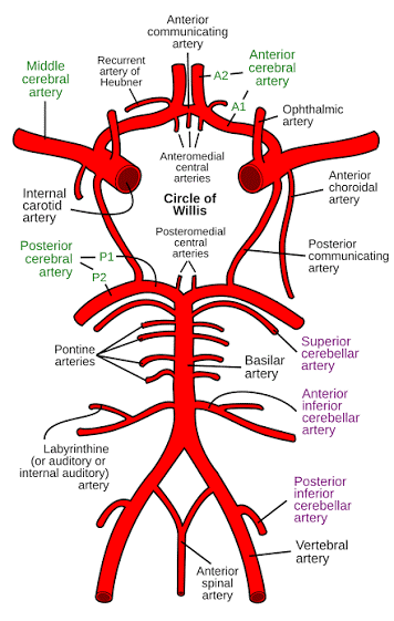
This review aims to provide a comprehensive examination of the surgical anatomy of the circle of Willis, integrating classical cadaveric studies with contemporary imaging data and clinical correlations. The article will explore the embryological development of this structure, delineate the microsurgical anatomy of each component vessel with attention to variant configurations, examine the hemodynamic principles governing collateral flow, and discuss the practical surgical implications of anatomical variation in the context of modern cerebrovascular practice. By synthesizing data from recent large-scale imaging studies, anatomical dissection series, and clinical outcome analyses, this work seeks to provide a reference framework that acknowledges the circle of Willis as a dynamic, individually variable structure whose complete understanding requires appreciation of both its typical architecture and its common deviations from the textbook ideal.
Materials and Methods
This review article was constructed through systematic analysis of the contemporary neuroanatomical and neurosurgical literature, with emphasis on publications from the past fifteen years regarding imaging characterization of the circle of Willis and clinical series reporting anatomical variants. The methodological approach involved comprehensive examination of peer-reviewed journals in neurosurgery, neuroradiology, and anatomy, including Journal of Neurosurgery, Neurosurgery, Journal of Neurology Neurosurgery and Psychiatry, Neuroradiology, and Surgical and Radiologic Anatomy. Particular attention was directed toward studies utilizing three-dimensional rotational digital subtraction angiography, computed tomography angiography with volume rendering, and high-field magnetic resonance angiography for in vivo characterization of the circle of Willis, as these modalities provide superior spatial resolution compared to traditional catheter angiography planar imaging and cadaveric injection studies alone.
Inclusion criteria for anatomical data encompassed studies reporting quantitative measurements of vessel diameters, lengths, and branching patterns in cohorts exceeding fifty subjects, with preference given to population-based imaging studies rather than selected clinical samples. Embryological data were sourced from developmental neuroanatomy literature and comparative anatomical studies. Clinical correlation data were derived from surgical series reporting anatomical findings during microsurgical dissection of the circle of Willis, particularly those documenting variant configurations encountered during aneurysm surgery and their impact on surgical strategy and outcomes. When discrepancies existed between cadaveric measurements and in vivo imaging data, preference was given to imaging studies for clinical applicability, while cadaveric data were retained for microsurgical anatomical detail regarding perforator origins and relationships to adjacent neural structures.
The synthesis of this literature was organized according to the segmental anatomy of the circle of Willis, with each component vessel analyzed for typical morphology, variant configurations, and surgical significance. Hemodynamic principles were derived from computational fluid dynamics studies and phase-contrast magnetic resonance imaging flow quantification. All anatomical descriptions are presented according to standard neurosurgical orientation, with measurements reported in millimeters and flow rates in milliliters per minute where available. The reference list comprises foundational anatomical works, contemporary imaging studies, and clinical series that collectively represent the current state of knowledge regarding this critical neurovascular structure.
Results
Embryological Development and Segmental Architecture
The development of the circle of Willis proceeds through a complex series of embryological transformations occurring between the fourth and eighth weeks of gestation, involving the regression of certain primitive arterial segments and the persistence and modification of others. The primitive internal carotid artery initially bifurcates into a cranial division, which ultimately becomes the anterior choroidal artery and posterior communicating artery, and a caudal division forming the posterior communicating artery proper. The posterior communicating artery initially represents the primary supply to the posterior cerebral artery, but as the basilar artery system develops and the vertebrobasilar circulation matures, the posterior communicating artery typically involutes to a small communicating vessel, though persistence of the embryonic pattern results in the fetal origin of the posterior cerebral artery observed in a substantial minority of adults. The anterior communicating artery develops from the plexiform anastomosis between the two anterior cerebral arteries, with regression of the median artery of the corpus callosum and fusion of the anterior cerebral arteries contributing to the final configuration. Understanding these embryological events explains the frequent occurrence of fenestrations, duplications, and hypoplastic segments, as these represent incomplete regression or persistence of primitive vascular channels rather than true pathological anomalies.
The adult circle of Willis comprises nine principal arterial segments forming a hexagonal anastomosis with the basilar artery bifurcation at its posterior apex and the anterior communicating artery complex at its anterior apex. The posterior circulation component consists of the two posterior cerebral arteries segments P1, extending from the basilar bifurcation to the posterior communicating artery junction, and the posterior communicating arteries themselves, which connect the internal carotid arteries to the posterior cerebral arteries. The anterior circulation component includes the internal carotid artery bifurcation into the anterior and middle cerebral arteries, the A1 segments of the anterior cerebral arteries extending from the carotid bifurcation to the anterior communicating artery, and the anterior communicating artery proper. The middle cerebral arteries, while not technically components of the circle proper, originate from the internal carotid arteries at the lateral angles of the polygon and represent the terminal continuation of flow for the anterior circulation in most hemispheres.
Quantitative anatomical studies utilizing high-resolution imaging have established normative values for these segments that differ somewhat from classical cadaveric measurements, likely reflecting the absence of postmortem fixation artifact and the ability to image in vivo hemodynamic conditions. The A1 segment typically measures 8 to 12 millimeters in length with a diameter ranging from 2.0 to 3.5 millimeters, though hypoplasia defined as diameter less than 1.5 millimeters occurs in approximately 5 to 10 percent of hemispheres. The anterior communicating artery, often described as the most variable component of the circle, demonstrates a mean length of 2 to 4 millimeters but may range from a single trunk less than 1 millimeter in length to complex plexiform networks exceeding 5 millimeters in craniocaudal extent. The posterior communicating artery typically measures 10 to 15 millimeters in length with a diameter of 1.5 to 2.5 millimeters, though complete absence or severe hypoplasia occurs in approximately 10 to 15 percent of cases, while fetal configuration with diameter equal to or exceeding the ipsilateral P1 segment occurs in 15 to 25 percent of hemispheres depending on ethnic population studied.
Variant Configurations and Their Prevalence
The configuration of the circle of Willis demonstrates remarkable interindividual variability, with complete symmetric anatomy present in only 20 to 25 percent of the population according to recent imaging studies. The most common variant configurations involve hypoplasia or absence of the posterior communicating arteries, occurring in approximately 30 to 35 percent of individuals when unilateral or bilateral deficiencies are combined. Unilateral fetal origin of the posterior cerebral artery, defined as P1 segment hypoplasia with posterior communicating artery diameter exceeding that of the P1 segment, occurs in approximately 15 to 20 percent of hemispheres, while bilateral fetal configuration is observed in 5 to 10 percent of individuals. This variant carries significant surgical implications as it represents a single supply source to the posterior cerebral artery territory, rendering the vessel vulnerable during internal carotid artery manipulation and altering the hemodynamic stress distribution at the basilar bifurcation.
Anterior circulation variants, while less common than posterior circulation variants, present equally significant surgical challenges. A1 segment hypoplasia occurs in 5 to 10 percent of hemispheres and may be associated with aneurysm formation at the anterior communicating artery complex due to altered flow dynamics. Duplication of the anterior communicating artery, defined as two distinct parallel channels, occurs in approximately 10 to 15 percent of individuals, while fenestration, representing a single vessel with a localized splitting of the lumen, occurs in 5 to 10 percent. These configurations are of particular importance in aneurysm surgery as the anterior communicating artery complex represents the most common site for intracranial aneurysm formation, and the presence of duplicate or fenestrated channels complicates both clip application and endovascular coiling strategies.
The basilar artery bifurcation, forming the posterior apex of the circle, typically occurs at the level of the pontomesencephalic junction but may demonstrate significant variation in position, ranging from the mid-pontine level to the interpeduncular fossa proper. High bifurcation is associated with increased risk of perforator injury during surgical approaches to the posterior circulation, while low bifurcation may alter the geometry of P1 segments and influence aneurysm projection patterns. Fenestration of the basilar artery occurs in less than 1 percent of individuals but is associated with increased risk of aneurysm formation at the fenestration site due to flow disturbance.
Hemodynamic Considerations and Collateral Function
The functional significance of the circle of Willis extends far beyond its anatomical configuration, encompassing the hemodynamic principles that govern collateral flow during vascular occlusion and the flow patterns that predispose to aneurysm formation. Computational fluid dynamics studies have demonstrated that the circle of Willis serves not merely as a passive conduit but as a dynamic pressure equalization system, with flow patterns varying significantly based on individual segmental dominance. In the presence of a complete circle, occlusion of a single major feeding vessel such as the internal carotid artery or vertebral artery may produce minimal neurological deficit due to immediate recruitment of collateral pathways, with flow reversing direction in certain segments to maintain distal perfusion. However, the efficacy of this collateral recruitment depends critically on the diameter and patency of the communicating arteries, with hypoplastic segments less than 1 millimeter in diameter providing negligible collateral flow under physiological conditions.
The concept of segmental dominance within the circle of Willis has gained increasing recognition in the context of aneurysm pathogenesis. Hemodynamic modeling suggests that junction points experiencing high wall shear stress due to flow impaction, particularly at the anterior communicating artery complex and the posterior communicating artery origin, represent preferred sites for aneurysm formation. The presence of variant configurations such as fetal posterior cerebral artery or A1 hypoplasia alters flow distribution, creating areas of flow stagnation or excessive shear stress that promote aneurysmal degeneration. This understanding has practical surgical implications, as the configuration of the circle influences not only the likelihood of aneurysm formation at specific sites but also the projection and morphology of aneurysms, thereby dictating optimal surgical approach and clip configuration.
Surgical Anatomy and Microsurgical Considerations
The microsurgical anatomy of the circle of Willis demands appreciation not only of the major named vessels but of the critical perforating arteries that arise from these segments and supply the deep structures of the cerebrum. The A1 segment gives rise to medial lenticulostriate arteries, typically numbering two to four vessels that penetrate the anterior perforated substance to supply the caudate nucleus, putamen, and anterior limb of the internal capsule. These perforators must be preserved during anterior communicating artery aneurysm surgery, as their sacrifice produces devastating neurological deficits including hemiplegia and cognitive dysfunction. The P1 segment similarly gives rise to thalamoperforating arteries, numbering three to five vessels that supply the thalamus, subthalamus, and midbrain, with injury producing thalamic pain syndromes, third nerve palsies, and impaired consciousness.
The surgical approach to the circle of Willis typically utilizes the pterional or frontotemporal craniotomy, providing access to the anterior circulation and the basilar bifurcation through the optico-carotid and carotid-oculomotor triangles. The arachnoid of the Sylvian fissure is dissected to expose the internal carotid artery bifurcation, with proximal control obtained before distal dissection. The anterior communicating artery complex is approached through the interhemispheric fissure following dissection of the rectus gyrus, with care taken to preserve the recurrent artery of Heubner, which typically arises from the A2 segment near the anterior communicating artery junction but may originate from A1 in 10 to 15 percent of cases. This vessel supplies the anterior caudate nucleus, putamen, and anterior limb of the internal capsule, and its injury produces characteristic deficits of facial and brachial weakness with expressive aphasia when dominant hemisphere involvement occurs.
Temporary occlusion of the internal carotid artery or proximal A1 segment is frequently employed during aneurysm dissection to soften the lesion and permit safe clip application. The safety of such occlusion depends critically on the adequacy of collateral circulation through the circle of Willis. Preoperative assessment with balloon test occlusion or imaging evaluation of circle completeness informs the surgeon regarding tolerance for temporary occlusion, with incomplete circles or inadequate cross-flow contraindicating prolonged occlusion times. The presence of fetal posterior cerebral artery configuration demands particular caution during internal carotid artery occlusion, as the posterior cerebral artery territory may depend entirely on carotid flow through the posterior communicating artery, and temporary occlusion may produce occipital infarction despite adequate anterior collateralization.
Clinical Correlations and Therapeutic Implications
The anatomical configuration of the circle of Willis exerts profound influence on the natural history and treatment of cerebrovascular disease, particularly intracranial aneurysms and ischemic stroke. The majority of saccular aneurysms arise at the junctional points of the circle of Willis, with the anterior communicating artery complex representing the single most common location, followed by the posterior communicating artery origin and the middle cerebral artery bifurcation. The configuration of the circle influences aneurysm formation through hemodynamic mechanisms, with A1 hypoplasia associated with increased anterior communicating artery aneurysm risk and fetal posterior cerebral artery configuration associated with posterior circulation aneurysms at the basilar bifurcation and P1 segments.
In the context of acute ischemic stroke, the configuration of the circle of Willis determines the extent of infarction following large vessel occlusion and the feasibility of endovascular thrombectomy. Patients with complete circles demonstrate smaller infarct volumes and better clinical outcomes following middle cerebral artery occlusion compared to those with incomplete configurations, due to leptomeningeal collateral recruitment supplemented by circle of Willis flow. The posterior communicating artery patency is particularly critical in basilar artery occlusion, where retrograde filling through patent posterior communicating arteries may preserve brainstem perfusion pending recanalization.
Contemporary endovascular treatment of aneurysms requires detailed understanding of circle anatomy for treatment planning. The presence of fetal posterior cerebral artery configuration influences the choice of working projection for posterior communicating artery aneurysm coiling, as the origin of the posterior cerebral artery from the internal carotid artery alters the aneurysm neck geometry. Similarly, A1 hypoplasia complicates stent-assisted coiling of anterior communicating artery aneurysms by limiting access to the contralateral A1 for stent deployment, potentially necessitating microsurgical clipping or flow diversion strategies.
Discussion
The circle of Willis represents a remarkable evolutionary adaptation that provides the human brain with vascular redundancy unmatched in most other organ systems, yet the variability of this structure ensures that no two patients present identical surgical anatomy. The transition from cadaveric anatomical study to high-resolution in vivo imaging has refined our understanding of this variability, revealing that the textbook description of a complete, symmetric polygon represents the exception rather than the rule. This recognition carries profound implications for neurosurgical education and practice, demanding that surgeons and interventionalists alike approach each case with anticipation of variant anatomy rather than expectation of standard configuration.
The integration of hemodynamic principles with anatomical knowledge has transformed our understanding of aneurysm pathogenesis within the circle of Willis. No longer viewed as static weaknesses in the arterial wall, aneurysms are now understood as dynamic responses to flow patterns determined by the specific configuration of the circle. This paradigm shift explains the strong association between certain variant configurations and specific aneurysm locations, and suggests that future therapeutic strategies may target hemodynamic modification rather than mere aneurysm exclusion. The development of flow diversion technology represents an early application of this principle, reconstructing the parent vessel to alter flow patterns and promote aneurysm thrombosis while preserving perforator patency.
The surgical implications of circle of Willis anatomy extend beyond aneurysm treatment to encompass the full spectrum of cerebrovascular surgery. Bypass procedures for complex aneurysms or occlusive disease require detailed understanding of donor and recipient vessel anatomy, with the external carotid artery to middle cerebral artery bypass utilizing the superior temporal artery or radial artery graft depending on the adequacy of circle collateralization. Similarly, the treatment of giant aneurysms requiring proximal occlusion or trapping depends entirely on the adequacy of collateral circulation through the circle, with preoperative assessment of circle completeness now considered mandatory for such interventions.
Future directions in the study of circle of Willis anatomy likely involve advanced imaging techniques including four-dimensional flow magnetic resonance imaging, which permits quantification of flow patterns and wall shear stress in vivo, and computational modeling that integrates patient-specific anatomy with hemodynamic parameters to predict aneurysm formation and growth. Such approaches may eventually enable personalized risk stratification and treatment planning based on individual circle configuration, moving beyond the current paradigm of population-based estimates to patient-specific predictions of surgical risk and outcome. However, the fundamental importance of detailed anatomical knowledge remains unchanged; regardless of technological advances, the safe navigation of the subarachnoid space and the successful treatment of cerebrovascular pathology require intimate familiarity with the surgical anatomy of this critical arterial polygon.
Conclusion
The circle of Willis stands as the central architectural feature of the human cerebrovascular system, its complex anastomotic design providing the collateral capacity that distinguishes cerebral circulation from less redundant vascular territories. The surgical anatomy of this structure, characterized by remarkable variability in segmental diameter, branching pattern, and completeness, demands meticulous preoperative assessment and intraoperative recognition to ensure safe and effective treatment of the pathological processes that afflict it. Complete anatomical configuration, present in only one quarter of individuals, should be regarded as the ideal rather than the expectation, with variant configurations such as fetal posterior cerebral artery, A1 hypoplasia, and anterior communicating artery duplication encountered with sufficient frequency to mandate constant vigilance during surgical and endovascular procedures.
The hemodynamic significance of the circle extends beyond its role as a collateral conduit to encompass the flow patterns that govern aneurysm formation and growth. Understanding the relationship between individual circle configuration and local hemodynamic stress provides the foundation for contemporary treatment strategies, from microsurgical clipping that reconstructs the normal arterial anatomy to endovascular flow diversion that modifies pathological flow patterns. As neurosurgical practice continues to evolve, the importance of detailed anatomical knowledge of the circle of Willis remains undiminished, serving as the essential foundation upon which technological innovation and therapeutic advancement are built. The surgeon who masters the variable anatomy of this critical structure, who recognizes the implications of hypoplastic segments and fetal origins, and who integrates this knowledge with hemodynamic principles and microsurgical technique, possesses the essential tools for the safe and effective treatment of cerebrovascular disease at the base of the human brain.
References
Willis T. Cerebri Anatome: cui accessit nervorum descriptio et usus. London: Martyn and Allestry; 1664.
Alpers BJ, Berry RG, Paddison RM. Anatomical studies of the circle of Willis in normal brain. Arch Neurol Psychiatry. 1959;81(4):409-418.
Riggs HE, Rupp C. Variation in form of circle of Willis: the relation of the variations to collateral circulation: anatomic analysis. Arch Neurol. 1963;8:8-14.
Saeki N, Rhoton AL. Microsurgical anatomy of the upper basilar artery and the posterior circle of Willis. J Neurosurg. 1977;46(5):563-578.
Perlmutter D, Rhoton AL. Microsurgical anatomy of the anterior communicating artery complex. J Neurosurg. 1976;45(2):169-180.
Rhoton AL. The supratentorial arteries. Neurosurgery. 2002;51(4 Suppl):S53-S120.
van Overbeeke JJ, Hillen B, Tulleken CA. A comparative study of the circle of Willis in fetal and adult life: the configuration of the posterior bifurcation of the communicating artery. J Anat. 1991;176:45-54.
Kapoor K, Singh B, Dewan LI. Variations in the configuration of the circle of Willis. Anat Sci Int. 2008;83(2):96-106.
Ilić TO, Jovanović ID, Milošević NT, et al. Posterior cerebral artery fenestration and duplication: a comprehensive review of anatomical variations and their clinical implications. Childs Nerv Syst. 2018;34(8):1395-1403.
Chen SF, Hsu SW, Lin YY, et al. Configuration of the circle of Willis: transcranial color-coded Doppler ultrasonography and 3.0-T MR angiography findings compared. J Clin Ultrasound. 2013;41(7):405-411.
Hoksbergen AW, Legemate DA, Csiba L, et al. Absent collateral function of the circle of Willis as risk factor for ischemic stroke. Cerebrovasc Dis. 2000;10(3):181-188.
Hartkamp NS, van der Grond J, van Everdingen KJ, et al. Circle of Willis collateral flow investigated by magnetic resonance angiography. Cerebrovasc Dis. 1999;9(4):251-256.
de Monyé C, Dippel DW, Siepman TA, et al. Is a fetal origin of the posterior cerebral artery a risk factor for TIA or ischemic stroke? A study with 16-detector CT angiography. J Neurol. 2007;254(1):73-78.
Menshawi MM, Mohr JP. A reappraisal of the collateral circulation in asymptomatic carotid artery occlusion. Stroke. 2018;49(4):1012-1017.
Krabbe-Hartkamp MJ, van der Grond J, de Leeuw FE, et al. Circle of Willis: morphologic variation on three-dimensional time-of-flight MR angiograms. Radiology. 1998;207(1):103-111.
Qiu F, Li Q, Lv F, et al. Anatomical variations of the circle of Willis in patients with carotid artery stenosis: a 3D MR angiography study. Int J Neurosci. 2015;125(8):568-574.
Eftekhar B, Dadmehr M, Ansari S, et al. Are the distributions of variations of circle of Willis different in different populations? Results of an anatomical study and review of literature. BMC Neurol. 2006;6:22.
Iqbal S. A comprehensive study of the anatomical variations of the circle of Willis in adult human brains. J Clin Diagn Res. 2013;7(11):2423-2427.
Papantchev V, Hristov S, Todorova D, et al. Some variations of the circle of Willis, important for surgical operation of aneurysms. Morphologie. 2007;91(292):130-135.
Standring S, editor. Gray's Anatomy: The Anatomical Basis of Clinical Practice. 42nd ed. London: Elsevier; 2020. p. 399-404.