
Surgical Anatomy of the Vermiform Appendix: Anatomical Variations and Their Importance in Appendectomy
1. Tanabaeva Aiperi
2. Pranav Avhad
3. Ahtesham Ali
4. Islamuddin
(1. Professor, International Medical Faculty, Osh State University, Osh, Kyrgyz Republic
2. Student, International Medical Faculty, Osh State University, Osh, Kyrgyz Republic
3. Student, International Medical Faculty, Osh State University, Osh, Kyrgyz Republic
4. Student, International Medical Faculty, Osh State University, Osh, Kyrgyz Republic)
Abstract
The vermiform appendix is a narrow, blind ended diverticulum arising from the caecum and represents one of the most frequently encountered structures in emergency abdominal surgery. Although appendectomy is commonly performed, successful operative management depends on a detailed understanding of appendiceal anatomy and its considerable anatomical variability. Differences in the position, length, orientation, mesoappendix, arterial supply, relationship with the terminal ileum and caecum, and occasionally the number of appendices can substantially influence the clinical presentation of appendicitis and the technical difficulty of appendectomy. The classical description of a right lower quadrant appendix is therefore insufficient for safe surgical practice.
This review examines the surgical anatomy of the vermiform appendix, with particular emphasis on anatomical variations and their implications for appendectomy. Relevant anatomical literature, contemporary surgical guidelines, systematic reviews, and recent studies published through 2026 were reviewed. The appendix is usually located in the right iliac fossa, but its tip may occupy retrocaecal, pelvic, subcaecal, pre-ileal, post-ileal, paracaecal, or less commonly subhepatic and left sided positions. The base is relatively constant because it arises at the convergence of the three taeniae coli, making this anatomical relationship one of the most dependable landmarks during surgery. In contrast, the tip has considerable mobility and is responsible for much of the variation encountered during appendectomy. The mesoappendix contains the appendicular artery and shows important variation in its length, position, and relationship with the appendix. Recent laparoscopic research has demonstrated that mesoappendiceal configuration can influence operative difficulty and the method used to control the appendicular vessels.
Laparoscopic appendectomy is currently considered the preferred surgical approach when appropriate expertise and equipment are available. Nevertheless, the operative technique must be adapted to anatomical variation. Retrocaecal, pelvic, retroileal, subhepatic, and left sided appendices may require modified port positioning, additional dissection, altered traction, or an alternative incision when an open approach is necessary. Recognition of the appendiceal base, careful identification of the mesoappendix and appendicular artery, preservation of the caecum and terminal ileum, and awareness of unusual positions are essential for preventing incomplete appendectomy, vascular bleeding, bowel injury, abscess formation, and missed appendicitis. A sound understanding of surgical anatomy therefore remains the foundation of safe appendectomy despite advances in imaging and minimally invasive surgery.
Keywords: vermiform appendix, surgical anatomy, appendicitis, anatomical variation, mesoappendix, appendicular artery, laparoscopic appendectomy, open appendectomy, caecum, ileocaecal region
1. Introduction
The vermiform appendix is a narrow tubular structure arising from the caecum near the ileocaecal junction. It has traditionally been described as a vestigial organ, but modern anatomical and immunological research has demonstrated that the appendix contains substantial lymphoid tissue and participates in mucosal immune function and interactions with the intestinal microbiome. Its importance in clinical medicine, however, is most commonly associated with acute appendicitis, one of the most frequent causes of acute abdominal pain requiring emergency surgical assessment.
Appendectomy is among the most commonly performed emergency surgical procedures. Despite its frequency, the operation cannot be regarded as anatomically uniform. The appendix exhibits substantial variation in its position, length, direction, mesenteric attachment, vascular anatomy, and relationship with adjacent organs. A recent systematic review of anatomical variations identified retrocaecal and pelvic positions as particularly common, while also documenting subcaecal, paracaecal, pre-ileal, post-ileal, intracaecal, subhepatic, and left sided locations. Variations in length, diameter, shape, and even duplication have also been reported.
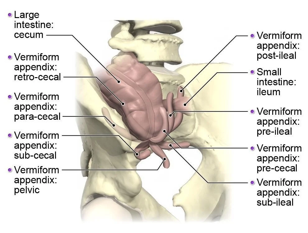
The clinical significance of this variation is considerable. The classical description of appendicitis often begins with poorly localized visceral pain followed by pain in the right lower quadrant. This pattern is partly dependent on the location of the appendix. A pelvic appendix may produce suprapubic or pelvic symptoms and may irritate the urinary bladder or rectum. A retrocaecal appendix may produce flank or back discomfort and may be associated with less pronounced anterior abdominal tenderness. A retroileal appendix can be closely related to the terminal ileum and may produce atypical symptoms. A subhepatic appendix can mimic cholecystitis or other right upper quadrant pathology. Rare left sided appendices can create a diagnostic challenge because the symptoms may not fit the expected anatomical pattern.
Anatomical variation is equally important during surgery. A surgeon approaching the caecum expecting to find a freely mobile appendix extending inferiorly may encounter a densely adherent retrocaecal appendix, an appendix lying behind the terminal ileum, or a long pelvic appendix extending deep into the pelvis. In each situation, the surgeon must adapt the dissection according to the anatomical relationships encountered. Failure to appreciate these variations can result in prolonged operative time, unnecessary tissue trauma, bleeding, bowel injury, incomplete removal of the appendix, or failure to identify the inflamed appendix.
The base of the appendix provides an important exception to this variability. Although the tip can occupy many different positions, the base usually originates at the posteromedial or medial aspect of the caecum at the point where the three taeniae coli converge. Following the taeniae coli toward their point of convergence is therefore one of the most reliable methods of identifying the appendiceal base during an operation.
The development of laparoscopic surgery has further increased the importance of anatomical knowledge. Laparoscopy provides a wide view of the abdominal cavity, making it possible to inspect the caecum, terminal ileum, pelvis, and right upper abdomen when necessary. However, visualization alone does not eliminate the challenges created by anatomical variation. The surgeon must understand the three dimensional relationships of the appendix and mesoappendix in order to interpret what is seen and determine the safest route of dissection.
The objective of this article is to provide a comprehensive review of the surgical anatomy of the vermiform appendix and to examine the major anatomical variations that influence appendectomy. Particular attention is given to the position of the appendix, mesoappendix, vascular anatomy, relationships with surrounding structures, and the practical implications of these variations for open and laparoscopic surgery.
2. Materials and Methods
This article was prepared as a narrative literature review focusing on the surgical anatomy of the vermiform appendix and the relevance of anatomical variation to appendectomy. Literature was identified using PubMed and other established biomedical literature sources, together with major international surgical guidelines and recent peer reviewed anatomical studies.
The search strategy focused on combinations of the terms vermiform appendix, appendix anatomy, appendiceal anatomical variation, appendicular artery, mesoappendix, appendectomy, laparoscopic appendectomy, open appendectomy, retrocaecal appendix, pelvic appendix, subhepatic appendix, left sided appendix, duplicated appendix, and appendicitis. Recent literature published through 2026 was prioritized when available. Particular attention was given to the 2024 systematic review of anatomical variations, the 2025 study describing mesoappendix position during laparoscopic appendectomy, recent anatomical studies of the appendicular vessels, and the 2025 edition of the World Society of Emergency Surgery Jerusalem Guidelines published in 2026.
The literature was assessed according to four major themes. The first was normal surgical anatomy, including the origin, structure, peritoneal relationships, vascular supply, lymphatic drainage, and neural supply of the appendix. The second was anatomical variation, with emphasis on appendiceal position, length, orientation, mesoappendiceal configuration, vascular patterns, and rare structural anomalies. The third was the relationship between anatomical variation and clinical manifestations of appendicitis. The fourth was the effect of anatomical variation on operative strategy, including laparoscopic and open appendectomy.
The purpose of this review was not to generate new patient data but to synthesize existing anatomical and surgical evidence into a clinically relevant description suitable for medical education. No human participants were recruited and no identifiable patient information was used.
3. Results
3.1 General anatomy of the vermiform appendix
The vermiform appendix is a true diverticulum of the caecum containing mucosa, submucosa, muscular layers, and serosa. It usually arises from the posteromedial aspect of the caecum a short distance below the ileocaecal valve. The adult appendix commonly measures approximately 8 to 10 cm, although considerable variation exists and lengths extending well beyond this range have been documented. Some anatomical references report a much wider overall range, emphasizing that appendiceal length should not be assumed during surgery.
The base of the appendix is relatively fixed compared with the tip. This difference is surgically important because the surgeon can usually identify the appendix by first locating the caecum and tracing the taeniae coli toward their convergence. Once the base has been identified, the appendix can generally be followed toward its tip even if the distal portion lies in an unusual position.
The lumen of the appendix communicates directly with the caecum. Its lumen is narrow and can become obstructed by lymphoid hyperplasia, faecaliths, foreign material, or other pathological processes. Obstruction can increase intraluminal pressure, impair venous and lymphatic drainage, and initiate the inflammatory process characteristic of acute appendicitis.
The wall contains abundant lymphoid tissue, particularly during childhood and adolescence. The appendix is therefore not simply a redundant tubular extension of the caecum. Its immunological characteristics help explain why inflammatory and infectious processes within the appendix can produce marked local and systemic responses.
3.2 The caecum as the principal surgical landmark
The caecum provides the starting point for anatomical identification of the appendix. It is usually situated in the right iliac fossa and is partly or completely covered by peritoneum. Its position, however, can also vary depending on the length of the ascending colon, mobility of the caecum, and developmental anatomy.
The three taeniae coli converge at the base of the appendix. This relationship is particularly valuable during appendectomy because the distal appendix may be hidden by the caecum, terminal ileum, adhesions, inflammatory tissue, or the greater omentum. In such situations, blindly searching for the appendix can cause unnecessary trauma. Identifying the caecum and following the taeniae toward their convergence provides a systematic method of localization.
The anterior and lateral surfaces of the caecum are generally accessible during laparoscopy. A retrocaecal appendix may, however, be hidden behind the caecum and ascending colon. The surgeon may need to mobilize the caecum to expose the appendix. This should be performed carefully because the inflammatory process can distort normal tissue planes.
3.3 Peritoneal relationship and mesoappendix
The appendix is generally intraperitoneal and is associated with a mesentery called the mesoappendix. The mesoappendix is a fold of peritoneum derived from the mesentery of the terminal ileum and contains the appendicular artery, accompanying veins, lymphatics, nerves, and connective tissue.
The mesoappendix is not uniform in length or configuration. It may extend along most of the appendix or terminate before reaching the tip. When the mesoappendix is short, the appendix may appear relatively fixed to adjacent structures. When it is long, the appendix can have greater mobility and may occupy the pelvis or other dependent areas.
Recent research has demonstrated that the position of the mesoappendix itself can vary during laparoscopic appendectomy. A 2025 study proposed a classification of mesoappendix position based on laparoscopic findings and emphasized that these configurations may influence operative difficulty, the need for additional ports, and the preferred strategy for controlling the mesoappendix.
This is clinically important because surgeons often describe appendiceal position but pay less attention to the mesoappendix. In practice, the mesoappendix determines the route by which the appendicular vessels are approached. A bulky, inflamed, or unusually positioned mesoappendix can obscure the base and make standard dissection more difficult.
3.4 Arterial supply
The principal arterial supply of the appendix is provided by the appendicular artery, usually a branch of the ileocolic artery, which itself arises from the superior mesenteric artery. The appendicular artery travels within the mesoappendix toward the appendix and gives branches along its length.
The arterial anatomy is clinically important because the appendicular artery must be controlled during appendectomy. Although the artery is often described as a single vessel, variations occur. Additional arterial branches and different patterns of entry into the mesoappendix have been reported.
A 2025 study involving 287 appendectomy patients demonstrated significant variation in mesoappendix extension and appendicular arterial patterns, highlighting the importance of identifying the vascular anatomy rather than assuming that every appendix possesses an identical vascular arrangement.
The arterial supply is especially relevant when the appendix is inflamed. Inflammation may increase tissue friability and obscure vascular planes. A surgeon who divides the mesoappendix without adequate visualization of the vessels can encounter bleeding that is difficult to control laparoscopically. Careful identification of the vascular pedicle therefore remains a central component of safe appendectomy.
3.5 Venous and lymphatic drainage
The appendicular veins accompany the arterial branches and ultimately drain into the ileocolic venous system and superior mesenteric venous circulation. Venous congestion can occur during acute appendicitis and contributes to edema and progression of inflammation.
Lymphatic drainage follows the vascular supply toward ileocolic lymph nodes and then into superior mesenteric lymphatic pathways. The abundant lymphoid tissue of the appendix contributes to its immunological role.
The anatomical relationship between lymphatic tissue and the lumen is clinically relevant in children and young adults because lymphoid hyperplasia may narrow the appendiceal lumen and contribute to obstruction. This is one reason why acute appendicitis is particularly common in younger age groups.
3.6 Neural supply
The appendix receives autonomic innervation through the superior mesenteric plexus. Visceral afferent fibers initially convey pain from appendiceal inflammation in a poorly localized manner, which contributes to the classical early periumbilical or central abdominal pain experienced by many patients.
As inflammation extends to the parietal peritoneum adjacent to the appendix, somatic sensory pathways become involved and pain becomes more localized. The eventual location of pain depends partly on the anatomical position of the appendix.
The neural anatomy therefore provides an important link between anatomy and clinical examination. A pelvic appendix can irritate pelvic structures and produce urinary or rectal symptoms, while a retrocaecal appendix can produce less typical abdominal tenderness. Anatomical variation should consequently be considered whenever the clinical presentation is inconsistent with the usual pattern of right lower quadrant appendicitis.
3.7 Variations in the position of the appendix
The most important anatomical variations involve the position of the appendiceal tip. The base is relatively consistent, but the appendix can extend in multiple directions from the caecum.
Retrocaecal appendix
The retrocaecal position is commonly reported as one of the most frequent anatomical configurations. The appendix lies posterior to the caecum and may extend superiorly or inferiorly along the posterior abdominal wall. It can also lie partly or completely behind the ascending colon.
The retrocaecal appendix has important implications for appendectomy. During an open operation through a conventional right lower quadrant incision, the appendix may not be immediately visible after entering the peritoneal cavity. The surgeon may see the caecum but find no obvious appendix extending from its anterior surface.
During laparoscopy, the retrocaecal appendix may similarly remain hidden until the caecum is mobilized. If the appendix is firmly adherent to the posterior abdominal wall, dissection must be performed carefully to avoid injury to the retroperitoneal structures.
Clinically, retrocaecal appendicitis may produce atypical symptoms. Because the inflamed appendix can lie close to the psoas muscle, pain may be associated with movement of the right hip. Anterior abdominal tenderness may be less pronounced than expected because the inflamed appendix is separated from the anterior parietal peritoneum by the caecum.
Pelvic appendix
The pelvic appendix extends inferiorly from the caecum and may descend toward the pelvic cavity. It is particularly important because its symptoms can differ substantially from those of a typical anterior appendix.
A pelvic appendix may lie close to the urinary bladder, rectum, sigmoid colon, uterus, fallopian tube, or ovary. Inflammation may therefore produce urinary frequency, dysuria, pelvic discomfort, rectal pressure, or diarrhea. In females, pelvic appendicitis can mimic gynecological conditions such as ovarian pathology, adnexal inflammation, or ectopic pregnancy.
During laparoscopic appendectomy, a pelvic appendix is often relatively easy to visualize once the small bowel and omentum are moved away from the pelvis. However, the mesoappendix may be long and mobile, and the appendix can become adherent to pelvic structures when inflammation is advanced. The surgeon must distinguish inflammatory adhesions from normal tissue and avoid injury to reproductive or urinary structures.
Subcaecal appendix
A subcaecal appendix extends inferiorly beneath the caecum. It may remain within the right iliac fossa and can be relatively close to the iliac vessels, ureter, or pelvic structures depending on its length.
Subcaecal appendicitis may resemble pelvic or retrocaecal disease. During surgery, the caecum may need to be elevated to expose the appendix. The relationship with the ileocaecal region should be carefully assessed before dividing the mesoappendix.
Pre-ileal appendix
In the pre-ileal position, the appendix lies anterior to the terminal ileum. This configuration can result in inflammatory contact with the small bowel and may cause symptoms that are less typical than those of a straightforward right lower quadrant appendix.
During laparoscopic surgery, the appendix may initially be hidden beneath the terminal ileum. Gentle mobilization of the small bowel can reveal the appendix. Excessive traction should be avoided because an inflamed appendix may be friable and may adhere strongly to the ileum.
Post-ileal or retroileal appendix
The post-ileal appendix lies posterior to the terminal ileum. This position can make the appendix difficult to identify because it is hidden behind the ileal loops.
The inflammatory process may also spread along the mesentery or produce localized ileal irritation. During appendectomy, the terminal ileum may need to be mobilized to expose the appendix, and careful assessment is required to distinguish the appendix from inflamed or adherent small bowel.
Paracaecal appendix
A paracaecal appendix lies alongside the caecum rather than directly anterior or posterior to it. Its location may make it relatively accessible laparoscopically, but inflammation can result in adhesions between the appendix and caecal wall.
This position is particularly relevant because an inflamed appendix can become closely attached to the caecum. Careless dissection may injure the caecal wall, particularly when severe inflammation or perforation is present.
Precaecal appendix
A precaecal appendix lies anterior to the caecum. This is less challenging to identify during surgery because it may be visible immediately after exposure of the caecum. However, an inflamed precaecal appendix can become adherent to the greater omentum or anterior abdominal wall.
Subhepatic appendix
A subhepatic appendix is an uncommon anatomical variant in which the appendix extends upward toward the liver. It may occur because of an unusually high caecum, incomplete descent of the caecum, intestinal malrotation, or a long ascending colon.
This position has major clinical importance because appendicitis can produce right upper quadrant pain and tenderness, potentially mimicking acute cholecystitis, hepatic disease, or other hepatobiliary conditions. Recent radiological literature emphasizes that unusual appendiceal positions can substantially alter both diagnosis and the appearance of inflammatory complications on imaging.
At surgery, a subhepatic appendix may require mobilization of the ascending colon or caecum. The surgeon must also be aware of the proximity of the gallbladder, duodenum, liver, and hepatic flexure.
Left sided appendix
A left sided appendix is rare but clinically significant. It can occur in association with intestinal malrotation, situs inversus, or an unusually mobile caecum and colon.
Patients may present with left lower quadrant pain or even more diffuse abdominal symptoms. If appendicitis is suspected despite left sided symptoms, imaging becomes particularly valuable. During surgery, the expected right sided location of the caecum cannot be assumed.
A left sided appendix demonstrates why appendicitis should remain in the differential diagnosis even when the pain location is atypical. Anatomical variation can alter the physical examination without changing the underlying disease.
3.8 Variations in appendiceal length and shape
Appendiceal length varies considerably between individuals. A short appendix may remain close to the caecum, whereas a long appendix may extend into the pelvis, behind the caecum, or toward the terminal ileum.
Length has practical surgical implications because a long appendix can cross anatomical compartments and become closely related to structures that are not normally encountered during appendectomy. A long pelvic appendix may extend deep into the pelvis, while a long retrocaecal appendix may extend upward toward the hepatic region.
The diameter of the appendix also varies. Enlargement may be physiological in certain contexts but is particularly relevant radiologically when acute inflammation, neoplasm, or other pathology is suspected.
The external shape may also differ. The appendix can appear straight, curved, coiled, or tortuous. A curved appendix may be more difficult to grasp and expose, particularly when inflammation causes adhesions.
3.9 Duplication and other rare anatomical anomalies
Although extremely uncommon, duplication of the appendix has been described. The duplicated structures may arise from a common caecal region or represent two separate appendiceal structures. Triple appendices are even rarer.
The surgical significance of duplication is considerable because failure to recognize a second appendix can result in persistent symptoms after apparently successful appendectomy. Surgeons should therefore examine the caecal region carefully when the anatomy appears unusual or when an apparent appendix does not fully explain the operative findings.
Other rare anomalies include intracaecal, intramural, and completely hidden appendices. These conditions are uncommon but reinforce the principle that the appendix should be identified from its base and anatomical relationships rather than solely by its superficial appearance. The systematic literature confirms that a broad range of unusual appendiceal configurations has been documented.
3.10 Mesoappendix variation and its operative significance
The mesoappendix is often treated as a simple anatomical structure, but recent evidence demonstrates that its configuration can vary substantially. Its length, attachment, vascular arrangement, and relationship with the appendix may all influence the technical steps of appendectomy.
A fully extending mesoappendix can provide a relatively straightforward route to the appendicular vessels. In contrast, a mesoappendix that does not extend to the tip may leave part of the appendix relatively exposed and mobile. Inflammation can further distort these relationships.
The 2025 laparoscopic study of mesoappendix positioning proposed a classification based on the position encountered during appendectomy and demonstrated that different configurations may require different operative strategies.
This finding has educational importance because it demonstrates that the difficulty of appendectomy is not determined solely by whether the appendix is retrocaecal or pelvic. The mesoappendix itself can determine how easily the appendix can be grasped, elevated, divided, and removed.
3.11 Anatomical basis of laparoscopic appendectomy
Laparoscopic appendectomy has become the preferred operative approach for acute appendicitis in contemporary guidelines when appropriate equipment and expertise are available. The 2025 WSES Jerusalem Guidelines, published in 2026, continue to identify laparoscopic appendectomy as the standard surgical approach and emphasize that clinical decision making should incorporate patient characteristics and available resources.
The principal anatomical advantage of laparoscopy is visualization of the entire abdominal cavity. The surgeon is not restricted to the region around a skin incision and can inspect the caecum, terminal ileum, pelvis, upper abdomen, and contralateral side when necessary.
The conventional three port approach generally provides access for visualization, tissue manipulation, and instrument control. Port positions may be modified depending on patient anatomy, body habitus, previous surgery, or the location of the appendix.
After establishing access, the surgeon identifies the caecum. The taeniae coli are followed to their convergence to identify the appendiceal base. The appendix is then traced toward its tip, taking into account its position and relationship to the terminal ileum and caecum.
The mesoappendix is subsequently exposed and the appendicular vessels are controlled. The appendix is divided close to its caecal origin according to the operative technique and institutional practice. The specimen is removed while minimizing contamination of the abdominal cavity.
Anatomical identification is more important than memorization of a rigid sequence. For example, a retrocaecal appendix may require mobilization of the caecum, whereas a pelvic appendix may require mobilization of small bowel and pelvic structures. A post-ileal appendix may require careful manipulation of the terminal ileum. The operation therefore changes according to anatomy.
3.12 Open appendectomy and anatomical considerations
Although laparoscopic appendectomy is currently preferred in most settings where appropriate expertise and equipment are available, open appendectomy remains an important procedure. It may be performed when laparoscopic facilities are unavailable, when patient factors make laparoscopy unsuitable, or when conversion becomes necessary.
The classical open approach uses an incision in the right lower quadrant. The exact incision may vary according to surgeon preference and the expected anatomical position of the appendix.
The principal challenge of open surgery is that the incision provides a limited field of view. If the appendix is not immediately identified, the surgeon must avoid unnecessary extension of the incision or excessive manipulation of the bowel.
The caecum is identified and delivered into the wound when possible. The taeniae coli can then be followed to the appendiceal base. If the appendix is retrocaecal, mobilization of the caecum may be necessary. If it is pelvic, the surgeon may need to follow the appendix downward into the pelvis.
Anatomical variation is therefore particularly important in open appendectomy because the operation depends heavily on the relationship between the incision and the location of the appendix.
3.13 Anatomical variation and clinical presentation
The location of the appendix has a direct effect on the clinical manifestations of appendicitis. The classical progression of pain from the central abdomen to the right lower quadrant occurs because visceral pain is initially poorly localized and later becomes localized when the parietal peritoneum is irritated.
However, this pattern is not universal. A pelvic appendix can cause suprapubic pain and urinary symptoms. A retrocaecal appendix can produce flank discomfort or pain on hip movement. A subhepatic appendix can produce right upper quadrant pain. A left sided appendix can produce left sided abdominal symptoms.
These variations create diagnostic challenges and increase the importance of imaging in patients whose clinical presentation is atypical.
Modern radiology also benefits from knowledge of anatomical variation. CT can identify the appendix in unusual positions and can demonstrate inflammatory changes extending into adjacent compartments. Recent radiological reviews emphasize that appendiceal location and inflammatory spread should be considered when interpreting atypical presentations and appendiceal abscesses.
3.14 Anatomical variation and operative complications
Anatomical variation can contribute to several complications during appendectomy. The most immediate concern is vascular bleeding, particularly when the appendicular artery or its branches are divided without adequate visualization.
Bowel injury is another concern. A retroileal appendix may be closely associated with the terminal ileum. A subhepatic appendix may lie near the duodenum or gallbladder. A pelvic appendix can be adjacent to the bladder, rectum, uterus, ovary, or other pelvic structures.
Caecal injury may occur when a densely inflamed appendix is adherent to the caecum. In severe appendicitis, the normal tissue planes may be destroyed, making separation difficult.
Another potential complication is incomplete appendectomy. This can occur if the base is not correctly identified or if a difficult appendix is divided without confirming its complete removal. The risk is particularly important in unusual anatomy and severe inflammation.
The most reliable protection against these complications is anatomical orientation. The surgeon should identify the caecum, locate the taeniae coli, confirm the appendiceal base, understand the mesoappendix, and determine the position of the appendix before undertaking extensive dissection.
4. Discussion
The vermiform appendix provides an excellent example of why surgical anatomy cannot be reduced to memorization of a standard diagram. The appendix has a relatively constant origin but a highly variable distal position. This combination explains both the reliability of the appendiceal base as a surgical landmark and the wide variety of operative findings encountered during appendectomy.
The convergence of the taeniae coli remains one of the most dependable anatomical landmarks. Regardless of whether the appendix is retrocaecal, pelvic, pre-ileal, post-ileal, subcaecal, or another less common position, its base generally remains connected to the caecum at the point of taenia convergence. This explains why an operation should begin with identification of the caecum rather than an attempt to locate the appendix tip blindly.
The importance of appendiceal position has been recognized for many years, but recent literature reinforces how extensive the variation actually is. A systematic review published in 2024 analyzed 40 relevant studies and demonstrated substantial differences in appendiceal location and morphology. This finding has direct educational relevance because students frequently learn the appendix as a structure located simply in the right iliac fossa. Such a description is adequate for basic anatomy but incomplete for surgical anatomy.
Among positional variants, retrocaecal and pelvic appendices are particularly important. The retrocaecal appendix may be hidden behind the caecum and can be difficult to expose through a small open incision. The pelvic appendix, by contrast, can extend into the pelvis and produce symptoms involving the urinary or gastrointestinal tract. Both variants can alter the physical examination and operative approach.
The clinical significance of a pelvic appendix is especially important in women. The appendix may lie near the ovary, fallopian tube, uterus, or urinary bladder, creating potential diagnostic confusion. In such patients, appendicitis may mimic gynecological disease. Conversely, gynecological conditions may mimic appendicitis. Anatomical awareness therefore contributes not only to surgical safety but also to preoperative diagnosis.
The retrocaecal appendix demonstrates another important principle. The absence of dramatic anterior right lower quadrant tenderness does not exclude appendicitis. The inflamed appendix may be separated from the anterior parietal peritoneum by the caecum, producing a less typical examination. This is a clear example of how anatomical relationships influence clinical findings.
Subhepatic and left sided appendices are rare but particularly important because they can lead to diagnostic delay. A subhepatic appendix can mimic acute gallbladder disease, while a left sided appendix can mimic diverticular or other left sided pathology. The 2026 radiological literature continues to emphasize the importance of recognizing unusual appendiceal positions when evaluating atypical abdominal pain.
The mesoappendix deserves greater attention in surgical education. Traditionally, students learn that the mesoappendix contains the appendicular artery, but recent surgical research demonstrates that its configuration itself varies. The 2025 study by Cheng and colleagues introduced a laparoscopic classification of mesoappendix position and showed that this anatomical variation can influence operative strategy.
This finding is important because operative difficulty is often incorrectly attributed solely to the position of the appendix. In reality, two appendices occupying the same anatomical region may present very different operative challenges depending on mesoappendix length, inflammation, adhesions, vascular anatomy, and the relationship with adjacent bowel.
The vascular anatomy is equally important. The appendicular artery is usually derived from the ileocolic arterial system, but variations in its branching pattern occur. A 2025 clinical anatomical study demonstrated variability in appendicular arterial patterns and mesoappendiceal extension among patients undergoing appendectomy. This supports the principle that the surgeon should identify the actual vascular anatomy rather than rely exclusively on the expected textbook pattern.
The transition from open to laparoscopic appendectomy has changed the way anatomical variation is managed. In open surgery, the surgeon is limited by the incision and may need to mobilize the caecum or extend the incision when the appendix is difficult to locate. Laparoscopy provides a much wider field and allows the surgeon to inspect multiple abdominal compartments. This is one reason why contemporary guidelines recommend laparoscopic appendectomy when appropriate equipment and expertise are available. The 2020 WSES guidelines already demonstrated advantages of laparoscopy over open appendectomy, including lower postoperative pain, fewer surgical site infections, shorter hospital stay, and faster recovery. The updated 2025 edition continues to recognize laparoscopic appendectomy as the standard surgical approach.
However, laparoscopy does not eliminate anatomical difficulties. A surgeon operating laparoscopically must still understand where the appendix can hide. A retrocaecal appendix may require mobilization of the caecum. A retroileal appendix may require careful movement of the terminal ileum. A pelvic appendix may require identification of pelvic organs. A subhepatic appendix may require examination of the upper right abdomen. Therefore, laparoscopic surgery increases visualization but does not replace anatomical knowledge.
This distinction is particularly important in surgical education. A trainee who learns only the standard appearance of the appendix may become disoriented when the appendix is not immediately visible. A trainee who understands the relationship between the caecum, taeniae coli, terminal ileum, mesoappendix, and surrounding compartments can approach the problem systematically.
The concept of systematic localization is therefore more useful than memorization of individual positions. The surgeon first identifies the caecum. The taeniae coli are followed to their convergence. The base of the appendix is identified. The direction of the appendix is then determined by following it away from the caecum. This method remains valid even when the appendix is in an unusual position.
The same principle applies to open surgery. When the appendix cannot be identified through a standard incision, the surgeon should not immediately assume that it is absent. The caecum should be identified, and the taeniae should be followed. Mobilization of the caecum may reveal a retrocaecal appendix. Mobilization of the terminal ileum may reveal a retroileal appendix. Examination of the pelvis may reveal a pelvic appendix.
The possibility of duplication, although extremely rare, further demonstrates the importance of complete anatomical assessment. If the operative appearance is inconsistent with the expected anatomy or if symptoms persist after apparently adequate appendectomy, an unusual appendiceal configuration should be considered. The literature has documented duplicated and even triplicated appendices, although these are exceptional findings.
Another important aspect of contemporary appendiceal surgery is that appendectomy is no longer the only possible treatment for every patient with appendicitis. Modern guidelines recognize antibiotic treatment as an option for selected patients with uncomplicated appendicitis. The 2025 WSES guidelines emphasize patient selection and clinical context when considering nonoperative treatment. Nevertheless, when appendectomy is chosen, the same anatomical principles remain fundamental.
The surgical anatomy also has implications for complicated appendicitis. Perforation, abscess, and inflammatory phlegmon can obliterate normal tissue planes. In these situations, anatomical variation may influence the route through which infection spreads. A pelvic appendix may produce a pelvic abscess, while a retrocaecal appendix may produce a retrocaecal or retroperitoneal inflammatory collection. Recent consensus work has emphasized anatomotopographic classification of appendiceal abscesses because their location influences imaging interpretation and therapeutic planning.
The anatomy of the appendix therefore has implications extending beyond routine appendectomy. It influences diagnosis, imaging, operative exposure, dissection, complication prevention, and management of advanced disease.
One of the most valuable lessons from recent anatomical research is that variation should be considered normal rather than exceptional. The surgeon should expect the appendix to vary. The retrocaecal and pelvic positions may be common enough to be anticipated routinely, while subhepatic, left sided, intracaecal, and duplicated appendices should be remembered as less common possibilities.
The importance of anatomical knowledge becomes even greater in minimally invasive surgery because the surgeon is working with instruments rather than directly palpating tissues. Laparoscopy provides an excellent visual field but reduces tactile feedback. Inflammation can obscure landmarks, and the appearance of tissues may differ substantially from the standard anatomical specimen. A strong understanding of normal anatomy allows abnormal findings to be recognized more accurately.
The development of newer technologies, including advanced imaging and robotic surgery, should therefore be regarded as complementary to anatomical knowledge rather than a replacement for it. Modern imaging can identify an unusual appendix before surgery, but the surgeon must still understand what the imaging finding means anatomically. Similarly, enhanced visualization can show the appendix clearly but does not automatically reveal the safest route for vascular control or dissection.
The contemporary approach to appendectomy is consequently based on a combination of anatomical orientation, careful tissue handling, appropriate imaging, minimally invasive techniques where suitable, and adaptation to the individual patient's anatomy.
5. Conclusion
The vermiform appendix is a small structure with substantial anatomical variability and considerable surgical importance. Although its base generally arises from the caecum at the convergence of the taeniae coli, the distal appendix may occupy numerous positions, including retrocaecal, pelvic, subcaecal, pre-ileal, post-ileal, paracaecal, precaecal, subhepatic, and, rarely, left sided or intracaecal locations. Variations in appendiceal length, shape, mesoappendix, and vascular supply further increase the anatomical diversity encountered during appendectomy.
These variations have direct clinical consequences. They can alter the location and character of abdominal pain, modify physical examination findings, mimic other abdominal or pelvic diseases, and complicate radiological diagnosis. They also have direct operative implications because the appendix may be hidden behind the caecum, terminal ileum, or other structures or may extend into anatomical compartments distant from its usual location.
The convergence of the taeniae coli remains one of the most dependable surgical landmarks for locating the appendiceal base. Once the base has been identified, the appendix can be traced toward its tip regardless of its position. Knowledge of the mesoappendix and appendicular vascular anatomy is equally important because these structures demonstrate clinically meaningful variation and determine the safest method of vascular control.
Laparoscopic appendectomy has become the preferred surgical approach in contemporary practice when suitable equipment and expertise are available. Its major anatomical advantage is the ability to inspect the entire abdominal cavity and adapt the operative strategy to unusual appendiceal positions. Nevertheless, laparoscopy does not remove the need for detailed anatomical knowledge. Instead, it requires the surgeon to understand three dimensional anatomy and recognize variations from a different visual perspective.
Open appendectomy remains an important procedure and requires particular attention to anatomical localization because the operative field is more restricted. In either approach, the surgeon must avoid blind dissection and should systematically identify the caecum, trace the taeniae coli, locate the appendiceal base, determine the orientation of the appendix, and understand the relationship of the mesoappendix to surrounding structures.
Current evidence confirms that anatomical variation is not merely an academic feature of the appendix. It directly influences diagnosis, imaging, operative difficulty, surgical approach, and complication risk. Recent studies of mesoappendiceal configuration and appendicular vascular anatomy further demonstrate that even apparently routine appendectomy involves considerable anatomical diversity.
Ultimately, safe appendectomy depends on the ability to adapt surgical technique to individual anatomy. Modern imaging and minimally invasive technology have improved the surgeon's ability to identify and treat appendicitis, but these technologies remain most effective when supported by a thorough understanding of the surgical anatomy of the vermiform appendix.
References
Podda M, Ceresoli M, De Simone B, et al. Diagnosis and Treatment of Acute Appendicitis: 2025 Edition of the World Society of Emergency Surgery Jerusalem Guidelines. JAMA Surgery. 2026;161(3):283-295. doi:10.1001/jamasurg.2025.6218.
Di Saverio S, Podda M, De Simone B, et al. Diagnosis and treatment of acute appendicitis: 2020 update of the WSES Jerusalem guidelines. World Journal of Emergency Surgery. 2020;15:27.
Hodge BD, Kashyap S, Khorasani-Zadeh A. Anatomy, Abdomen and Pelvis: Appendix. StatPearls. Updated 2023.
Appendectomy. StatPearls. National Center for Biotechnology Information. Updated 2025.
Sakellariadis A, Sofou F, Chrysikos D, Sampsakos-Mariolis T, Schizas D, Troupis T, Filippou D. Anatomical Variations of the Vermiform Appendix. Acta Medica Academica. 2024;53(3). doi:10.5644/ama2006-124.461.
Cheng E, Shamavonian R, Mui J, et al. Mesoappendix position variations in laparoscopic appendicectomy: a new anatomical classification to guide surgical strategy. Updates in Surgery. 2025;77:835-842. doi:10.1007/s13304-025-02172-7.
[Authors]. Anatomical and clinical variations in the mesoappendix and appendicular arteries: implications for appendicitis and surgical outcomes. ANZ Journal of Surgery. 2025;95(12):2599-2604. doi:10.1111/ans.70090.
[Authors]. Anatomical Variations of the Vermiform Appendix. Systematic review of anatomical variations in position and morphology. 2024.
Carr NJ. Normal appendix: anatomy, specimen dissection and histology relevant to pathological practice. 2024. doi:10.1002/9781119423195.ch28.
Pavan Amaral AH, Bonetto Nicolau Bignotto C, Alves Ferreira Martins E, et al. When the appendix hides: the radiologist's guide to appendix variants and mimics. Abdominal Radiology. 2026. doi:10.1007/s00261-026-05665-0.
[Authors]. Imaging-guided classification and diagnostic pathways for appendiceal abscesses: results from the 2025 Italian Society of Research in Surgery and Italian Society of Emergency and Trauma Surgery consensus conference. Surgery. 2026.
Andersson RE. Meta-analysis of the clinical and epidemiological evidence concerning the development and pathogenesis of acute appendicitis. British Journal of Surgery. 2004;91:28-37.
Bhangu A, Søreide K, Di Saverio S, Assarsson JH, Drake FT. Acute appendicitis: modern understanding of pathogenesis, diagnosis, and management. The Lancet. 2015;386:1278-1287.
Gorter RR, Eker HH, Gorter-Stam MAW, et al. Diagnosis and management of acute appendicitis: EAES consensus development conference. Surgical Endoscopy. 2016;30:4668-4690.
Flum DR, Davidson GH, Monsell SE, et al. A randomized trial comparing antibiotics with appendectomy for appendicitis. New England Journal of Medicine. 2020;383:1907-1919.
CODA Collaborative. A randomized trial comparing antibiotics with appendectomy for appendicitis. New England Journal of Medicine. 2020;383:1907-1919.
Sallinen V, Akl EA, You JJ, et al. Meta-analysis of antibiotics versus appendicectomy for non-perforated acute appendicitis. British Journal of Surgery. 2016;103:656-667.
Jaschinski T, Mosch CG, Eikermann M, Neugebauer EAM, Sauerland S. Laparoscopic versus open surgery for suspected appendicitis. Cochrane Database of Systematic Reviews. 2018;11:CD001546.
Mason RJ, Moazzez A, Moroney JR, Katkhouda N. Laparoscopic vs open appendectomy in patients with acute appendicitis and a BMI greater than 30. Surgical Endoscopy. 2007;21:1591-1594.
Swank HA, Eshuis EJ, Ubbink DT, et al. Routine histopathology of appendectomy specimens: a systematic review. Surgical Endoscopy. 2011;25:1676-1685.
Humes DJ, Simpson J. Acute appendicitis. BMJ. 2006;333:530-534.
Petroianu A. Diagnosis of acute appendicitis. International Journal of Surgery. 2012;10:115-119.
Birnbaum BA, Wilson SR. Appendicitis at the millennium. Radiology. 2000;215:337-348.
Rao PM, Rhea JT, Novelline RA, Mostafavi AA, McCabe CJ. Effect of computed tomography of the appendix on treatment of patients and use of hospital resources. New England Journal of Medicine. 1998;338:141-146.
Addiss DG, Shaffer N, Fowler BS, Tauxe RV. The epidemiology of appendicitis and appendectomy in the United States. American Journal of Epidemiology. 1990;132:910-925.
Flum DR. Acute appendicitis: appendectomy or the "antibiotics first" strategy. New England Journal of Medicine. 2015;372:1937-1943.
Livingston EH, Woodward WA, Sarosi GA, Haley RW. Disconnect between incidence of nonperforated and perforated appendicitis. Annals of Surgery. 2007;245:886-892.
Carr NJ. The pathology of acute appendicitis. Annals of Diagnostic Pathology. 2000;4:46-58.
Jones MW, Lopez RA, Deppen JG. Appendicitis. StatPearls. Treasure Island, Florida: StatPearls Publishing.
Humes DJ, Simpson J. Acute appendicitis. BMJ. 2006;333:530-534.