
Topographical Anatomy of the Femoral Triangle and its Importance in Vascular Surgery: Analysis of Regional Architecture, Access Strategies, and Complication
1. Tanabaeva Aiperi
2. Kanchan Kakde
3. Prajakta Markad
4. Vaishnavi Thavare
(1. Professor, International Medical Faculty, Osh State University, Osh, Kyrgyz Republic
2. Student, International Medical Faculty, Osh State University, Osh, Kyrgyz Republic
3. Student, International Medical Faculty, Osh State University, Osh, Kyrgyz Republic
4. Student, International Medical Faculty, Osh State University, Osh, Kyrgyz Republic)
Abstract
The femoral triangle represents the most critical anatomical gateway to the arterial and venous systems of the lower extremity, serving as the predominant access site for diagnostic and therapeutic endovascular procedures, open vascular reconstructions, and hemodynamic monitoring. Despite its superficial location and apparent simplicity, this anatomical region encompasses a complex neurovascular arrangement where the femoral artery, vein, and nerve traverse a confined fascial compartment bounded by muscular and ligamentous structures, presenting significant risks of hemorrhage, nerve injury, lymphatic complications, and access site morbidity when surgical anatomy is not respected. This comprehensive review examines the topographical anatomy of the femoral triangle through the lens of contemporary vascular surgical practice, synthesizing classical anatomical knowledge with recent advances in ultrasound-guided access, percutaneous closure devices, and minimally invasive techniques. The article presents detailed analysis of the triangle's boundaries, contents, and fascial relationships, with particular attention to the anatomical variations of the femoral artery bifurcation, the course of the femoral nerve and its branches, and the lymphatic drainage patterns that influence wound healing and infection risk. Recent epidemiological data indicate that over five million femoral access procedures are performed annually worldwide, with vascular access complications representing a leading cause of morbidity and healthcare expenditure, underscoring the continued relevance of precise topographical knowledge. The review further explores the surgical implications of variant anatomy including high and low arterial bifurcation, accessory saphenous veins, and anomalous femoral nerve branching, alongside the critical distinction between common femoral artery and superficial femoral artery cannulation that defines the safety of percutaneous interventions. By integrating classical cadaveric studies with contemporary imaging data, ultrasound guidance protocols, and clinical outcome analyses, this article provides vascular surgeons, interventionalists, and anatomists with a robust framework for understanding the femoral triangle not merely as a conduit for catheterization, but as a complex anatomical region where millimeter-scale precision determines patient outcomes including limb salvage, quality of life, and procedural success.
Introduction
The femoral triangle, first described in systematic anatomical detail by the Italian anatomist Bartolomeo Eustachi in the sixteenth century and later refined by the British surgeon and anatomist Astley Cooper in his monumental work on hernias and arterial anatomy, remains the most frequented anatomical region in vascular surgery, serving as the primary access point to the arterial system for the majority of endovascular interventions performed globally. This triangular depression, located in the upper anterior thigh extending from the inguinal ligament to the apex where the sartorius and adductor longus muscles converge, contains within its fascial boundaries the femoral nerve, artery, and vein arranged in a lateral to medial sequence that has become memorized by generations of medical students through the mnemonic NAVY, yet whose true surgical complexity extends far beyond this simplified arrangement. The importance of this region to vascular surgery cannot be overstated: it is here that the common femoral artery, the largest arterial access site in the body with a diameter typically ranging from six to ten millimeters, provides the conduit through which catheters, balloons, stents, and endografts are deployed to treat aortic aneurysms, peripheral arterial disease, and cerebrovascular pathology.
The evolution of vascular surgery from the era of open surgical exposure with extensive incisions and ligation of multiple vessels, through the transition to percutaneous Seldinger technique introduced in 1953, to the contemporary era of ultrasound-guided micropuncture access and percutaneous closure devices has paradoxically increased rather than diminished the importance of detailed topographical anatomy. While early vascular surgeons relied on direct visualization of anatomical structures through open exposure, the modern interventionalist frequently accesses the femoral artery through percutaneous puncture guided by fluoroscopy and ultrasound, requiring mental reconstruction of the underlying anatomy from surface landmarks and imaging data. This shift has created new challenges: the incidence of access site complications including pseudoaneurysm formation, arteriovenous fistula, retroperitoneal hemorrhage, and femoral nerve injury persists at rates of two to six percent despite technological advances, with certain complications directly attributable to anatomical ignorance or misidentification of vascular structures.
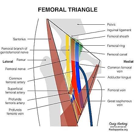
The femoral triangle presents a region of concentrated anatomical hazard where the femoral artery lies superficial and vulnerable to injury, where the femoral nerve traverses the lateral aspect subject to compression and traction injuries, and where the femoral vein and lymphatics occupy the medial aspect at risk of damage during dissection or catheterization. The common femoral artery represents the optimal site for arterial access due to its superficial location, relatively large diameter, and compressibility against the femoral head, yet the precise location of the arterial puncture relative to the inguinal ligament and the femoral head determines the risk of complications. Punctures above the inguinal ligament risk retroperitoneal hemorrhage due to the absence of bony support for compression, while punctures below the bifurcation into the superficial femoral and profunda femoris arteries risk pseudoaneurysm formation, thrombosis, and nerve injury due to the smaller caliber and deeper location of these vessels.
Contemporary vascular practice has witnessed an exponential increase in the volume and complexity of endovascular procedures, with transcatheter aortic valve replacement, endovascular aneurysm repair, and mechanical thrombectomy for stroke now performed routinely through femoral access. This expansion has been accompanied by increasing patient complexity including obesity, peripheral arterial disease, and previous groin interventions that distort normal anatomy and increase technical difficulty. The adoption of ultrasound guidance for femoral access has been shown to reduce complications by up to fifty percent in randomized trials, yet this technology requires interpretation of anatomical structures in cross-sectional imaging, demanding integration of topographical knowledge with sonographic anatomy.
This review aims to provide a comprehensive examination of the topographical anatomy of the femoral triangle, integrating classical anatomical descriptions with contemporary vascular surgical practice, imaging studies, and complication prevention strategies. The article will explore the boundaries and contents of the triangle, delineate the critical relationships between neurovascular structures, examine anatomical variations that influence surgical approach, and discuss the clinical importance of precise topographical knowledge in preventing access site complications and optimizing patient outcomes in modern vascular surgery.
Materials and Methods
This review article was constructed through systematic analysis of the contemporary vascular surgical and anatomical literature, with emphasis on publications from the past two decades regarding femoral access techniques, anatomical variations, and complication prevention strategies. The methodological approach involved comprehensive examination of peer-reviewed journals in vascular surgery, interventional radiology, and anatomy, including Journal of Vascular Surgery, European Journal of Vascular and Endovascular Surgery, Journal of Endovascular Therapy, Annals of Vascular Surgery, and Clinical Anatomy. Particular attention was directed toward studies utilizing duplex ultrasound characterization of femoral triangle anatomy, computed tomography angiography for preoperative mapping, and cadaveric dissection series providing quantitative measurements of neurovascular relationships.
Inclusion criteria for anatomical data encompassed studies reporting quantitative measurements of vessel diameters, depths, and branching patterns in cohorts exceeding fifty subjects, with preference given to imaging studies performed in vivo rather than cadaveric measurements alone. Clinical correlation data were derived from surgical series reporting access site complication rates, ultrasound guidance outcomes, and closure device performance, with specific attention to studies comparing anatomical landmark-guided versus ultrasound-guided access. Embryological data were sourced from developmental anatomy literature to explain variant configurations including arterial bifurcation patterns and nerve anomalies. When discrepancies existed between classical anatomical descriptions and contemporary surgical findings, preference was given to intraoperative observations and imaging data that reflect live surgical anatomy.
The synthesis of this literature was organized according to the topographical regions encountered during femoral access and open surgical exposure, progressing from the superior boundary at the inguinal ligament to the apex of the triangle and the underlying muscular floor. All anatomical descriptions are presented according to standard surgical orientation, with measurements reported in millimeters and centimeters. The reference list comprises foundational anatomical works, contemporary vascular surgical series, and technological studies that collectively represent the current state of knowledge regarding femoral triangle anatomy and its surgical importance.
Results
Boundaries and Superficial Architecture
The femoral triangle, also known by the eponym Scarpa's triangle in reference to the Italian anatomist Antonio Scarpa, is a subfascial space located in the upper third of the anterior thigh, shaped as an inverted triangle with its base superior at the inguinal ligament and its apex inferior where the medial border of the sartorius muscle crosses the medial border of the adductor longus. The superior boundary is formed by the inguinal ligament, which extends from the anterior superior iliac spine laterally to the pubic tubercle medially, representing the inferior edge of the external oblique aponeurosis and the point of transition between the external iliac and femoral vessels. The lateral boundary is formed by the medial border of the sartorius muscle, the longest muscle in the body originating from the anterior superior iliac spine and descending obliquely across the thigh. The medial boundary is formed by the medial border of the adductor longus muscle, which originates from the pubic bone and descends to insert on the linea aspera of the femur. The floor of the triangle is formed by the iliopsoas muscle laterally and the pectineus muscle medially, with the adductor longus contributing to the inferomedial floor.
The roof of the femoral triangle consists of skin, subcutaneous tissue, and the superficial fascia containing the superficial inguinal lymph nodes, the great saphenous vein, and cutaneous nerves. The superficial inguinal lymph nodes, typically numbering eight to ten, are arranged in a horizontal chain parallel to the inguinal ligament and a vertical chain along the terminal great saphenous vein, draining the lower extremity, perineum, and lower abdominal wall. These lymph nodes are surgically significant as their presence in the subcutaneous tissue of the femoral triangle necessitates careful dissection during open exposure to avoid lymphatic leak and subsequent lymphocele or lymphorrhea, complications that can delay wound healing and predispose to infection. The great saphenous vein, the longest vein in the body, ascends in the superficial fascia on the medial side of the thigh and passes through the saphenous opening or fossa ovalis to drain into the femoral vein approximately three to four centimeters inferior to the inguinal ligament. This junction represents a critical landmark as the superficial epigastric, superficial circumflex iliac, and superficial external pudendal veins join the great saphenous vein near its termination, creating a confluence that must be preserved during dissection to avoid venous hypertension and varicosities.
The deep fascia of the thigh, known as the fascia lata, forms the fascial roof of the femoral triangle proper, splitting to enclose the femoral vessels and creating the femoral sheath, a funnel-shaped fascial tube extending approximately three to four centimeters inferior to the inguinal ligament. The femoral sheath is divided into three compartments: the lateral compartment containing the femoral artery, the intermediate compartment containing the femoral vein, and the medial compartment known as the femoral canal containing lymphatics and a deep inguinal lymph node, with the femoral septum closing the canal inferiorly. The femoral canal is clinically significant as the site of femoral hernias, where abdominal contents may protrude through the femoral ring into the canal, potentially compressing the femoral vein and causing venous congestion. The femoral sheath is thickest anteriorly over the artery, providing a palpable pulse, and is intimately adherent to the adventitia of the vessels, requiring sharp dissection during open exposure.
Neurovascular Contents and Relationships
The contents of the femoral triangle are arranged from lateral to medial in the order of femoral nerve, femoral artery, femoral vein, and femoral canal, remembered by the mnemonic NAVY or NAVEL when including the lymphatics. The femoral nerve, the largest branch of the lumbar plexus arising from the posterior divisions of the second, third, and fourth lumbar nerves, enters the triangle beneath the inguinal ligament lateral to the femoral artery, lying outside the femoral sheath in the groove between the iliacus and psoas major muscles. The nerve immediately divides into anterior and posterior divisions, with the anterior division providing motor branches to the sartorius and pectineus muscles and sensory branches including the intermediate and medial cutaneous nerves of the thigh, while the posterior division provides motor branches to the quadriceps femoris and sensory innervation via the saphenous nerve, the longest cutaneous branch supplying the medial aspect of the leg and foot. The femoral nerve is positioned lateral to the femoral artery at the level of the inguinal ligament, typically separated by a distance of one to two centimeters, but its branches cross anterior to the artery to reach their destinations, placing them at risk during surgical dissection and retraction.
The femoral artery represents the continuation of the external iliac artery distal to the inguinal ligament, entering the triangle lateral to the femoral vein and medial to the femoral nerve. The artery gives off several superficial branches within the triangle including the superficial epigastric artery, superficial circumflex iliac artery, and superficial external pudendal artery, which supply the skin and subcutaneous tissues of the lower abdominal wall and perineum. The common femoral artery then bifurcates into the superficial femoral artery and the profunda femoris artery or deep femoral artery, typically occurring two to five centimeters distal to the inguinal ligament, though this bifurcation demonstrates significant anatomical variation with high bifurcation occurring in ten to twenty percent of individuals and low bifurcation in five to ten percent. The superficial femoral artery continues distally through the adductor canal to become the popliteal artery, while the profunda femoris artery descends posterolaterally to supply the thigh musculature through its circumflex femoral and perforating branches.
The femoral vein ascends medial to the femoral artery within the femoral sheath, receiving the great saphenous vein and the deep femoral vein before passing posterior to the inguinal ligament to become the external iliac vein. The vein is initially medial to the artery at the level of the apex of the triangle, but becomes posterior to the artery at the inguinal ligament due to the spiral course of the vessels. This relationship is important during venous access procedures, as the femoral vein can be accessed medial to the arterial pulsation, with ultrasound guidance confirming the relative positions and patency. The femoral vein is valveless in its terminal portion, permitting retrograde flow during placement of inferior vena cava filters and venous interventions.
Anatomical Variations and Surgical Hazards
The anatomical variations within the femoral triangle present significant surgical hazards that must be recognized to prevent complications. The most clinically relevant variation involves the level of the femoral artery bifurcation, which occurs proximal to the inguinal ligament in approximately ten to twenty percent of individuals, resulting in the superficial femoral artery and profunda femoris artery originating within the pelvis or at the level of the inguinal ligament. This high bifurcation places the bifurcation at risk during percutaneous access based on surface landmarks, as a puncture intended for the common femoral artery may inadvertently enter the superficial femoral artery, increasing the risk of pseudoaneurysm, thrombosis, and difficult compression due to the smaller caliber and deeper location of the vessel. Conversely, low bifurcation occurring more than five centimeters distal to the inguinal ligament may result in a lengthy common femoral segment that permits safe access but requires awareness of the distance from the inguinal ligament to avoid punctures that are too distal.
The profunda femoris artery origin demonstrates variation in its relationship to the superficial femoral artery, arising posterolaterally in the majority of cases but occasionally arising posteromedially or directly posteriorly, with duplicate profunda origins occurring in five to ten percent of individuals. The lateral circumflex femoral artery may arise directly from the common femoral artery rather than from the profunda in ten to fifteen percent of cases, crossing anterior to the superficial femoral artery and placing it at risk during dissection. Medial circumflex femoral artery variations are less common but may influence the blood supply to the femoral head in cases of profunda occlusion.
The femoral nerve demonstrates variations including accessory femoral nerves arising from the lumbar plexus and entering the triangle lateral to the main nerve, and bifid femoral nerves splitting around the femoral artery in rare cases. The lateral femoral cutaneous nerve, arising from the second and third lumbar nerves, enters the thigh medial to the anterior superior iliac spine and may pass through the inguinal ligament or over it, at risk during lateral dissection for iliac exposure. Injury to this nerve produces meralgia paresthetica, characterized by burning pain and paresthesia over the lateral thigh.
Clinical Importance in Vascular Surgery
The femoral triangle serves as the primary access site for the majority of endovascular procedures, with the common femoral artery representing the preferred location due to its large caliber, superficial location, and compressibility against the femoral head. The ideal puncture site is the anterior wall of the common femoral artery overlying the middle of the femoral head, typically located one to two centimeters distal to the inguinal ligament, allowing effective manual compression against the bony prominence to achieve hemostasis. Punctures above the inguinal ligament enter the external iliac artery within the retroperitoneum, where bleeding may not be tamponaded by manual compression and may result in life-threatening retroperitoneal hemorrhage. Punctures below the bifurcation into the superficial femoral artery risk pseudoaneurysm formation due to the smaller vessel caliber and the mobility of the vessel within the thigh, with reported pseudoaneurysm rates of five to fifteen percent following superficial femoral artery puncture compared to less than two percent for common femoral artery puncture.
The use of ultrasound guidance for femoral access has become the standard of care in contemporary practice, with randomized trials demonstrating a fifty percent reduction in access site complications including pseudoaneurysm, arteriovenous fistula, and retroperitoneal hemorrhage. Ultrasound allows real-time visualization of the femoral artery bifurcation, identification of the inferior epigastric artery origin which marks the level of the inguinal ligament, and detection of variant anatomy including high bifurcation and accessory vessels. The micropuncture technique utilizing a twenty-one gauge needle and zero-inch guidewire reduces the size of the arterial defect and facilitates precise access, particularly valuable in anticoagulated patients and those with peripheral arterial disease.
Open surgical exposure of the femoral triangle remains necessary for complex endovascular procedures requiring large-bore access, for femoral endarterectomy, and for management of access site complications. The standard oblique incision is made approximately two centimeters below and parallel to the inguinal ligament, extending from the lateral border of the femoral pulse medially for approximately five to six centimeters. Dissection proceeds through subcutaneous tissue with ligation of superficial veins and preservation of lymphatics where possible, division of the fascia lata in the direction of its fibers, and opening of the femoral sheath anterior to the artery. The common femoral artery is exposed from the inguinal ligament to the bifurcation, with vessel loops placed around the artery proximal and distal to the intended puncture site. Care must be taken to preserve the femoral nerve branches crossing the field, particularly the anterior division branches to the sartorius, and to avoid injury to the femoral vein medially and the profunda femoris artery posterolaterally.
Closure of the femoral artery following open exposure or percutaneous access requires attention to the anatomical layers to prevent pseudoaneurysm and arteriovenous fistula formation. Primary repair with polypropylene suture is standard for open exposure, while percutaneous closure devices including suture-mediated systems and collagen plugs have been developed to facilitate hemostasis. However, these devices require deployment within the common femoral artery above the bifurcation and below the inguinal ligament, necessitating precise knowledge of the arterial anatomy to avoid complications including device deployment in the superficial femoral artery causing stenosis or occlusion.
Discussion
The femoral triangle, despite its superficial accessibility, represents a region of concentrated anatomical complexity where arterial, venous, neural, and lymphatic structures converge in a confined space, presenting hazards that have persisted despite technological advances in imaging and access techniques. The transition from blind percutaneous access based on surface landmarks to ultrasound-guided micropuncture technique represents a significant advance in patient safety, yet complications including retroperitoneal hemorrhage, pseudoaneurysm, and nerve injury continue to occur, often attributable to anatomical variations not recognized preoperatively or during the procedure.
The importance of understanding the relationship between surface landmarks and underlying anatomy cannot be overstated. The inguinal skin crease, commonly used as a surface landmark, does not reliably correspond to the inguinal ligament, particularly in obese patients where the crease may lie several centimeters distal to the ligament, leading to punctures that are too low and enter the superficial femoral artery. The femoral head, palpable fluoroscopically, represents the most reliable landmark for determining the optimal puncture site, with the ideal location being over the middle third of the femoral head where the common femoral artery overlies bone and can be effectively compressed.
The anatomical variations of the femoral artery bifurcation, occurring in up to one-third of individuals, mandate preoperative imaging assessment for complex endovascular procedures. Computed tomography angiography with three-dimensional reconstruction allows identification of high bifurcation, tortuosity, and calcification that may influence access strategy, while duplex ultrasound provides real-time assessment at the bedside. The recognition that the superficial femoral artery, despite its name, is a major conduit to the lower leg and not merely a superficial vessel, has led to efforts to rename it the femoral artery to avoid confusion and prevent inappropriate ligation or puncture.
The lymphatic anatomy of the femoral triangle, often neglected in surgical teaching, has gained recognition as a determinant of wound complications following vascular access and lymph node dissection. The preservation of lymphatic channels during groin dissection, or deliberate ligation with suture ligation rather than electrocautery, reduces the incidence of lymphocele and lymphorrhea that can delay healing and predispose to prosthetic graft infection. The superficial inguinal lymph nodes, located in the subcutaneous tissue overlying the femoral vessels, may require excision for malignancy staging or treatment, with subsequent lymphatic drainage alterations causing chronic lymphedema of the lower extremity.
Future directions in femoral access likely involve increasing utilization of alternative access sites including radial artery access for coronary and peripheral interventions, transaxillary access for structural heart procedures, and percutaneous femoral access with advanced closure devices allowing same-day discharge. However, the femoral triangle will remain the workhorse access site for complex aortic interventions, large-bore device delivery, and emergent procedures where the large caliber of the common femoral artery is essential. The integration of fusion imaging combining preoperative computed tomography with real-time ultrasound may further enhance precision in access, while artificial intelligence algorithms for anatomical recognition may assist novice operators in identifying optimal puncture sites.
Conclusion
The femoral triangle represents the most critical anatomical region in vascular surgery, serving as the primary gateway to the arterial system for endovascular interventions while presenting hazards of hemorrhage, nerve injury, and lymphatic complications that demand precise topographical knowledge. The common femoral artery, with its optimal location overlying the femoral head and its capacity for manual compression, remains the access site of choice for the majority of vascular procedures, yet its variable bifurcation patterns and relationship to adjacent structures require constant vigilance. The adoption of ultrasound-guided access has significantly reduced complications, but this technology supplements rather than replaces fundamental understanding of the triangle's boundaries, contents, and variations. As vascular surgery continues to evolve toward increasingly complex endovascular procedures requiring larger access sheaths and percutaneous techniques, the importance of femoral triangle anatomy remains undiminished, ensuring that future generations of vascular surgeons and interventionalists must master this region not merely as a technical exercise, but as a prerequisite for safe and effective patient care.
References
Eustachi B. Opuscula anatomica. Venice: Vincentius Luchinus; 1564.
Cooper A. The anatomy and surgical treatment of inguinal and congenital hernia. London: Longman; 1804.
Seldinger SI. Catheter replacement of the needle in percutaneous arteriography: a new technique. Acta Radiol. 1953;39(5):368-376.
Scarpa A. Sull'ernie. Milan: Reale Accademia di Scienze, Lettere ed Arti; 1809.
Tisi PV, Callam MJ. Type of incision for groin access in arterial operations. Cochrane Database Syst Rev. 2006;(4):CD006435.
Dudeck O, Teichgräber U, Podrabsky P, et al. A randomized trial assessing the value of ultrasound-guided puncture of the femoral artery for interventional investigations. Int J Cardiovasc Imaging. 2004;20(5):363-368.
Abu-Fadel MS, Sparling JM, Zacharias SJ, et al. High-risk patients for vascular access complications undergoing coronary procedures: comparison of ultrasound-guided access versus conventional access. J Invasive Cardiol. 2009;21(12):657-661.
Sherev DA, Shaw RE, Brent BN. Angiographic predictors of femoral access site complications: implication for planned percutaneous coronary intervention. Catheter Cardiovasc Interv. 2005;65(2):196-202.
Garrett PD, Eckart RE, Bauch TD, et al. Fluoroscopic localization of the femoral head as a landmark for common femoral artery cannulation. Catheter Cardiovasc Interv. 2005;65(2):205-207.
Schnyder G, Sawhney N, Whisenant B, et al. Common femoral artery anatomy is influenced by demographics and comorbidity: implications for cardiac and peripheral invasive studies. Catheter Cardiovasc Interv. 2001;53(3):289-295.
Grier D, Hartnell G. Percutaneous femoral artery puncture: practice with the assistance of a Doppler probe. Clin Radiol. 1990;41(5):325-327.
Spijkerboer AM, Scholten FG, Mali WP, et al. Antegrade puncture of the femoral artery: morphologic study. Radiology. 1990;176(1):57-60.
Gabriel M, Pawlaczyk K, Waliszewski K, et al. Location of femoral artery puncture site and the risk of postcatheterization pseudoaneurysm formation. Int J Cardiol. 2007;120(2):167-171.
Toursarkissian B, Allen BT, Petrinec D, et al. Spontaneous closure of selected iatrogenic pseudoaneurysms and arteriovenous fistulae. J Vasc Surg. 1997;25(5):803-808.
Kent KC, McArdle CR, Kennedy B, et al. A prospective study of the clinical outcome of femoral pseudoaneurysms and arteriovenous fistulas induced by arterial puncture. J Vasc Surg. 1993;17(1):125-131.
Ricci MA, Trevisani GT, Pilcher DB. Vascular complications of cardiac catheterization. Am J Surg. 1994;167(4):375-378.
Skillman JJ, Kim D, Baim DS. Vascular complications of percutaneous femoral cardiac interventions: incidence and operative repair. Arch Surg. 1988;123(10):1207-1212.
Kelm M, Perings SM, Jax T, et al. Incidence and clinical outcome of iatrogenic femoral arteriovenous fistulas: implications for risk stratification and treatment. J Am Coll Cardiol. 2002;39(2):291-297.
Tsetis D. Endovascular treatment of complications of femoral arterial access. Cardiovasc Intervent Radiol. 2010;33(3):457-468.