
Topographical Anatomy of the Inguinal Canal: Surgical Approaches to Inguinal Hernia
1. Tanabaeva Aiperi
2. Atiqur Rahman
3. Amir Matin
4. Mohd Arif Khan
(1. Professor, International Medical Faculty, Osh State University, Osh, Kyrgyz Republic
2. Student, International Medical Faculty, Osh State University, Osh, Kyrgyz Republic
3. Student, International Medical Faculty, Osh State University, Osh, Kyrgyz Republic
4. Student, International Medical Faculty, Osh State University, Osh, Kyrgyz Republic)
Abstract
Inguinal hernia remains one of the most frequently encountered conditions in general surgery and represents a clinically important interaction between abdominal wall anatomy, mechanical forces, and surgical technique. Successful treatment depends not only on recognition of the hernia but also on a detailed understanding of the topographical anatomy of the inguinal region. The inguinal canal is a short oblique passage through the lower anterior abdominal wall containing the spermatic cord in males and the round ligament of the uterus in females. Its boundaries are formed by several layers of the abdominal wall, while its relationship with the inferior epigastric vessels, femoral vessels, pubic tubercle, Cooper's ligament, iliopubic tract, and nerves of the groin determines the anatomical classification of hernias and influences the choice of operative approach.
This review examines the topographical anatomy of the inguinal canal and its relevance to contemporary surgical management of inguinal hernia. A literature-based methodology was used to synthesize anatomical studies, international guidelines, randomized controlled trials, systematic reviews, and recent meta-analyses concerning open, laparoscopic, and robotic hernia repair. Particular attention was given to the anatomical distinctions between indirect and direct inguinal hernias, the myopectineal orifice of Fruchaud, important neurovascular structures, and the principles underlying anterior and posterior approaches.
Current evidence supports mesh-based repair for most adult patients requiring surgery, with laparo-endoscopic repair providing important advantages in appropriately selected patients, particularly for bilateral hernias and recurrent hernias after anterior repair. Open anterior mesh repair remains an important and widely applicable technique, particularly when laparoscopy is unsuitable or unavailable. Totally extraperitoneal and transabdominal preperitoneal approaches provide broad visualization of the entire myopectineal orifice and can reduce postoperative pain and accelerate recovery in experienced hands. Robotic repair offers technical and ergonomic advantages, although current randomized evidence has not demonstrated consistent superiority over conventional laparoscopy and its greater cost remains an important consideration. Anatomical knowledge remains central to minimizing recurrence, vascular injury, nerve injury, chronic postoperative inguinal pain, and damage to the spermatic cord structures.
Keywords: inguinal canal, inguinal hernia, topographical anatomy, indirect hernia, direct hernia, Lichtenstein repair, TEP, TAPP, robotic hernia repair, myopectineal orifice, chronic postoperative inguinal pain
1. Introduction
Inguinal hernia is a protrusion of intra-abdominal tissue through a weakness or defect in the groin region and is among the most common conditions requiring surgical treatment worldwide. Groin hernia surgery accounts for millions of procedures annually, with contemporary estimates exceeding 20 million repairs worldwide each year. Although the operation is common, it is not anatomically simple. The surgeon works within a region containing several overlapping muscular, fascial, vascular, lymphatic, and neural structures, many of which show considerable anatomical variation. The success of repair therefore depends on understanding the three-dimensional relationships of the inguinal region rather than simply identifying and closing the visible hernia defect.
The inguinal canal is traditionally described as an approximately 4 cm oblique passage in the lower anterior abdominal wall extending from the deep inguinal ring to the superficial inguinal ring. Its orientation is not merely an anatomical curiosity. The obliquity of the canal, the arrangement of the surrounding abdominal musculature, and the relationship between the deep ring, inferior epigastric vessels, and femoral region form the anatomical basis for understanding how different types of groin hernia develop. The canal also provides a pathway through which important structures pass, including the spermatic cord in males and the round ligament of the uterus in females.
The clinical classification of inguinal hernias into indirect and direct types is based largely on their relationship to the inferior epigastric vessels. An indirect inguinal hernia passes through the deep inguinal ring and lies lateral to the inferior epigastric vessels. A direct inguinal hernia protrudes through the posterior wall of the inguinal canal in the region of Hesselbach's triangle and lies medial to the inferior epigastric vessels. Femoral hernia is anatomically distinct but clinically important because it occupies the same general groin region and may coexist with an inguinal hernia. The distinction is especially important in women because femoral hernias are relatively more frequent and have a greater risk of emergency presentation. Recent evidence continues to support a low threshold for operative management of symptomatic groin hernias in women and emphasizes the value of approaches that adequately visualize the femoral canal.
The modern concept of groin hernia repair has evolved considerably. Historical procedures attempted to reinforce or reconstruct the posterior wall of the inguinal canal using sutures, often under tension. The introduction and refinement of prosthetic mesh shifted the emphasis toward tension-free reinforcement. Lichtenstein's tension-free hernioplasty became one of the most influential open techniques and remains a major reference procedure in contemporary surgery.
The development of minimally invasive surgery subsequently changed the anatomical perspective of hernia repair. Rather than approaching the defect from the anterior surface of the abdominal wall, laparoscopic techniques enter the preperitoneal space and place mesh behind the entire myopectineal orifice. The transabdominal preperitoneal approach and totally extraperitoneal approach therefore provide a posterior view of the groin anatomy. This has important implications for bilateral disease, recurrent hernia, femoral hernia, and the recognition of occult defects.
The objective of this review is to provide a detailed university-level account of the topographical anatomy of the inguinal canal and to explain how this anatomy determines the principles, advantages, limitations, and complications of contemporary surgical approaches to inguinal hernia.
2. Materials and Methods
This article was prepared as a narrative review based on contemporary surgical and anatomical literature. Literature was identified using PubMed-indexed publications, international surgical guidelines, systematic reviews, meta-analyses, randomized controlled trials, and established anatomical references. Particular emphasis was placed on publications addressing inguinal canal anatomy, abdominal wall anatomy, inguinal hernia classification, open mesh repair, non-mesh repair, laparoscopic TEP and TAPP repair, robotic repair, mesh fixation, chronic postoperative inguinal pain, recurrent hernia, and management in women.
The literature synthesis incorporated the 2018 international HerniaSurge guidelines and their subsequent update, which incorporated evidence available through 2022 and provided updated recommendations for contemporary groin hernia management. More recent systematic reviews and meta-analyses published during 2024, 2025, and 2026 were also considered to reflect current evidence regarding open versus minimally invasive surgery, TEP versus TAPP, robotic surgery, mesh fixation, and non-mesh techniques.
The review focused primarily on adult inguinal hernia because the anatomical and surgical principles in adults differ from those of pediatric hernia. Pediatric hernia is largely related to persistence of the processus vaginalis and therefore requires a different conceptual approach.
The evidence was synthesized according to four principal domains: topographical anatomy of the inguinal region, pathoanatomical classification of hernia, surgical approaches based on anatomical planes, and contemporary outcomes and complications. Because this is a narrative review and not a new clinical investigation, no human participants were recruited, no patient-identifiable data were used, and institutional ethical approval was not required.
3. Results
3.1 Topographical anatomy of the inguinal region
The inguinal region occupies the lower anterior abdominal wall immediately superior to the medial half of the inguinal ligament. It represents a transition zone between the abdomen and the lower limb and contains several anatomical structures that must be considered simultaneously during hernia surgery. The region is formed by layers of skin, superficial fascia, external oblique aponeurosis, internal oblique muscle, transversus abdominis muscle, transversalis fascia, extraperitoneal tissue, and parietal peritoneum.
The inguinal canal is an oblique passage through the lower anterior abdominal wall. Its deep opening, known as the deep inguinal ring, is an aperture in the transversalis fascia situated lateral to the inferior epigastric vessels. The canal extends anteromedially to the superficial inguinal ring, which is an opening in the aponeurosis of the external oblique muscle. The superficial ring lies superior and slightly lateral to the pubic tubercle.
The direction of the canal is clinically important. The deep ring is located more laterally and posteriorly, whereas the superficial ring lies more medially and anteriorly. This oblique arrangement allows the abdominal wall muscles to contribute to a dynamic mechanism that can narrow the canal during increased intra-abdominal pressure. When the abdominal muscles contract, the internal oblique and transversus abdominis muscles move toward the inguinal ligament and contribute to reinforcement of the posterior wall. Failure or attenuation of this mechanism, combined with defects in the posterior wall or persistence of the processus vaginalis, contributes to hernia formation.
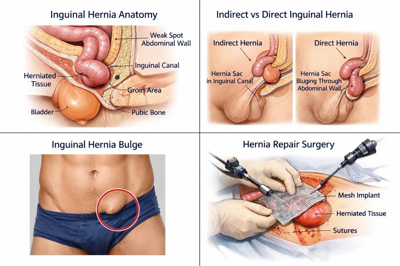
The four classical boundaries of the inguinal canal are the anterior wall, posterior wall, roof, and floor. The anterior wall is formed primarily by the external oblique aponeurosis. Laterally, it receives additional reinforcement from the internal oblique muscle. The posterior wall is principally formed by the transversalis fascia, with medial reinforcement from the conjoint tendon and related aponeurotic structures. The roof is formed by the arching fibers of the internal oblique and transversus abdominis muscles. The floor is formed principally by the inguinal ligament, with medial contribution from the lacunar ligament.
This classical description is useful for learning anatomy, but modern hernia surgery requires a broader three-dimensional interpretation. The posterior surface of the inguinal region contains the transversalis fascia, preperitoneal tissues, inferior epigastric vessels, iliopubic tract, Cooper's ligament, and the femoral canal. Consequently, the surgeon must understand both the anterior inguinal canal and the posterior preperitoneal space.
The spermatic cord is the principal male structure traversing the inguinal canal. It contains the ductus deferens, testicular artery, cremasteric vessels, artery of the ductus deferens, pampiniform venous plexus, genital branch of the genitofemoral nerve, lymphatic vessels, and associated connective tissue. The cord acquires coverings as it passes through the abdominal wall. The internal spermatic fascia is derived from the transversalis fascia, the cremasteric fascia and muscle are associated with the internal oblique layer, and the external spermatic fascia is derived from the external oblique aponeurosis.
In females, the inguinal canal transmits the round ligament of the uterus. The canal is generally less prominent than in males, but its surgical importance remains substantial because groin hernias in women have a relatively higher association with femoral hernias and therefore require careful evaluation of the entire myopectineal region.
3.2 Deep and superficial inguinal rings
The deep inguinal ring is an opening in the transversalis fascia and represents the entrance to the inguinal canal. It is located lateral to the inferior epigastric vessels. The spermatic cord in males and the round ligament in females pass through this opening.
The relationship between the deep ring and inferior epigastric vessels is one of the most important landmarks in inguinal hernia surgery. An indirect hernia enters the canal through the deep ring and therefore lies lateral to the inferior epigastric vessels. The hernia may remain within the canal or progress through the superficial ring and extend into the scrotum in males.
The superficial inguinal ring is a triangular opening in the external oblique aponeurosis. Its medial and lateral crura surround the opening, and intercrural fibers help maintain its configuration. The ring represents the exit of the inguinal canal and lies close to the pubic tubercle.
During open anterior repair, the superficial ring is encountered after incision of the external oblique aponeurosis. During laparoscopic posterior repair, the superficial ring is not directly approached because the operation is performed from the preperitoneal aspect.
3.3 Hesselbach's triangle
Hesselbach's triangle is a clinically important region of the posterior inguinal wall. It is bounded medially by the lateral border of the rectus abdominis muscle, laterally by the inferior epigastric vessels, and inferiorly by the inguinal ligament. A direct inguinal hernia protrudes through the posterior wall within this region and therefore lies medial to the inferior epigastric vessels.
Direct hernias usually occur as acquired defects or areas of weakness in the posterior abdominal wall. They tend to occur in older adults, although they can also occur in younger individuals. The hernia sac generally pushes forward through the transversalis fascia rather than passing through the deep inguinal ring.
The distinction between direct and indirect hernias is not merely descriptive. It provides an essential intraoperative landmark. When the inferior epigastric vessels are identified during laparoscopic surgery, a hernia lateral to them is interpreted as indirect, whereas a defect medial to them is interpreted as direct. This relationship becomes particularly important when the surgeon is operating from the posterior aspect.
3.4 The myopectineal orifice of Fruchaud
A broader anatomical concept that has become central to modern hernia surgery is the myopectineal orifice of Fruchaud. This region represents the potential area through which groin hernias can occur and includes the inguinal and femoral regions.
The myopectineal orifice is bounded superiorly by the arching fibers of the internal oblique and transversus abdominis muscles, medially by the rectus sheath and pubic region, laterally by the iliopsoas muscle, and inferiorly by the pectineal region and superior pubic structures. It is divided by the inguinal ligament into an upper inguinal compartment and a lower femoral compartment.
This concept explains an important principle of modern mesh repair. A mesh placed only over the visible hernia defect may not adequately protect the entire potential hernia-producing region. Posterior preperitoneal mesh repair is designed to cover the myopectineal orifice broadly, thereby reinforcing the areas through which indirect, direct, and femoral hernias may develop.
3.5 Iliopubic tract and Cooper's ligament
The iliopubic tract is a thickened band of the transversalis fascia that runs approximately parallel to the inguinal ligament. It is particularly important during posterior and laparoscopic approaches. The tract forms an important landmark separating the internal inguinal region from the femoral region and is related to the deep inguinal ring and femoral vessels.
Cooper's ligament, also called the pectineal ligament, is a dense fibrous structure located along the superior pubic ramus and pectineal line. It provides a strong fixation point during selected hernia repairs, especially when dealing with large medial defects or femoral hernias. However, the vascular structures surrounding the pubic and pectineal region require careful identification before fixation.
The surgeon operating in the preperitoneal space must also recognize the lacunar ligament, pectineal ligament, femoral canal, external iliac vessels, and obturator region. The anatomy is especially important because inappropriate fixation in the wrong area can cause vascular injury or chronic postoperative pain.
3.6 Inferior epigastric vessels
The inferior epigastric artery usually arises from the external iliac artery shortly above the inguinal ligament and ascends medially toward the rectus sheath. It is accompanied by veins and becomes a critical anatomical landmark in hernia surgery.
The inferior epigastric vessels divide the inguinal region into lateral and medial areas from the perspective of hernia classification. Indirect hernias occur lateral to these vessels, while direct hernias occur medial to them.
During TAPP and TEP repair, visualization of the inferior epigastric vessels allows the surgeon to orient the entire groin. The surgeon must also recognize the deep circumflex iliac vessels, external iliac vessels, gonadal vessels, and other vascular structures within the preperitoneal space. The relationship of these structures to the mesh and fixation points is essential for safe surgery.
3.7 Nerve anatomy and chronic postoperative inguinal pain
The nerves of the groin are of particular importance because nerve injury or entrapment may result in persistent postoperative pain. The principal nerves relevant to open inguinal hernia repair are the ilioinguinal nerve, iliohypogastric nerve, and genital branch of the genitofemoral nerve.
The ilioinguinal nerve originates from the first lumbar spinal nerve and travels through the abdominal wall before entering the inguinal region. It is commonly encountered during open anterior repair and typically lies near the spermatic cord. It provides sensory innervation to the upper medial thigh and external genital region.
The iliohypogastric nerve usually travels superior to the ilioinguinal nerve and is located between the internal oblique and transversus abdominis muscles before reaching the anterior abdominal wall. Its location makes it vulnerable during dissection and suturing in the upper portion of the operative field.
The genital branch of the genitofemoral nerve travels with the spermatic cord in males and contributes to the cremasteric reflex. It is particularly important during cord dissection.
Modern anatomical studies emphasize that these nerves do not follow completely predictable pathways. A 2025 scoping review of cadaveric studies demonstrated considerable anatomical variation, including variations in the position and relationships of the ilioinguinal, iliohypogastric, and genitofemoral nerves and frequent nerve fusion. This variability helps explain why a purely textbook-based assumption about nerve position can be unsafe during surgery.
Chronic postoperative inguinal pain is a significant outcome after hernia repair. Its causes may include direct nerve injury, entrapment by sutures or mesh fixation, inflammatory reaction, mesh-related fibrosis, or persistent neuropathic pain. The modern approach therefore emphasizes careful identification and handling of nerves, avoidance of unnecessary traumatic dissection, and appropriate selection of mesh fixation methods.
3.8 Indirect and direct inguinal hernias
Indirect inguinal hernias are associated anatomically with the processus vaginalis and enter through the deep inguinal ring. They pass lateral to the inferior epigastric vessels and may extend through the entire inguinal canal. Large indirect hernias may descend into the scrotum.
Direct inguinal hernias occur through the posterior wall of the inguinal canal, usually in the region of Hesselbach's triangle. They lie medial to the inferior epigastric vessels and typically result from weakness of the posterior abdominal wall.
The distinction is especially useful during laparoscopic repair because both hernia types can be visualized from the posterior surface. However, modern surgery does not simply close the specific defect. Instead, the objective of mesh repair is broad reinforcement of the entire myopectineal orifice.
3.9 Surgical principles
The central surgical principle in modern inguinal hernia repair is durable reinforcement of the groin while minimizing tissue trauma and chronic pain. The operation must address the hernia defect, protect surrounding anatomical structures, and provide adequate coverage of potential sites of recurrence.
The choice between open and posterior minimally invasive repair depends on the patient's characteristics, hernia type, previous surgical history, surgeon expertise, available equipment, anesthesia considerations, and local resources. The updated HerniaSurge guidelines emphasize individualized selection rather than the assumption that one technique is universally superior.
3.10 Open anterior repair
The Lichtenstein operation remains one of the most established open mesh techniques. The incision is made in the groin region, followed by division of the external oblique aponeurosis and exposure of the inguinal canal. The spermatic cord or round ligament is mobilized, and the hernia sac is identified and treated according to its type. A flat synthetic mesh is then positioned to reinforce the posterior wall.
The strength of the Lichtenstein technique lies in its relative simplicity, reproducibility, and applicability under local, regional, or general anesthesia. It does not require pneumoperitoneum or specialized laparoscopic equipment and therefore remains important in many surgical environments.
However, because the operation occurs in the anterior plane, the surgeon must work close to the ilioinguinal, iliohypogastric, and genitofemoral nerves. Careful dissection is therefore necessary to reduce the risk of chronic pain.
The original tension-free concept introduced by Lichtenstein and colleagues represented an important change in hernia surgery by avoiding excessive tension on weakened tissues. Contemporary evidence continues to support mesh-based open repair as an effective treatment for many adult patients.
3.11 Shouldice and other non-mesh repairs
The Shouldice repair is the best-established non-mesh tissue repair for adult inguinal hernia. It reconstructs the posterior wall using a multilayered suture technique. Its success depends heavily on meticulous surgical technique and appropriate patient selection.
Recent evidence demonstrates an important trade-off between Shouldice and Lichtenstein repair. A 2024 meta-analysis of 14 randomized controlled trials involving 2,784 patients found a lower recurrence rate with Lichtenstein repair compared with Shouldice repair, although recurrence after Shouldice remained relatively low in selected patients. More recent evidence suggests that Shouldice may produce less chronic postoperative pain in some populations but may have a higher long-term recurrence risk.
The Desarda technique is another non-mesh approach that uses a strip of external oblique aponeurosis to reinforce the posterior wall. A 2024 meta-analysis of randomized trials reported favorable results for several postoperative outcomes, although the evidence does not establish it as universally superior to mesh repair.
These techniques remain relevant when mesh is contraindicated, declined, unavailable, or when an experienced surgeon considers tissue repair appropriate. Nevertheless, patient selection and surgeon expertise are fundamental.
3.12 Laparoscopic posterior repair
Laparoscopic inguinal hernia repair is based on a fundamentally different anatomical concept. Instead of approaching the defect from the anterior surface, the surgeon enters the preperitoneal space and positions mesh behind the abdominal wall. The mesh then covers the entire myopectineal orifice.
Two principal approaches are used: TAPP and TEP.
In TAPP repair, the surgeon enters the peritoneal cavity using laparoscopic access. The peritoneum overlying the groin is opened, the preperitoneal space is dissected, the hernia sacs are reduced, and a large mesh is placed over the myopectineal orifice. The peritoneum is subsequently closed over the mesh.
TAPP offers broad visualization of the abdominal cavity and preperitoneal anatomy. This may be particularly useful when the diagnosis is uncertain, when an occult contralateral hernia is suspected, or when complex anatomy is anticipated. However, it requires entry into the peritoneal cavity and therefore introduces risks associated with intraperitoneal access.
TEP repair avoids entering the peritoneal cavity. The surgeon creates the preperitoneal working space directly and dissects the retropubic and lateral spaces. The hernia defect is reduced and mesh is placed over the myopectineal orifice.
The anatomical advantage of both techniques is broad coverage of the potential hernia-producing area rather than treatment of only the visible defect.
3.13 TAPP versus TEP
The choice between TAPP and TEP has been debated for many years. Contemporary evidence indicates that both techniques are effective when performed by appropriately trained surgeons.
A 2024 Cochrane review concluded that evidence comparing TAPP and TEP does not demonstrate a consistent major clinical advantage of one technique across all important outcomes. More recent evidence continues to support broadly comparable outcomes.
A 2026 systematic review and meta-analysis of seven studies involving 986 patients found no significant differences between TAPP and TEP in operative time, hospital stay, or early postoperative pain, although some differences in postoperative complications were observed. A separate 2026 analysis focusing on bilateral hernia repair similarly found broadly comparable outcomes and concluded that the choice should depend mainly on surgeon expertise, patient characteristics, and available resources.
The practical distinction therefore lies more in operative anatomy and surgeon familiarity than in a universal superiority of one technique. TAPP provides direct intraperitoneal access and broad visualization, whereas TEP preserves the peritoneal cavity and works entirely within the preperitoneal plane.
3.14 Important posterior anatomical landmarks
During laparoscopic posterior repair, several anatomical structures have particular significance. The pubic symphysis and pubic tubercle provide medial orientation. Cooper's ligament provides an important inferior medial landmark. The inferior epigastric vessels identify the distinction between direct and indirect hernia regions. The external iliac vessels lie laterally and inferiorly, while the femoral vessels must be protected.
The vas deferens and gonadal vessels form important structures in the male pelvis. Their relationship produces a characteristic arrangement sometimes described in terms of the "triangle of doom" and "triangle of pain." The triangle of doom lies between the vas deferens and gonadal vessels and contains major vascular structures. The triangle of pain lies more laterally and contains important nerves. Although these terms are useful surgical teaching concepts, the underlying principle is more important than memorization: fixation devices should not be placed indiscriminately in these danger zones.
3.15 Mesh placement and fixation
Adequate mesh size and positioning are central to successful posterior repair. The mesh should provide broad overlap beyond the margins of the hernia defect and cover the entire myopectineal orifice. Insufficient coverage or folding of the mesh can contribute to recurrence.
Mesh fixation has historically been used to prevent migration, but modern evidence questions whether fixation is necessary for every laparoscopic repair. A 2026 meta-analysis of 16 randomized controlled trials involving 1,608 patients undergoing TEP repair found that mesh fixation was associated with a higher risk of chronic postoperative inguinal pain without a statistically significant reduction in recurrence. Fixation was also associated with small increases in operative time and early postoperative pain.
The appropriate strategy therefore depends on the defect and technique. Small lateral indirect defects may not require extensive fixation, while large medial defects may require selective fixation because of the potential for mesh displacement. The guiding principle is to achieve stable mesh placement while minimizing unnecessary trauma to tissues and nerves.
3.16 Recurrent inguinal hernia
Recurrent hernia requires careful assessment of the previous operative approach. One of the major principles of contemporary hernia surgery is that recurrence should preferably be repaired through an anatomical plane that has not previously been extensively dissected.
For example, after a previous anterior repair, a posterior laparo-endoscopic repair can provide access through relatively undisturbed tissue planes. Conversely, after a failed posterior repair, an anterior approach may be considered. Re-entering heavily scarred tissue can increase the risk of nerve, vascular, and cord injury.
The choice must nevertheless be individualized according to the type of recurrence, previous mesh position, patient factors, surgeon expertise, and available resources. The updated HerniaSurge recommendations specifically address recurrent hernia management as an important area requiring tailored surgical planning.
3.17 Inguinal hernia in women
Although inguinal hernia is more common in men, groin hernias in women deserve special attention. The presence of a femoral hernia can be difficult to recognize clinically, and the risk of emergency presentation is important.
Recent evidence from a 2026 meta-analysis involving more than 100,000 women with inguinal hernia and more than 18,000 with femoral hernia found substantially greater morbidity associated with emergency repair than elective surgery. Minimally invasive approaches were associated with lower recurrence rates than anterior repair in the analyzed literature.
This reinforces the anatomical value of posterior preperitoneal visualization in women. A laparo-endoscopic approach allows the surgeon to inspect the femoral canal as well as the inguinal region and can therefore reduce the likelihood of overlooking a femoral defect.
3.18 Watchful waiting
Not every inguinal hernia requires immediate surgery. Watchful waiting can be appropriate for selected men with asymptomatic or minimally symptomatic hernias who understand the possibility of future progression.
Long-term evidence, however, demonstrates that many patients eventually undergo surgery. In a randomized study of men aged 50 years or older followed for a median of 12 years, approximately 64% of those initially assigned to watchful waiting eventually crossed over to surgery. Incarceration remained relatively uncommon, occurring in approximately 4% of the watchful-waiting group.
A 2024 individual participant data meta-analysis similarly demonstrated that watchful waiting remains a reasonable option in selected men but that symptoms and patient preference frequently lead to later surgery.
The decision should therefore be individualized rather than based solely on the anatomical presence of a hernia.
3.19 Robotic inguinal hernia repair
Robotic surgery represents a further development of the posterior preperitoneal concept. Most robotic inguinal hernia repairs use a TAPP-type approach. The robotic platform provides three-dimensional visualization, articulated instruments, improved wrist-like movements, and potentially greater precision during dissection and suturing.
These technical advantages are particularly attractive for complex dissections, large defects, recurrent hernias, and situations requiring extensive reconstruction. However, technological advantages do not automatically translate into superior patient outcomes.
A 2026 systematic review and meta-analysis restricted to randomized controlled trials included seven trials involving 691 patients and found no statistically significant difference between robotic and laparoscopic repair in postoperative complications, recurrence, or same-day discharge. The certainty of evidence was low to very low.
Another 2026 systematic review found that robotic repair generally requires longer operative time and higher costs, while patient outcomes remain broadly comparable with conventional laparoscopy. Current evidence therefore supports robotic repair as a valuable technical option rather than a universally superior replacement for conventional laparoscopy.
4. Discussion
The anatomy of the inguinal canal provides the foundation upon which all modern inguinal hernia operations are built. Although the classical description of the canal as a short oblique passage remains useful, contemporary surgical anatomy requires a broader three-dimensional understanding of the entire groin. The surgeon must appreciate the relationships between the inguinal canal, posterior abdominal wall, femoral canal, pubic region, iliac vessels, nerves, and preperitoneal spaces.
One of the most important conceptual developments has been the transition from thinking about a hernia as an isolated hole to considering the myopectineal orifice as a functional anatomical unit. This concept explains why modern mesh repairs generally use sufficiently large prostheses to cover the direct, indirect, and femoral regions rather than simply placing material over the visible defect. The concept also explains the anatomical advantage of posterior repairs, because the surgeon can directly visualize and reinforce the entire potential hernia-producing area.
The relationship between the inferior epigastric vessels and the hernia sac remains a fundamental diagnostic landmark. In an indirect hernia, the sac arises lateral to the vessels and enters through the deep inguinal ring. In a direct hernia, the defect occurs medial to the vessels. This distinction is particularly valuable during TAPP and TEP repair, where the surgeon views the anatomy from behind rather than from the anterior surface.
The choice of surgical approach has increasingly shifted from a procedure-centered decision to a patient-centered decision. Contemporary guidelines do not support the idea that every patient should receive one particular repair. Instead, the surgeon must consider whether the hernia is unilateral or bilateral, primary or recurrent, direct or indirect, uncomplicated or incarcerated, and whether the patient has undergone previous repair.
For a primary unilateral inguinal hernia, open Lichtenstein repair remains a highly established option. It can be performed under local or general anesthesia and requires relatively limited equipment. It is therefore particularly valuable when laparo-endoscopic surgery is unavailable, contraindicated, or inappropriate. The technique also has a long history of clinical use and extensive evidence supporting its effectiveness.
At the same time, minimally invasive surgery has important advantages. Laparoscopic repair avoids a large anterior dissection and allows broad posterior mesh placement. Evidence from randomized trials and meta-analyses indicates favorable postoperative recovery, particularly with respect to early pain and return to normal activity in appropriately selected patients. A randomized comparison of TEP and Lichtenstein repair found broadly similar medium-term outcomes but identified short-term advantages for TEP regarding recovery and foreign body sensation.
The value of laparo-endoscopic surgery becomes even more apparent in bilateral hernia. Two anterior repairs require separate incisions and separate dissections, whereas a posterior laparoscopic approach allows both sides to be assessed and treated through the same operative field. Recent systematic reviews continue to demonstrate broadly comparable outcomes between TEP and TAPP for bilateral disease, reinforcing the importance of surgeon experience and individualized selection rather than rigid preference for one technique.
Chronic postoperative inguinal pain represents one of the major challenges in modern hernia surgery. The problem cannot be attributed to a single mechanism. Injury or entrapment of the ilioinguinal, iliohypogastric, or genitofemoral nerves may contribute, but chronic pain may also result from inflammatory changes, mesh-related fibrosis, fixation devices, or altered sensory processing.
This makes detailed anatomical knowledge more important than ever. Anatomical studies have demonstrated that groin nerves exhibit considerable variation. Therefore, surgeons should avoid relying on a single expected anatomical pathway. The operative field should be explored carefully, and unnecessary nerve manipulation should be avoided.
Mesh fixation represents another example of how anatomical understanding and evidence-based surgery interact. Traditional fixation methods were intended to prevent migration and recurrence. However, excessive fixation can introduce unnecessary trauma, particularly when sutures or tacks are placed near nerves or vascular structures. Recent evidence indicates that routine fixation in TEP may increase chronic postoperative pain without clearly reducing recurrence in all patients. This supports selective rather than indiscriminate fixation.
The evidence concerning non-mesh repairs is similarly nuanced. Shouldice repair remains an important tissue-based operation, especially when performed by experienced surgeons in appropriately selected patients. However, randomized evidence demonstrates a recurrence disadvantage compared with Lichtenstein repair in broad patient populations. Therefore, the continued role of non-mesh surgery should not be interpreted as evidence that mesh is unnecessary. Rather, it demonstrates that multiple technically valid approaches remain available when appropriately matched to the patient.
The situation is particularly important in young patients, patients with small hernias, patients with concerns about foreign material, and patients in settings where mesh is unsuitable. In such situations, the surgeon must balance recurrence risk against potential mesh-related complications and chronic pain.
Women require separate consideration because the anatomical distribution of groin hernias differs from that seen in men. A clinically diagnosed "inguinal hernia" in a woman may coexist with or actually represent a femoral hernia. Since the femoral canal is situated inferior to the inguinal ligament, anterior approaches may not provide the same broad visualization of the femoral region as posterior preperitoneal approaches. Current evidence therefore favors minimally invasive repair in appropriately selected women, particularly because of the importance of identifying and treating femoral defects.
Robotic surgery is an interesting development because it emphasizes the difference between technological capability and clinical necessity. Robotic platforms provide excellent visualization and instrument dexterity, but current randomized evidence has not demonstrated consistent superiority over laparoscopic surgery in recurrence or overall complications. The increased financial cost and operative time also remain relevant. Therefore, robotics should currently be viewed as an extension of minimally invasive surgical capability rather than an obligatory next step in hernia treatment.
The most important lesson from contemporary evidence is that surgical success depends more on correct anatomical understanding and appropriate technique than on the technological sophistication of the operating platform. An experienced surgeon performing a well-selected open repair may achieve an excellent outcome, while an inexperienced surgeon using an advanced minimally invasive platform may encounter significant complications.
The same principle applies to recurrent hernia. Reoperation in a previously dissected anatomical plane increases technical difficulty because normal tissue planes are replaced by scar tissue. The choice of an alternative plane can therefore reduce dissection difficulty and avoid previously implanted mesh. This is a direct example of anatomy influencing operative strategy.
The evolution of inguinal hernia surgery can consequently be understood as a progression from closure of a defect toward restoration of the mechanical integrity of the entire groin. Early tissue repairs concentrated on suturing weakened structures. Lichtenstein repair introduced broad tension-free prosthetic reinforcement. Posterior laparo-endoscopic surgery subsequently extended the concept by reinforcing the entire myopectineal orifice. Robotic surgery has further refined the technical ability to perform this posterior dissection, although evidence has not yet established clear superiority over standard laparoscopy.
Current evidence also supports careful consideration of conservative management. Watchful waiting is reasonable for selected men with minimal symptoms, but long-term studies demonstrate that a substantial proportion eventually require surgery. Thus, watchful waiting should be understood as active clinical observation rather than permanent avoidance of surgery.
Overall, the contemporary approach to inguinal hernia should integrate anatomical classification, patient factors, surgeon expertise, available resources, and evidence regarding postoperative outcomes. The surgeon must be able to move conceptually between the anterior inguinal canal and the posterior preperitoneal anatomy. Without this understanding, surgical techniques become a sequence of memorized steps. With it, the operation becomes an anatomically reasoned reconstruction of the groin.
5. Conclusion
The inguinal canal is a complex anatomical passage whose surgical importance extends far beyond its relatively small dimensions. Its relationships with the inferior epigastric vessels, inguinal ligament, pubic region, femoral canal, abdominal wall muscles, transversalis fascia, spermatic cord, and groin nerves determine the development and classification of inguinal hernia and directly influence surgical management.
The distinction between indirect and direct hernias remains fundamentally based on the relationship of the hernia to the inferior epigastric vessels. However, modern surgical anatomy increasingly emphasizes the myopectineal orifice as a unified region containing the potential sites of inguinal and femoral herniation. This anatomical concept explains the effectiveness of broad preperitoneal mesh placement during TEP and TAPP repair.
Open Lichtenstein repair remains an important and reliable operation, particularly when minimally invasive surgery is inappropriate or unavailable. Shouldice and other non-mesh techniques remain useful in selected patients and experienced hands, although contemporary evidence generally favors mesh repair for many adult patients because of lower recurrence rates.
Laparoscopic TEP and TAPP repair provide important advantages through posterior visualization and broad coverage of the myopectineal orifice. Current evidence does not demonstrate a universal superiority of TAPP over TEP or vice versa. Instead, surgeon experience, patient characteristics, previous operations, and local resources should guide the choice. Robotic surgery offers improved visualization and instrument dexterity but has not yet demonstrated consistent clinical superiority over conventional laparoscopy and remains associated with greater cost.
The prevention of chronic postoperative inguinal pain depends heavily on anatomical awareness. Variations in the ilioinguinal, iliohypogastric, and genitofemoral nerves mean that surgeons must avoid assuming that every patient has identical anatomy. Similarly, careful identification of vascular structures and appropriate mesh placement are essential for preventing serious complications.
Ultimately, successful inguinal hernia surgery is an application of topographical anatomy. Understanding the spatial relationships of the inguinal canal, posterior abdominal wall, preperitoneal spaces, nerves, vessels, and femoral region allows the surgeon to select the appropriate operative plane, perform safe dissection, position the mesh effectively, and minimize recurrence and chronic pain. The modern surgical approach therefore does not replace anatomy with technology. Rather, contemporary technology is most effective when it is guided by a precise understanding of anatomy.
References
HerniaSurge Group. International guidelines for groin hernia management. Hernia. 2018;22(1):1-165. doi:10.1007/s10029-017-1668-x. PubMed
Stabilini C, van Veenendaal N, Aasvang E, et al. Update of the international HerniaSurge guidelines for groin hernia management. BJS Open. 2023;7(5):zrad080. doi:10.1093/bjsopen/zrad080. PubMed
Lichtenstein IL, Shulman AG, Amid PK, Montllor MM. The tension-free hernioplasty. American Journal of Surgery. 1989;157(2):188-193. doi:10.1016/0002-9610(89)90526-6. PubMed
Aiolfi A, Cavalli M, Micheletto G, et al. Treatment of inguinal hernia: systematic review and updated network meta-analysis of randomized controlled trials. Annals of Surgery. 2021. PubMed
Andresen K, Rosenberg J. Transabdominal preperitoneal versus totally extraperitoneal laparoscopic techniques for inguinal hernia repair. Cochrane Database of Systematic Reviews. 2024;7:CD004703. doi:10.1002/14651858.CD004703.pub3. PubMed
Pompeu BF, Pasqualotto E, et al. Shouldice versus Lichtenstein inguinal hernia repair: a meta-analysis of randomized controlled trials. World Journal of Surgery. 2024;48(11):2604-2614. doi:10.1002/wjs.12352. Wiley Online Library
Ahmadinejad I, Jalali A, Ahmadinejad M, et al. Inguinal hernia: Lichtenstein versus Shouldice technique repair: a randomized controlled trial. Surgical Open Science. 2024;17:70-74. doi:10.1016/j.sopen.2024.01.001. PubMed
Yeow M, Aiolfi A, Lomanto D, et al. Watchful waiting to surgery in men with mildly symptomatic or asymptomatic inguinal hernia: an individual participant data meta-analysis of long-term follow-up of randomized controlled trials. Hernia. 2024. doi:10.1007/s10029-024-03118-5. PubMed
Huerta S, Garza AM. A systematic review of open, laparoscopic, and robotic inguinal hernia repair: management of inguinal hernias in the 21st century. Journal of Clinical Medicine. 2025;14(3):990. doi:10.3390/jcm14030990. PubMed
Tigora A, Radu PA, Garofil DN, et al. Modern perspectives on inguinal hernia repair: a narrative review on surgical techniques, mesh selection and fixation strategies. Journal of Clinical Medicine. 2025;14(14):4875. PubMed
Delgado LM, Pompeu BFP, Magalhães CM, et al. Shouldice versus TAPP for inguinal hernia repair: a systematic review and meta-analysis of randomized controlled trials. World Journal of Surgery. 2025;49(4):859-867. doi:10.1002/wjs.12514. PubMed
Schena CA, Laterza V, Rosa F. Robotic versus laparoscopic inguinal hernia repair: a systematic review and meta-analysis of randomized controlled trials. Surgical Endoscopy. 2026;40(9):7411-7425. doi:10.1007/s00464-026-13253-y. PubMed
Naim N, Reza S. Transabdominal preperitoneal versus totally extraperitoneal repair for inguinal hernia: a systematic review and meta-analysis. Annals of the Royal College of Surgeons of England. 2026. doi:10.1308/rcsann.2026.0003. PubMed
Carneiro AO, Calmet Rocca WR, Gonçalves LB, et al. Laparoscopic totally extraperitoneal versus transabdominal preperitoneal techniques in bilateral inguinal hernia repair: a systematic review and meta-analysis. Hernia. 2026;30(1):59. doi:10.1007/s10029-025-03515-4. PubMed
Tzeng WJ, Liu YW, Liu YY, et al. Laparoscopic totally extraperitoneal versus transabdominal preperitoneal techniques in bilateral inguinal hernia repair: a systematic review and meta-analysis. Surgical Endoscopy. 2026. doi:10.1007/s00464-026-13206-5. PubMed
Huber AK, Kaiser A, Gerdes S, et al. Management of inguinal and femoral hernias in women: meta-analysis of current practices and review of international guidelines. Updates in Surgery. 2026. doi:10.1007/s13304-026-02659-x. PubMed
Schena CA, Laterza V, Rosa F. Robotic versus laparoscopic inguinal hernia repair: a systematic review and meta-analysis of randomized controlled trials. Surgical Endoscopy. 2026. doi:10.1007/s00464-026-13253-y. Springer
Pompeu BFP, Pasqualotto E, et al. Chronic pain after Shouldice versus Lichtenstein inguinal hernia repair: a systematic review and meta-analysis. Hernia. 2026. doi:10.1007/s10029-026-03702-x. PubMed Central (PMC)
Valinoti AC, Aramburu S, Principe J, et al. Long-term follow-up in asymptomatic or oligosymptomatic inguinal hernias: watchful or watchless waiting? Surgical Endoscopy. 2026. doi:10.1007/s00464-026-13475-0. PubMed
Andersen KG, van der Meer TWK, van Veenendaal N, et al. A European algorithm and strategy for management of chronic postoperative inguinal pain. Journal of Abdominal Wall Surgery. 2026;5:16944. doi:10.3389/jaws.2026.16944. PubMed
A systematic review of anatomical variations of the ilioinguinal, iliohypogastric, and genitofemoral nerves based on cadaver studies. PubMed-indexed review. 2025. PubMed
Anatomy, Abdomen and Pelvis: Inguinal Region and Inguinal Canal. StatPearls, National Center for Biotechnology Information. NCBI
Aiolfi A, et al. Randomized and network evidence comparing Lichtenstein, TAPP, and TEP inguinal hernia repair. Annals of Surgery. 2021. PubMed
Recent systematic evidence comparing open, laparoscopic, and robotic approaches to adult inguinal hernia repair. Journal of Clinical Medicine. 2025. PubMed