
Rhombencephalon and The Fourth Ventricle: Structure, Development, and Clinical Correlations
1. Zarina Zhamaldinovna Toichieva
2. Priyadharshini Ramesh
3. Jothi Basu Kasinathan
4. Thirushanth Ramesh
(1. Lecturer, International Medical Faculty, Osh State University, Osh, Kyrgyz Republic
2. Student, International Medical Faculty, Osh State University, Osh, Kyrgyz Republic
3. Student, International Medical Faculty, Osh State University, Osh, Kyrgyz Republic
4. Student, International Medical Faculty, Osh State University, Osh, Kyrgyz Republic)
Abstract
The Rhombencephalon (hindbrain) is a critical area of the brain that regulates various bodily functions, including respiration, balance, and coordination. This structure includes the metencephalon and myelencephalon, which give rise to the pons, cerebellum, and medulla oblongata. One of the significant structures in the hindbrain is the Fourth Ventricles, a part of the ventricular system filled with cerebrospinal fluid. The fourth ventricles are found between the cerebellum and the brainstem and are critical in cerebrospinal fluid circulation between the brain and the spinal cord. Understanding the anatomy and function of the rhombencephalon and fourth ventricle is essential for clinical and neurological studies
Keywords: Rhombencephalon, hindbrain, metencephalon, myelencephalon, cerebellum, pons, medulla oblongata, fourth ventricle, ventricular system, cerebrospinal fluid, brainstem, neural development, central nervous system, hydrocephalus
Introduction
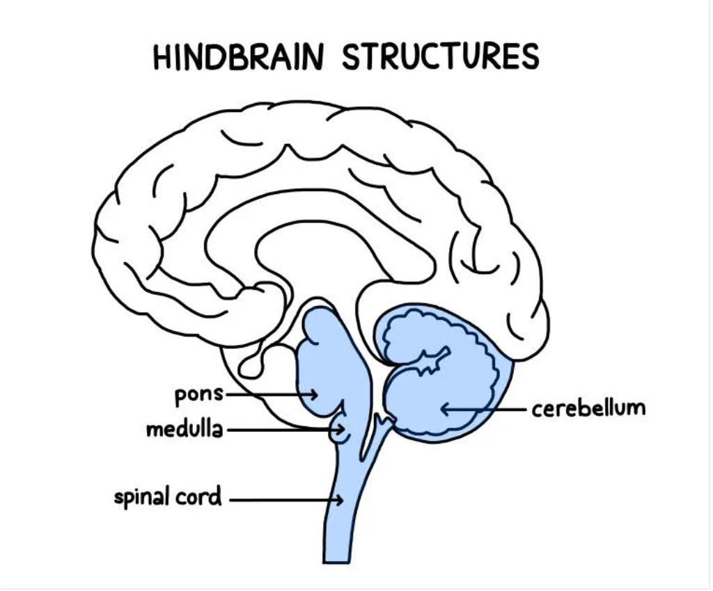
The brain of the human is an incredibly complex organ, functioning to regulate and control numerous body processes. Among those is the rhombencephalon, also called the hindbrain. It performs many significant roles in the maintenance of crucial body functions such as balancing and coordination, respiration, and control of the cardiovascular system. The division of the rhombencephalon leads to the appearance of three main structures – the cerebellum, the pons, and the medulla oblongata.
An important structure close to the rhombencephalon is the fourth ventricle. It belongs to the brain’s ventricular system and contains cerebrospinal fluid. The location of the fourth ventricle is between the cerebellum and brainstem. As a component of the circulatory system of cerebrospinal fluid, this structure performs a vital function for brain health. Understanding the structure and function of the rhombencephalon and fourth ventricle helps in learning about physiological brain processes and related diseases.
General feature and functions of Rhombencephalon
Rhombencephalon constitutes the posterior portion of the brain and is directly connected to the spinal cord at the bottom. Rhombencephalon is an important component that develops from the caudal portion of the neural tube of the embryo. This organ plays an essential role in brain structure formation.
Rhombencephalon ensures vital processes of the human organism including regulation of breathing, cardiac activity and blood pressure that are performed by the medulla oblongata. Additionally, this brain region controls maintenance of posture and coordination of body movement through the cerebellum whereas the pons acts as a mediator between various portions of the brain.
Moreover, rhombencephalon performs reflex functions of the body such as swallowing, coughing and sneezing.
Structure of Rhombencephalon
Rhombencephalon is divided into two main parts that include:
● Metencephalon: forms cerebellum and pons
● Myelencephalon: develops into medulla oblongata.
Metencephalon and myelencephalon coordinate actions to ensure control of voluntary and involuntary processes of the body.
1. Metencephalon
Metencephalon is the upper division of rhombencephalon. It differentiates into two main parts:
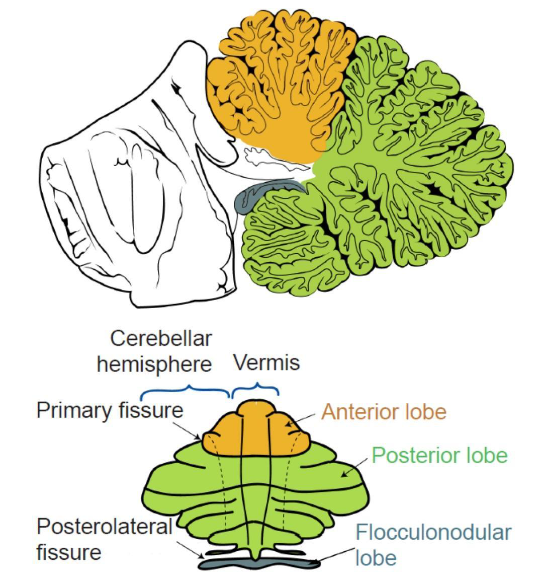
Cerebellum
Cerebellum is a highly folded structure situated behind the pons and medulla oblongata. It is concerned with the regulation of voluntary motor movements, balance, posture, and motor learning. The afferent inputs received from the cerebral cortex, spinal cord, and vestibular apparatus are integrated by it to improve the control over motor output. Cerebellum is made up of two hemispheres linked by vermis and divided into lobes and zones of function (i.e., vestibulocerebellum, spinocerebellum, and cerebrocerebellum).
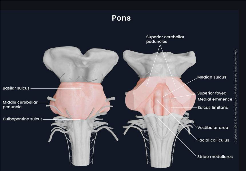
Pons
It is found above the medulla oblongata and below the cerebellum. It acts as a relay center between cerebrum and cerebellum through its nuclei and pontocerebellar transverse fibers. It also possesses nuclei of cranial nerves V-VIII and regulates breathing through pneumotaxic and apneustic centers.
2. Myelencephalon
Myelencephalon is the most caudally positioned division of the Rhombencephalon that develops into the Medulla oblongata. The Medulla oblongata is a crucial part of the brainstem that is found within the posterior cranial fossa and is contiguous inferiorly with the spinal cord through the foramen magnum.
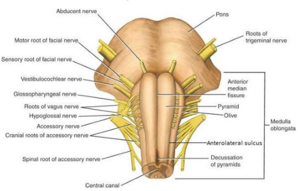
Medulla Oblongata
Medulla oblongata is a long cylindrical part located between the lower margin of the pons and the spinal cord. It extends over a distance of 3 cm and is the inferior region of the brainstem. This structure is made up of gray matter (nuclei) and white matter (tracts), which are both well organized.
External Features
At the anterior surface of the medulla oblongata, there is a pair of columns referred to as pyramids. The pyramids are composed of corticospinal tracts responsible for voluntary motor control. Most of these motor tracts are crossed in the pyramidal decussation towards the opposite side, thus explaining contralateral motor control. Lateral to the pyramids lie the olives, which contain the inferior olivary nuclei that assist in coordinating movement and learning processes.
Internal Structure
The structure within the medulla is made up of various nuclei and tracts:
Cranial nerve nuclei: Medulla has the nuclei of cranial nerves IX, X, XI, and XII, which control swallowing, voice production, and movement of tongue muscles.
Ascending tracts: Sensory tracts, including the medial lemniscus, that carry sensations like fine touch and proprioception to the brain.
Descending tracts: Tracts from the cortex like corticospinal tracts that play an important role in voluntary activities.
Function
Medulla oblongata contains life-supporting autonomic centers:
Cardiac center: Controls heart rate and cardiac muscle strength
Vasomotor center: Regulates the diameter of blood vessels and hence blood pressure
Respiratory centers: Control breathing pattern and volume
Moreover, it has reflex centers that regulate protective and visceral reflexes, including swallowing, coughing, sneezing, and vomiting.
Fourth Ventricles
Fourth ventricles are critical parts of the ventricular system of the brain. They are found in the posterior part of the cranial cavity and contain cerebrospinal fluid (CSF).
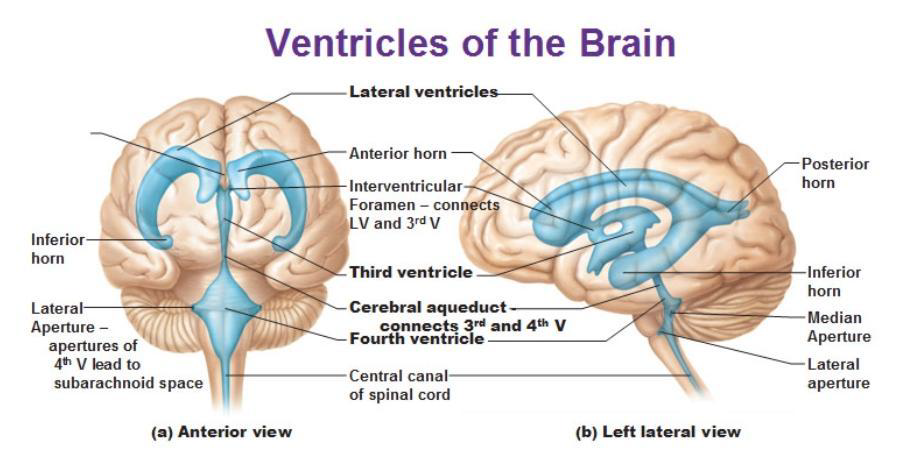
Location
The fourth ventricles lie posteriorly to the cerebellum and anteriorly to the brainstem, particularly the pons and Medulla oblongata. The structure lies dorsally to the pons and upper medulla but is found ventrally to the cerebellum. The structure communicates with the cerebral aqueduct dorsally and terminates at the central canal of the spinal cord ventrally.
Shape and Structure
Traditionally, the fourth ventricles have been described as diamond or tent-shaped cavities. Cross-sectionally, the structure is triangular, while in its posterior section, it assumes the shape of a rhomboid (rhomboid fossa). Such unique shapes result from the divergent nature of the cerebellum from the brainstem.
Boundaries of the Fourth Ventricle
Boundaries of the fourth ventricle are very distinct and play an important role in its anatomy and clinical relevance.
1. Roof
The roof of the fourth ventricle resembles a tent and consists of membranous and neural elements.
● Superior (upper) part:
Consists of superior medullary velum, which is a thin lamina of white matter spanning between the superior cerebellar peduncles.
● Inferior (lower) part:
It is made up of inferior medullary velum and tela choroidea containing choroid plexus, which is responsible for producing CSF.
The roof of the ventricle also has some openings, such as Median aperture (foramen of Magendie) and lateral apertures (foramina of Luschka), through which CSF is able to pass into the subarachnoid space
2. Floor (Rhomboid Fossa)
Floor of the fourth ventricle, or rhomboid fossa, is a diamond-shaped depression, formed by posterior surfaces of:
● Pons (superior portion)
● Medulla oblongata (inferior portion)
Important structures
1. Median sulcus – divides floor into two parts
2. Sulcus limitans – separates motor area from sensory
3. Facial colliculus – raised area due to facial nerve fibers
4. Hypoglossal and vagal trigones
3. Lateral boundaries
Consist of structures, connecting cerebellum to brainstem:
● Superior cerebellar peduncles (upper part)
● Inferior cerebellar peduncles (lower part)
Clinical Application of the Rhombencephalon and the Fourth Ventricle
The Rhombencephalon and the Fourth Ventricle are two interrelated structures whose damage may lead to neurological problems, since this region is responsible for numerous autonomic functions, the activities of many cranial nerves and CSF drainage.
1. Hydrocephalus
● Hydrocephalus develops due to obstruction of CSF passage through or around the fourth ventricle.
● Blockage at the medial and lateral apertures causes inability to pass into the subarachnoid space.
● Obstruction of the Cerebral aqueduct leads to expansion of the third and lateral ventricles.
● Increased intracranial pressure develops, causing Ventricle dilatation.
Clinical signs: headache, nausea, vomiting, papilledema, altered state of consciousness, and macrocrania in infants.
2. Brainstem Lesion
● As mentioned above, the rhombencephalon develops into the brainstem (pons and medulla). Therefore, any lesion at this level affects critical brain functions.
● Involves the Pons and Medulla oblongata
● Nuclei of cranial nerves V-XII
● Ascending and descending tracts
Clinical findings: Dysphagia, Dysarthria, Facial paresis
3. Tumors in the Fourth Ventricle
1. Ependymomas, medulloblastomas and other tumors may develop at this site.
2. These tumors cause compression of the cerebellum and brainstem
3. CSF passage obstruction, secondary hydrocephalus
Symptoms:
● Headache and nausea
● Cerebellar symptoms: ataxia and imbalance
REFERENCES
1. Vishram Singh. Textbook of Anatomy: Head, Neck and Brain, Elsevier.
2. Richard S. Snell. Clinical Neuroanatomy, Wolters Kluwer.
3. Wikipedia – Articles on Rhombencephalon, Fourth Ventricle, and Brainstem