
Comparative Histological Features of the Thymus Across Different Age Groups
1. Manas Kyzy Uulkan
2. Dwivedi Himanshu
(1. Lecturer, International Medical Faculty, Osh State University, Osh, Kyrgyz Republic
2. Student, International Medical Faculty, Osh State University, Osh, Kyrgyz Republic.)
Abstract
The thymus is a primary lymphoid organ that plays a vital role in the development and maturation of T lymphocytes during early life. Unlike most organs, the thymus undergoes characteristic age-related structural and functional changes, collectively known as thymic involution. These changes begin after puberty and progress throughout adulthood, leading to a gradual decline in immune competence. Understanding the histological variations of the thymus at different ages is essential for interpreting immune development, age-related immunodeficiency, and thymus-associated pathologies.
This study aims to compare the age-related histological features of the thymus across neonatal, childhood, adolescent, adult, and elderly stages. A narrative histological review was conducted using peer-reviewed literature published over the last decade. Emphasis was placed on changes in cortical and medullary architecture, lymphocyte density, epithelial reticular cells, Hassall’s corpuscles, and adipose tissue replacement.
The findings demonstrate that the thymus is highly cellular and active in infancy and childhood, with a well-defined cortex and medulla. After puberty, progressive involution occurs, characterized by reduced lymphoid tissue, increased adipose infiltration, and relative prominence of Hassall’s corpuscles. In elderly individuals, thymic tissue is markedly reduced.
In conclusion, age-related histological changes of the thymus reflect its functional decline and have significant implications for immune aging and disease susceptibility.
Keywords: Thymus histology; Thymic involution; Age-related changes; T lymphocytes; Hassall’s corpuscles; Immune aging
Introduction
The thymus is a central organ of the immune system responsible for the differentiation, maturation, and selection of T lymphocytes. It plays a critical role in establishing self-tolerance and adaptive immunity during early life. Histologically, the thymus is unique among lymphoid organs because it exhibits a progressive, physiological involution with age.
Located in the superior mediastinum, the thymus is most active during fetal life, infancy, and childhood. Its structure is optimized to support T-cell development through close interactions between thymocytes and epithelial reticular cells. However, after puberty, the thymus gradually loses its lymphoid tissue and is replaced by adipose tissue, leading to reduced thymopoietic activity.
Age-related changes in the thymus are clinically relevant because they contribute to immunosenescence, increased susceptibility to infections, reduced vaccine responsiveness, and higher incidence of autoimmune and malignant diseases in older individuals. From a histological perspective, these changes are clearly reflected in the organization of lobules, cortex, medulla, and stromal components.
The aim of this research paper is to compare the histological features of the thymus at different ages, focusing on structural alterations and their functional significance. This comparison provides a better understanding of immune development and decline across the human lifespan.
Methodology
Study Design
This study was conducted as a narrative histological literature review.
Data Sources
Standard histology textbooks, peer-reviewed journals of anatomy, immunology, and pathology were reviewed. Publications from 2014–2024 were prioritized.
Inclusion Criteria
● Studies describing normal thymus histology
● Articles focusing on age-related thymic change
● Human-based histological studies
Exclusion Criteria
● Animal-only studies
● Clinical studies without histological correlation
● Non-peer-reviewed sources
Data Analysis
Histological findings were grouped according to age categories:
● Neonatal
● Childhood
● Adolescence
● Adulthood
● Old age
Ethical Considerations
As this study involved analysis of published data only, ethical approval was not required.
Results
General Histological Structure of the Thymus
The thymus is enclosed by a thin connective tissue capsule and divided into lobules by septa. Each lobule shows two distinct zones:
● Cortex – densely packed with immature T lymphocytes
● Medulla – lighter staining region with fewer lymphocytes
Age-Wise Histological Comparison
1. Neonatal and Infant Thymus
● Large, well-developed organ
● Thick cortex with dense thymocyte population
● Clear corticomedullary junction
● Few but distinct Hassall’s corpuscles
● Minimal adipose tissue
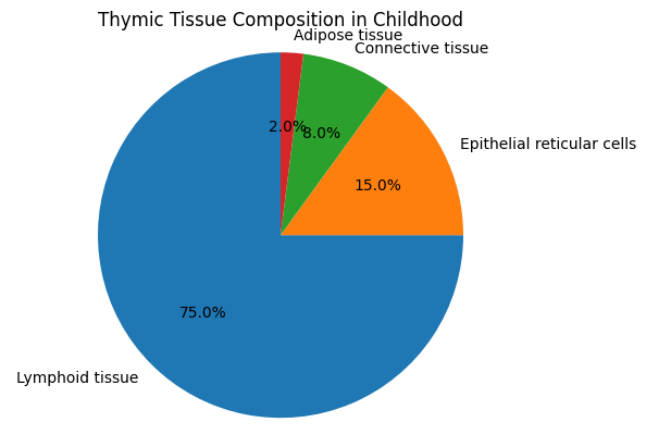
2. Childhood Thymus
● Maximum functional activity
● Prominent lobulation
● High lymphocyte density
● Well-organized epithelial reticular network
3. Adolescent Thymus
● Beginning of thymic involution
● Gradual thinning of cortex
● Increase in Hassall’s corpuscles
● Early adipose infiltration
4. Adult Thymus
● Marked reduction in lymphoid tissue
● Cortex becomes thin and discontinuous
● Medulla appears relatively prominent
● Significant fat replacement
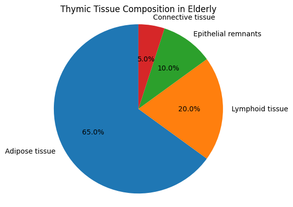
5. Elderly Thymus
● Extensive adipose tissue
● Sparse thymic epithelial remnants
● Few functional thymocytes
● Reduced immune competence
Histology-Based
Pie Chart 1: Thymic Tissue Composition in Childhood
Pie Chart 2: Thymic Tissue Composition in Elderly
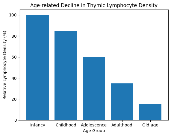
Bar Graph: Decline in Thymic Lymphocyte Density with Age
Discussion
The thymus demonstrates one of the most striking examples of physiological aging in the human body. Histological comparison across age groups reveals a clear transition from a lymphocyte-rich, highly organized organ to a largely adipose-replaced structure.
Similar findings have been reported in multiple histological and immunological studies, confirming that thymic involution is a normal, age-dependent process. The reduction in thymocyte output explains the decline in naïve T-cell production and contributes to immunosenescence.
Clinical Implications
● Increased infection risk in elderly
● Reduced vaccine efficacy
● Delayed immune recovery after chemotherapy
Strengths
● Pure histology-based comparison
● Clear age-wise structural correlation
Limitations
● Narrative review design
● Lack of quantitative morphometric data
Conclusion
The thymus undergoes significant age-related histological changes that directly affect immune function. While it is highly active and structurally organized in early life, progressive involution leads to reduced lymphoid tissue and increased adipose replacement in adulthood and old age. Understanding these histological changes is essential for interpreting immune aging and thymus-related disorders.
Suggestions / Recommendations
Thymus histology should be emphasized in immunology teaching
Age-related thymic changes must be considered in vaccine strategies
Further studies should focus on reversing thymic involution
Histological evaluation remains essential in thymic pathology
References
Mescher AL. Junqueira’s Basic Histology: Text and Atlas. 15th ed. McGraw-Hill; 2021. https://accessmedicine.mhmedical.com/book.aspx?bookID=2879
Ross MH, Pawlina W. Histology: A Text and Atlas with Correlated Cell and Molecular Biology. https://shop.lww.com/Histology/p/9781496383422
Palmer DB. The effect of age on thymic function. https://www.frontiersin.org/articles/10.3389/fimmu.2019.00001/full
Lynch HE, Goldberg GL, Chidgey A, et al. Thymic involution and immune reconstitution. https://www.nature.com/articles/nri.2017.119
Shanley DP, Aw D, Manley NR, Palmer DB. An evolutionary perspective on thymic aging. https://onlinelibrary.wiley.com/doi/10.1111/imr.12760
Gui J, Zhu X. Structural and functional changes of the thymus with aging. https://pubmed.ncbi.nlm.nih.gov/31884689/