
Skin Epidermis and Dermis
1. Manas Kyzy Uulkan
2. Chindhalore Sejal
(1. Lecturer, International Medical Faculty, Osh State University, Osh, Kyrgyz Republic
2. Student, International Medical Faculty, Osh State University, Osh, Kyrgyz Republic.)
Abstract
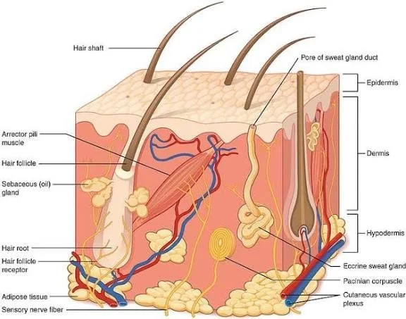
The skin is the largest organ of the human body and serves as a critical protective interface between the internal environment and external surroundings. It performs multiple vital functions, including mechanical protection, thermoregulation, sensation, immune defense, and metabolic activity. Structurally, the skin is composed of two principal layers: the epidermis and the dermis, each with distinct histological characteristics and specialized cellular components. Understanding the microscopic architecture of these layers is fundamental to clinical medicine, dermatopathology, and biomedical research.
This study aims to provide a comprehensive histological analysis of the epidermis and dermis, emphasizing their cellular composition, structural organization, and functional correlations. A narrative literature review was conducted using peer-reviewed histology and dermatology sources published within the last decade. Data were synthesized to describe epithelial stratification, dermal connective tissue organization, vascular supply, and neural components.
The findings highlight the complex stratification of the epidermis, dominated by keratinocytes, and the supportive, vascularized connective tissue framework of the dermis rich in collagen and elastic fibers. Structural variations across different skin regions were also noted. These histological features explain the skin’s remarkable resilience and regenerative capacity.
In conclusion, detailed knowledge of epidermal and dermal histology is essential for diagnosing skin disorders, interpreting biopsies, and understanding disease pathogenesis at the microscopic level.
Keywords: Skin histology; Epidermis; Dermis; Keratinocytes; Connective tissue; Skin layers; Dermatopathology
Introduction
The skin is a multifunctional organ that plays a central role in maintaining homeostasis and protecting the body from physical, chemical, and biological insults. From a histological perspective, the skin represents a highly specialized structure in which epithelial and connective tissues interact closely to perform protective and regulatory functions. Disorders affecting the skin often manifest as microscopic alterations long before clinical symptoms become apparent, highlighting the importance of histological understanding.
The skin consists of two main layers: the epidermis, an avascular stratified squamous epithelium, and the dermis, a vascular connective tissue layer that provides structural support and nourishment. Each layer contains specialized cells and extracellular components adapted to specific functional demands. Variations in thickness, cellular composition, and connective tissue density account for differences between thick and thin skin.
Histological study of the skin is clinically relevant in diagnosing inflammatory diseases, infections, autoimmune disorders, and neoplasms. Many dermatological conditions, including psoriasis, eczema, and skin cancers, are defined by characteristic microscopic changes in the epidermis and dermis.
The aim of this research paper is to present a detailed histological description of the epidermis and dermis, correlating microscopic structure with physiological function and clinical relevance. This work is intended to serve as a foundational reference for medical students and healthcare professionals studying skin histology.
Methodology
Study Design
This study was conducted as a narrative literature review focusing exclusively on the histology of the epidermis and dermis.
Data Sources
Peer-reviewed textbooks of histology, dermatology journals, and online medical databases were consulted. Emphasis was placed on sources published between 2014 and 2024 to ensure updated scientific accuracy.
Inclusion Criteria
● Studies describing microscopic structure of epidermis and dermis
● Articles focused on normal skin histology
● English-language peer-reviewed publications
Exclusion Criteria
● Studies focused solely on clinical dermatology without histological analysis
● Animal-only studies
● Non-peer-reviewed sources
Data Analysis
Information was systematically extracted and synthesized under thematic headings, including epidermal layers, dermal organization, cell types, and functional correlations.
Ethical Considerations
As this study involved secondary data analysis of published literature, no ethical approval was required.
Results
Histology of the Epidermis
The epidermis is composed of stratified squamous keratinized epithelium and lacks blood vessels. Its nutrition is derived by diffusion from the underlying dermis. The epidermis consists primarily of keratinocytes arranged in distinct layers.
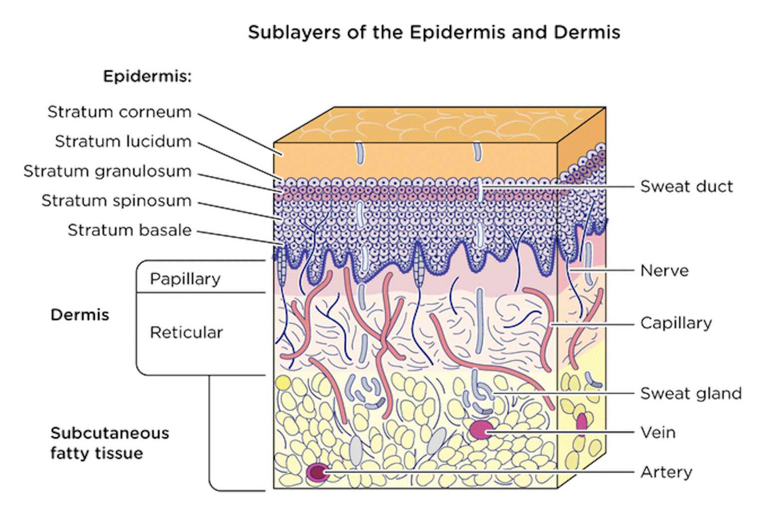
Epidermal Layers (Deep to Superficial)
Stratum basale – Single layer of cuboidal cells with high mitotic activity
Stratum spinosum – Polyhedral cells connected by desmosomes
Stratum granulosum – Cells with keratohyalin granules
Stratum lucidum – Present only in thick skin
Stratum corneum – Flattened, anucleated keratinized cells
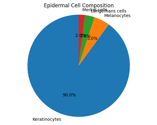
Keratinocytes account for approximately 90% of epidermal cells, while melanocytes, Langerhans cells, and Merkel cells provide pigmentation, immune surveillance, and sensory function respectively.
Histology of the Dermis
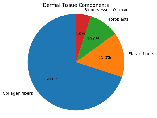
The dermis lies beneath the epidermis and consists of dense connective tissue rich in collagen and elastic fibres. It provides mechanical strength, elasticity, and metabolic support to the epidermis.
Dermal Layers
● Papillary dermis: Loose connective tissue with capillaries
● Reticular dermis: Dense irregular connective tissue with thick collagen bundles
Fibroblasts are the predominant cell type, responsible for extracellular matrix synthesis. Mast cells, macrophages, blood vessels, and nerve endings are also present.
Discussion
The histological organization of the skin reflects a highly efficient protective system. The stratified nature of the epidermis allows continuous renewal while maintaining a robust barrier. Keratinization is essential for preventing dehydration and mechanical damage.
Comparative studies indicate that thick skin contains a well-developed stratum lucidum, whereas thin skin lacks this layer. The dermis, through its collagen-elastic fiber network, provides tensile strength and flexibility, enabling the skin to withstand mechanical stress.
Alterations in epidermal turnover or dermal connective tissue composition are central to many skin disorders. For example, excessive keratinocyte proliferation characterizes psoriasis, while collagen degradation contributes to skin aging.
Strengths
● Comprehensive histological focus
● Correlation of structure with function
Limitations
● Narrative review design
● Lack of primary microscopic measurements
Conclusion
The epidermis and dermis together form a structurally complex and functionally integrated organ. Histological examination reveals specialized layers, cells, and connective tissue components essential for protection, sensation, and metabolic regulation. Understanding normal skin histology is crucial for recognizing pathological changes and improving diagnostic accuracy in dermatology and pathology.
Suggestions / Recommendations
Histology teaching should emphasize structure-function correlation
Routine skin biopsies must be interpreted with strong histological knowledge
Future research should focus on molecular-histological integration
Advanced imaging techniques should be incorporated into dermatopathology training
References
Ross MH, Pawlina W. Histology: A Text and Atlas with Correlated Cell and Molecular Biology.
https://shop.lww.com/Histology/p/9781496383422Mescher AL. Junqueira’s Basic Histology: Text and Atlas. 15th ed.
https://accessmedicine.mhmedical.com/book.aspx?bookID=2879Kanitakis J. Anatomy, histology and immunohistochemistry of normal human skin.
https://pubmed.ncbi.nlm.nih.gov/27331638/Chu DH, et al. Development and structure of skin.
https://www.jaad.org/article/S0190-9622(18)32542-9/fulltextProksch E, Brandner JM, Jensen JM. The skin: an indispensable barrier.
https://onlinelibrary.wiley.com/doi/10.1111/exd.13550Elder DE, et al. Lever’s Histopathology of the Skin. 11th ed.
https://shop.lww.com/Lever-s-Histopathology-of-the-Skin/p/9781496387925Tobin DJ. Introduction to skin aging.
https://www.sciencedirect.com/science/article/pii/S0965206X16300260