
Beyond Serum Creatinine: Urinary Extracellular Vesicle as Early Biomarkers of Kidney Injury
1. Kebekova Aizat Ashimzhanovna
2. Thangadurai Renugadevi
3. Malik Alshifa Parveen
4. Rabik Hussain Sabitha Maryam
(1. Lecturer, International Medical Faculty, Osh State University, Osh, Kyrgyz Republic
2. Student, International Medical Faculty, Osh State University, Osh, Kyrgyz Republic
3. Student, International Medical Faculty, Osh State University, Osh, Kyrgyz Republic
4. Student, International Medical Faculty, Osh State University, Osh, Kyrgyz Republic)
Abstract
Serum creatinine has served as the cornerstone of kidney function assessment for more than a century, yet its shortcomings as a biomarker of acute kidney injury (AKI) are now impossible to ignore. Creatinine is a functional marker rather than a structural one; it rises late, is confounded by muscle mass, hydration, diet, and drugs, and remains silent during the crucial early window in which tubular injury is still reversible. Over the past two decades, urinary extracellular vesicles (uEVs) — nanoscale, membrane-bound particles released by every epithelial cell along the nephron — have emerged as a compelling alternative window into renal pathology. Because uEVs are shed directly from the cells that constitute the injured tissue, their protein, lipid, and nucleic acid cargo reflects real-time, segment-specific events within the kidney itself. This article reviews the limitations of serum creatinine, the biology and isolation of uEVs, the current landscape of uEV biomarker candidates in AKI and chronic kidney disease (CKD), the methodological and translational barriers to clinical adoption, and the future directions that may finally move uEV diagnostics from the research bench to the bedside. The central argument advanced here is that uEVs do not merely replace creatinine as "another marker," but offer a fundamentally different category of information: a non-invasive molecular biopsy of the kidney.
Keywords: acute kidney injury; urinary extracellular vesicles; exosomes; biomarkers; nephrology; serum creatinine
1. Introduction
Few numbers in clinical medicine carry as much weight — and as little nuance — as the serum creatinine. Every day, physicians around the world make decisions about fluid resuscitation, drug dosing, renal replacement therapy, and prognosis based on this single, inexpensive measurement. The Kidney Disease: Improving Global Outcomes (KDIGO) consensus definition of AKI, which superseded the earlier RIFLE and AKIN classifications, rests almost entirely on changes in serum creatinine and urine output (Bufkin and Varughese, 2024). Yet every nephrologist has met the patient in whom creatinine told a comfortable lie: the septic, volume-overloaded patient whose creatinine stayed deceptively low while acute tubular necrosis progressed, or the cachectic elderly woman whose "normal" creatinine of 0.8 mg/dL masked the loss of half her nephron mass.
The fundamental problem is conceptual. Creatinine is a surrogate for glomerular filtration, and glomerular filtration is a late event in the injury cascade. In most forms of AKI, the renal tubular epithelial cells are the primary locus of damage, and a measurable reduction in glomerular filtration rate (GFR) is a downstream, insensitive development (Klein and Forni, 2024). The serum half-life of creatinine lengthens from approximately four hours to as much as 24–72 hours as GFR falls, meaning the concentration may not rise until 24–36 hours after the initiating insult — precisely the window in which early intervention might change the trajectory of the disease. Compounding this delay, the relationship between GFR and creatinine is non-linear: large losses of GFR can produce changes in creatinine that remain within the reference range and are dismissed as noise. Extrarenal factors further erode its fidelity — nutritional status, age, muscle mass, fluid resuscitation, corticosteroid therapy, and drugs such as trimethoprim and cimetidine that compete for tubular secretion all distort the signal (Bufkin and Varughese, 2024). A post hoc analysis of the Fluid and Catheter Treatment Trial illustrated the scale of the problem: AKI was revealed or reclassified in up to 18% of patients once creatinine values were adjusted for net fluid balance, and those misclassified patients had mortality rates comparable to those with overt AKI (Klein and Forni, 2024).
Against this backdrop, the search for earlier, more specific biomarkers of kidney injury has been one of the most active fronts in nephrology research. The Acute Disease Quality Initiative (ADQI) consensus conference on AKI biomarkers has recommended that functional and damage markers be interpreted together within a structured kidney health assessment, acknowledging that no single analyte can capture the complexity of the syndrome (Ostermann et al., 2020). Novel markers such as NGAL, KIM-1, TIMP-2/IGFBP7, and urinary dickkopf-3 have each made inroads — the last of these predicted postoperative AKI after cardiac surgery with an AUROC of 0.78 — yet none has fully displaced creatinine in routine practice (Ostermann et al., 2020). It is within this gap, between the ideal and the available, that urinary extracellular vesicles have found their rationale.
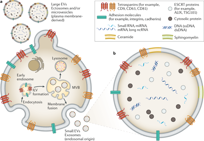
Extracellular vesicles (EVs) are membrane-bound particles released by all cell types. In urine, they originate almost exclusively from cells lining the genitourinary tract, with the overwhelming majority derived from the nephron itself — podocytes, proximal tubule, loop of Henle, distal tubule, and collecting duct. Every day, on the order of a trillion such vesicles pass into the urine, each one carrying a protected cargo of proteins, lipids, mRNA, and microRNA sampled from its cell of origin at the moment of release. Because the vesicle membrane shields its contents from the hostile, protease-rich urinary environment, uEV cargo arrives at the collection tube in remarkably good condition. The implication is profound: a urine sample, centrifuged appropriately, becomes a liquid biopsy of the entire nephron, offering segment-resolved molecular information that no blood test can supply. This article examines how far that promise has been realized, what evidence supports the leading uEV biomarker candidates, and what must happen before uEV analysis enters routine clinical care.
2. Methods
This article was written as a narrative review informed by a structured survey of the peer-reviewed literature. PubMed, Scopus, and Google Scholar were searched for publications through late 2025 using combinations of the terms "urinary extracellular vesicles," "exosomes," "acute kidney injury," "biomarker," "fetuin-A," "nephrin," "aquaporin," "microRNA," and "chronic kidney disease." Preference was given to original human and translational studies, large-scale proteomic analyses, consensus statements such as the MISEV (Minimal Information for Studies of Extracellular Vesicles) guidelines, and systematic reviews. Animal studies were included where they provided mechanistic insight directly relevant to biomarker kinetics. Landmark earlier studies were retained where they established foundational concepts. Sources were selected on the basis of methodological transparency, validation in independent cohorts, and citation impact. All synthesis and interpretation presented here are original to this article; quantitative findings are attributed to their primary sources and referenced accordingly.
3. Results
3.1 The biology of urinary extracellular vesicles: why they matter
Extracellular vesicles are classified broadly by size and biogenesis into exosomes (roughly 40–160 nm, formed within multivesicular bodies and released by exocytosis), microvesicles (100–1000 nm, budding directly from the plasma membrane), and larger apoptotic bodies. In urine, small EVs of the exosomal type predominate, and they are released constitutively from the apical membranes of every nephron segment. Miranda and colleagues demonstrated in 2010 that urinary exosomes contain nucleic acids — including intact mRNA and microRNA — that are protected from urinary RNases and stable enough for analysis, establishing the feasibility of transcriptomic biomarker discovery from urine (Miranda et al., 2010). This finding opened the door to what might be called the "vesicle hypothesis" of urinary diagnostics: that each uEV is a time-stamped snapshot of the intracellular state of a specific nephron segment at the moment of its release.
Large-scale proteomic work has since validated this hypothesis at scale. Wu and colleagues performed a comprehensive mass-spectrometric assessment of uEV proteins and demonstrated that the uEV proteome reliably mirrors protein changes occurring within the kidney tissue itself, including segment-specific channel and transporter abundance (Wu et al., 2021). This correspondence is the critical property that distinguishes uEV markers from conventional plasma or whole-urine markers: a rise in an uEV-borne proximal tubule protein such as NHE3 or aquaporin-1 localizes the injury anatomically, something neither creatinine nor plasma NGAL can do.
3.2 Protein cargo: the first generation of uEV biomarkers
The protein biomarker story began with a study that remains one of the most cited in the field. Zhou and colleagues applied two-dimensional difference gel electrophoresis and mass spectrometry to urinary exosomes isolated from cisplatin-treated rats, then validated candidates across ischemia-reperfusion, volume depletion, and human ICU cohorts (Zhou et al., 2008). Of the proteins identified, exosomal fetuin-A emerged as the standout: it increased 31.6-fold within two to eight hours of ischemia-reperfusion injury, rose 52.5-fold by day two of cisplatin nephrotoxicity — a full day before serum creatinine changed — and, critically, did not increase in prerenal azotemia (Zhou et al., 2008). This last point deserves emphasis, because the distinction between functional prerenal states and structural tubular injury is precisely where creatinine fails most conspicuously. The same group subsequently identified activating transcription factor 3 (ATF3) in urinary exosomes, which was detectable up to two days before creatinine elevation or histological damage in both ischemic and nephrotoxic models, with similar early kinetics observed in AKI patients (Chen et al., 2014).
Other protein candidates have accumulated steadily. The sodium-hydrogen exchanger NHE3, a proximal tubule apical transporter, is elevated in urinary small EVs across cisplatin-, ischemia-, diuretic-, ARB-, and sepsis-associated AKI, and shows greater sensitivity than creatinine changes in comparative analyses (reviewed in Lv et al., 2025). Aquaporin dynamics add temporal resolution: in cisplatin injury, uEV aquaporin-1 rises modestly within 24 hours while aquaporin-2 falls precipitously, and the combined AQP1/AQP2 ratio signals damage before any hematological marker moves. Podocyte injury, historically invisible to urinary biochemistry, becomes detectable through uEV-borne WT-1 and nephrin, permitting early recognition of glomerular damage in conditions such as diabetic kidney disease and lupus nephritis (Alves et al., 2025).
The most technically striking recent advance is single-vesicle proteomics. In 2025, Li and colleagues reported a single-uEV proteomic platform that identified complement receptor CD35 as a biomarker for sepsis-associated AKI, demonstrating that analysis at the resolution of individual vesicles can extract diagnostically useful signals that bulk measurements dilute (Li et al., 2025). This work matters beyond its specific finding: it suggests that the heterogeneity of uEV populations, long treated as noise, may itself be a readable signal.
3.3 Nucleic acid cargo: microRNAs as kinetic fingerprints
If proteins report what the cell was doing, microRNAs report what it is preparing to do. uEV microRNA profiles exhibit stage-specific choreography that no static protein marker can match. In ischemia-reperfusion AKI, miR-16, miR-24, and miR-200c rise during the acute injury phase, whereas miR-9a, miR-141, miR-200a, and miR-429 characterize early recovery; these miRNAs converge on the Zeb1/2 axis implicated in TGF-β-driven fibrosis, hinting at a molecular bridge between acute injury and chronic scarring (reviewed in Lv et al., 2025). Urinary exosomal miR-29c correlates with the histological extent of renal fibrosis and has been proposed as an early marker of the AKI-to-CKD transition — arguably the single most consequential prognostic question in the field, given that survivors of AKI carry substantially elevated long-term risks of CKD, cardiovascular morbidity, and death (Klein and Forni, 2024). In scrub typhus-associated AKI, Yun and colleagues showed that uEV miR-21 levels are markedly elevated and inversely correlate with both white cell count and estimated GFR, providing an etiology-specific diagnostic signal (reviewed in Yang and Camussi, 2025).
The metabolomic dimension is younger but growing. Proteomic surveys have catalogued 251 proteins in uEVs from patients with vancomycin-induced AKI, and metabolomic analyses in cisplatin- and aminoglycoside-injured rats have identified 65 candidate metabolites reflecting renal cellular damage — an untapped layer of the cargo that may eventually complement protein and RNA panels (Lv et al., 2025).
3.4 uEVs beyond AKI: chronic kidney disease and glomerular pathology
The uEV framework extends naturally to chronic disease, where the diagnostic problem is not speed but sensitivity to subclinical progression. In chronic kidney disease and its complications, EV surface proteins including KIM-1, WNT4, LCN2 (NGAL), and TNFR1 have been catalogued as early-detection candidates (Garcia et al., 2024). Ugarte and colleagues demonstrated that LCN2 within uEVs detects tubular injury in type 1 diabetes before conventional markers of diabetic nephropathy appear. More recently, podocyte-derived EVs carrying CKAP4 have been proposed as non-invasive diagnostic markers for diabetic nephropathy, and uEV analysis has revealed early inflammatory and damage signals in Fontan-associated single-ventricle physiology, a population at high risk for insidious kidney injury (Grange et al., 2025; Zhang et al., 2025). In lupus nephritis, podocyte EVs combined with immune mediators distinguished active renal involvement from quiescent disease (Alves et al., 2025). These studies collectively suggest that uEV analysis may eventually serve not merely to detect injury earlier, but to phenotype it — distinguishing glomerular from tubular pathology, inflammatory from ischemic insults, and reversible stress from irreversible fibrosis.
4. Discussion
4.1 The conceptual case: a liquid biopsy versus a functional proxy
The deepest argument for uEV biomarkers is not that they rise earlier than creatinine, though they do. It is that they answer a different question. Creatinine asks, "how much filtration capacity remains?" uEVs ask, "what is happening inside the nephron, right now, and where?" This distinction has practical consequences. Early AKI is a syndrome, not a single disease; sepsis-associated AKI, ischemic ATN, and drug nephrotoxicity involve different mechanisms, different prognoses, and potentially different therapies. A diagnostic that can only detect the common functional endpoint of these processes — a late rise in a filtration surrogate — is structurally incapable of supporting precision nephrology. A diagnostic built on segment-specific molecular cargo is not. The ADQI consensus has already moved toward a framework in which damage biomarkers complement functional ones and inform etiology, prognosis, and treatment response (Ostermann et al., 2020); uEV panels are arguably the most information-dense implementation of that framework currently imaginable, because they carry not one marker but the coordinated expression signature of the injured cell.
There is also a human dimension that deserves acknowledgment. AKI is frequently asymptomatic, and its late recognition has been directly linked to excess mortality: delayed diagnosis means missed windows for intervention during the period when injury remains reversible (Bufkin and Varughese, 2024). Every hour of diagnostic lead time that a fetuin-A, ATF3, or NHE3 measurement buys is an hour in which a nephrotoxin can be stopped, volume status corrected, or hemodynamics optimized. The promise of uEV diagnostics is ultimately measured not in fold-changes but in those recovered hours.
4.2 Barriers to translation
Against this optimism must be set an honest accounting of the obstacles. The first is methodological standardization. uEV isolation remains dominated by differential ultracentrifugation — slow, equipment-intensive, and operator-dependent — with precipitation kits, size-exclusion chromatography, ultrafiltration, and immunoaffinity capture each trading purity against yield in different ways. Urine itself is a hostile preanalytical variable: hydration state, pH, proteinuria, and Tamm–Horsfall protein polymer entrapment all affect vesicle recovery, and normalization strategies (to urine creatinine, to excretion rate, to particle count) are not harmonized across studies. The MISEV guidelines have established reporting standards, and efforts toward laboratory validation frameworks continue, but no uEV assay yet exists as a certified in vitro diagnostic with defined analytical performance specifications (reviewed in Diagnostics, 2026).
The second barrier is cohort scale. Many of the foundational studies were small: the original fetuin-A human validation included three ICU patients with AKI (Zhou et al., 2008). Two decades later, large, prospective, multicenter cohorts remain scarce. Biomarkers that perform brilliantly in homogeneous animal models or small single-center series frequently attenuate in the messy heterogeneity of real critical care populations, as the history of urinary NGAL in adults painfully demonstrated. The third barrier is the reference-standard paradox: uEV biomarkers are usually validated against creatinine-defined AKI, which embeds the very limitations they are designed to overcome. True structural validation requires correlation with histology, imaging, or adjudicated clinical outcomes — expensive, slow, and ethically constrained.
The fourth consideration is one the research community discusses too little: cost and workflow. A creatinine costs pennies and returns in minutes. An uEV assay requiring ultracentrifugation, RNA extraction, or single-particle proteomics cannot compete on those terms and should not try to. The realistic clinical niche is targeted deployment in high-risk, high-stakes settings — post-cardiac surgery, nephrotoxin exposure, transplant monitoring, sepsis — where the marginal cost of early information is trivial against the cost of a missed AKI episode. Biomarker combinations may also prove pragmatically important: the pairing of the furosemide stress test with urinary CCL14, for example, predicted progression to RRT indications with an AUC of 0.87, illustrating how uEV-adjacent markers may ultimately function within multi-analyte panels rather than as standalone replacements (Klein and Forni, 2024).
4.3 Limitations of this review
This article is a narrative synthesis rather than a systematic review, and therefore shares the susceptibility of the genre to selection bias. The literature on uEV biomarkers is characterized by publication bias toward positive findings, small sample sizes, heterogeneous isolation methods, and inconsistent normalization. Where animal data dominate, as in the kinetic characterization of several candidates, extrapolation to human disease carries inherent uncertainty. Quantitative comparisons across studies were avoided deliberately because methodological heterogeneity makes pooled effect estimates misleading. These caveats notwithstanding, the directional consistency of findings across independent groups, species, and injury models strengthens confidence in the underlying biology.
5. Conclusion
Serum creatinine will not disappear from clinical practice, nor should it; as a cheap, ubiquitous functional marker it remains useful for population screening and longitudinal monitoring. But the evidence assembled here supports a clear conclusion: creatinine alone is an inadequate sentinel for kidney injury, and the field has known this for a generation. Urinary extracellular vesicles offer the most biologically coherent solution yet proposed to this problem. Because they are shed directly from the injured tissue, their cargo — fetuin-A, ATF3, NHE3, the aquaporins, stage-specific microRNAs, podocyte proteins, and the complement receptors emerging from single-vesicle proteomics — reports structural injury earlier, localizes it anatomically, distinguishes prerenal from intrinsic states, and may forecast the transition from acute injury to chronic disease. What stands between this promise and the clinic is not the biology, which is increasingly solid, but the engineering: standardized isolation, harmonized normalization, prospective validation in large and diverse cohorts, and assays designed for the laboratory bench rather than the ultracentrifuge. If those problems are solved — and the trajectory of the last five years suggests they will be — the routine urine sample may finally deliver what nephrology has always wanted: a painless molecular biopsy of the kidney, available before the damage is done.
References
Bufkin KB, Varughese JG. Review of the limitations of current biomarkers in acute kidney injury clinical practices. Cureus. 2024;16(1):e52777.
Klein SJ, Forni LG. Biomarkers in acute kidney injury. Ann Intensive Care. 2024;14:144.
Ostermann M, Zarbock A, Goldstein S, et al. Recommendations on acute kidney injury biomarkers from the Acute Disease Quality Initiative consensus conference. JAMA Netw Open. 2020;3(10):e2019209.
Zhou H, Pisitkun T, Aponte A, et al. Exosomal fetuin-A identified by proteomics: a novel urinary biomarker for detecting acute kidney injury. Kidney Int. 2008;74(6):809–821.
Chen HH, Lai PF, Lan YF, et al. Exosomal ATF3 RNA attenuates pro-inflammatory gene MCP-1 transcription in renal ischemia-reperfusion. J Cell Physiol. 2014;229(9):1202–1211.
Miranda KC, Bond DT, McKee M, et al. Nucleic acids within urinary exosomes/microvesicles are potential biomarkers for renal disease. Kidney Int. 2010;78(2):191–199.
Wu Q, Poulsen SB, Murali SK, et al. Large-scale proteomic assessment of urinary extracellular vesicles highlights their reliability in reflecting protein changes in the kidney. J Am Soc Nephrol. 2021;32(9):2195–2209.
Li N, Tang TT, Gu M, et al. Single urinary extracellular vesicle proteomics identifies complement receptor CD35 as a biomarker for sepsis-associated acute kidney injury. Nat Commun. 2025;16:6960.
Lv LL, Cao Y, Liu D, et al. Emerging frontiers in acute kidney injury: the role of extracellular vesicles. J Transl Med (PMC11880721). 2025.
Yang Y, Camussi G. Extracellular vesicles in acute kidney injury. Biomed Pharmacother (PMC12094478). 2025.
Garcia NA, et al. Comprehensive strategy for identifying extracellular vesicle surface proteins as biomarkers for chronic kidney disease. Front Physiol. 2024;15:1328362.
Grange C, Deorsola L, Degiovanni B, et al. Urinary extracellular vesicle analysis reveals early signs of kidney inflammation and damage in single ventricle paediatric patients after Fontan operation. Int J Nanomedicine. 2025;20:5907–5922.
Zhang H, Lou K, Qian L, et al. CKAP4 in extracellular vesicles derived from podocyte serves as a non-invasive diagnostic biomarker for diabetic nephropathy and promotes vascular calcification. J Extracell Vesicles. 2025;14:e70208.
Alves L, Lemos AP, Martins J, et al. Podocyte extracellular vesicles and immune mediators as urinary biomarkers in active lupus nephritis. Sci Rep. 2025;15:28630.
Urinary extracellular vesicles biomarkers in chronic kidney disease: from discovery to clinical implementation. Diagnostics (Basel). 2026;16(14):2181.
Urinary exosomes: a promising biomarker of drug-induced nephrotoxicity. Front Med (Lausanne). 2023;10:1251839.
Tőkés-Füzesi M, Woth G, Ernyey B, et al. Microparticles and acute renal dysfunction in septic patients. J Crit Care. 2013;28(2):141–147.