
Early Recognition of Respiratory Distress in Children Clinical Assessment, Warning Signs and Evidence-Based Approaches
1. Altynai Nuralieva
2. Bhavani Mohan Dharaneesh
(1. Lecturer, International Medical Faculty, Osh State University, Osh, Kyrgyz Republic
2. Student, International Medical Faculty, Osh State University, Osh, Kyrgyz Republic.)
Abstract
Background: Respiratory illness remains one of the commonest reasons children are brought to hospital and one of the leading causes of death in children under five. Many of these deaths are not caused by a lack of treatment but by a late realisation that the child was seriously unwell.
Aim: To bring together current evidence on how respiratory distress is recognised in children aged one month to 18 years, from bedside examination to monitoring tools and early warning systems, and to consider how this works in settings with and without abundant resources.
Methods: This narrative review draws on peer-reviewed studies, systematic reviews, World Health Organization (WHO) guidance and international paediatric guidelines, identified through searches of PubMed/MEDLINE, Embase, the Cochrane Library and WHO databases.
Main findings: Careful observation of respiratory rate, work of breathing and mental state remains the most reliable foundation of assessment. Pulse oximetry detects hypoxaemia that clinical signs often miss, but is unavailable in many settings. Scoring systems such as the Paediatric Early Warning Score (PEWS) and the Paediatric Respiratory Assessment Measure (PRAM) support decision-making, though evidence that they improve outcomes is mixed. Delayed care-seeking, staff shortages and limited oxygen supply are the main barriers in low-resource settings.Early recognition depends less on technology than on trained eyes, repeated observation and a low threshold to escalate. Standardised, context-appropriate assessment protocols, supported by affordable monitoring tools, offer the most realistic route to fewer preventable deaths.
Keywords: respiratory distress; children; paediatric early warning score; pulse oximetry; hypoxaemia; work of breathing; respiratory failure
1. Introduction
1.1 What we mean by respiratory distress
Respiratory distress is what we see when a child has to work harder than normal to breathe. It is a clinical picture rather than a diagnosis: faster breathing, flaring nostrils, the chest pulling in with each breath, a child who is restless, tired or simply too quiet. Respiratory failure is the point at which that effort is no longer enough to keep oxygen up and carbon dioxide down. The skill of paediatric practice lies in noticing the first, so that the second never arrives.
1.2 A very common problem
Pneumonia, bronchiolitis, asthma and related conditions account for a large share of paediatric admissions worldwide. Acute lower respiratory infection is among the leading infectious causes of death in children under five, and the great majority of those deaths occur in low- and middle-income countries [1,2]. Even where mortality is low, respiratory illness drives emergency attendances, intensive care admissions and a great deal of parental anxiety.
1.3 Why timing matters
Children compensate remarkably well, until they cannot. A healthy infant can maintain normal oxygen levels through rapid breathing and extra muscle effort for hours, and then deteriorate within minutes once reserves run out. Missing the early phase means treating a child who is already exhausted, hypoxic and acidotic, when simple measures such as oxygen, positioning or a bronchodilator would have been enough hours earlier.
1.4 Consequences of delay
Delayed recognition is linked with avoidable intensive care admission, cardiorespiratory arrest, longer hospital stay and death. In resource-limited settings, children frequently arrive after several days of illness and after visits to more than one provider, so that the window for simple intervention has already narrowed [3]. Delay also has quieter costs: family distress, lost days of school and work, and long-term lung effects in some children.
1.5 Aim and scope
This review examines how respiratory distress is recognised in children from one month to 18 years. It covers relevant anatomy and physiology, common causes, bedside assessment, warning signs, monitoring tools, severity scoring, early management and the particular challenges of low-resource settings. Neonatal respiratory distress is outside our scope.
2. How Children Differ: Anatomy and Physiology
2.1 Upper and lower airways
The upper airway runs from the nose and mouth through the pharynx to the larynx. In children it is narrow, and in young infants who breathe mainly through the nose even mild congestion can matter. The lower airway, from the trachea through the bronchi to the alveoli, continues to grow and branch well into childhood, with most alveoli formed after birth.
2.2 Why a child is not a small adult
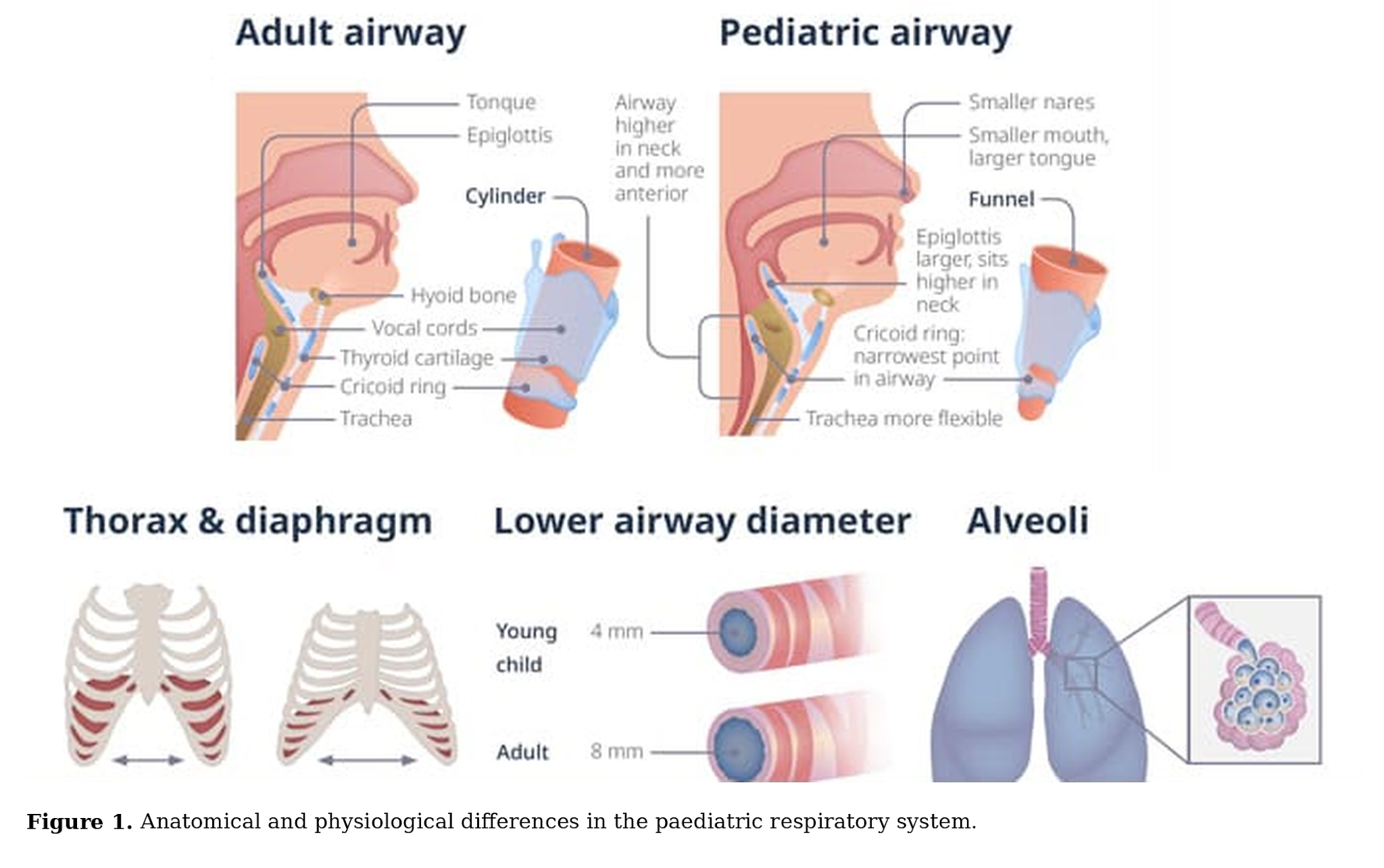
Several differences explain why children deteriorate so quickly:
• Small airway calibre. Airway resistance rises with the fourth power of the reduction in radius, so a millimetre of swelling that an adult would barely notice can halve an infant's airflow.
• Large tongue and floppy upper airway, which obstruct easily, particularly in a drowsy child.
• Compliant chest wall. The ribs are largely cartilaginous and sit more horizontally, so the chest wall sinks in under negative pressure. This is why retractions are such a visible sign in children.
• Reliance on the diaphragm. Infants depend on diaphragmatic movement, which is easily impaired by a distended abdomen, and the diaphragm fatigues sooner.
Less respiratory reserve. Fewer, smaller alveoli and lower functional residual capacity leave little buffer when ventilation falls.
2.3 Oxygen demand
Children consume more oxygen per kilogram than adults, roughly two to three times as much in infancy. A higher demand combined with a smaller reserve means that oxygen saturation can fall quickly when ventilation is compromised, and fever, crying or feeding make this worse.
2.4 Respiratory rate across childhood
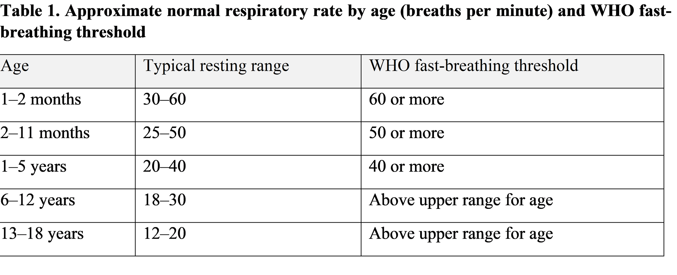
Normal breathing rate falls steadily with age, and any assessment has to be read against the child's age rather than a single adult-style cut-off. Large observational data show wide normal ranges and a lot of overlap between healthy and unwell children, which is a reminder that a number alone is never the full story [4]. WHO uses simple age-based thresholds for fast breathing in pneumonia assessment [3].
3. Why Children Get Into Difficulty: Aetiology
3.1 Infectious causes
Pneumonia is the largest single cause of respiratory deaths in young children. Fast breathing and chest indrawing are the key clinical markers in WHO guidance, and hypoxaemia is the strongest predictor of death [3,5].
Bronchiolitis affects infants, usually in winter, most often from respiratory syncytial virus. Illness peaks around days three to five, with feeding difficulty, wheeze and crackles. Management is largely supportive, and guidelines advise against routine bronchodilators or steroids [6,7].
Croup presents with a barking cough, hoarse voice and inspiratory stridor, typically in children aged six months to three years. A single dose of corticosteroid reduces symptoms and return visits [8].
Pertussis can be subtle in young infants, who may have apnoea or cyanosis with little cough. It is a reminder to think of it in unimmunised or incompletely immunised babies.
3.2 Non-infectious causes
Asthma is the commonest chronic cause. Severe exacerbations may present as a "silent chest", which is a warning of near-total airflow obstruction rather than reassurance [9].
Foreign body aspiration should be considered in any toddler with sudden onset coughing, choking or wheeze, especially if unilateral or not responding to treatment.
Congenital abnormalities such as tracheomalacia, vascular rings or congenital heart disease may first declare themselves with an apparently routine respiratory infection.
Pulmonary oedema, whether cardiogenic or from other causes, causes breathlessness, crackles and sometimes frothy secretions.
3.3 Systemic causes
Not every breathless child has a lung problem. Metabolic acidosis (for example in diabetic ketoacidosis) produces deep, laboured breathing. Severe anaemia, sepsis, raised intracranial pressure, poisoning and neuromuscular disease can all alter breathing. Looking for these protects against the mistake of treating the lungs while the real problem lies elsewhere.
4. Clinical Assessment at the Bedside
4.1 Start by listening to the story
A short, focused history often points to the cause and the urgency. Useful questions include how long the child has been unwell, whether the onset was sudden, how feeding and urine output have changed, whether there has been choking or exposure to smoke, and what the child's past history, immunisation status and medication use look like. Parents who say "he is just not himself" deserve to be taken seriously; the parental sense that something is wrong is itself a sign.
4.2 General look before you touch
Some of the most valuable information comes before the stethoscope. From the doorway, ask: Is the child alert and interactive? How is the colour? Are they sitting up in a position of comfort, or tripoding? Are they able to speak, feed or cry normally? An anxious child who is refusing to lie down or an infant who has stopped feeding is giving you information.
4.3 Inspection, palpation and percussion
With the chest exposed, look for chest wall movement, symmetry, nasal flaring, intercostal, subcostal and suprasternal retractions, and head bobbing in infants. Palpation checks tracheal position, chest expansion and, in older children, vocal fremitus. Percussion is dull over consolidation or effusion and hyper-resonant in pneumothorax or hyperinflation, although in a distressed small child it is often limited by cooperation.
4.4 Auscultation
Listen for air entry, wheeze, crackles, bronchial breathing and stridor, and compare sides. Reduced air entry in a distressed child is more worrying than loud wheeze, because it can signal that too little air is moving to produce a sound. Auscultation findings vary between observers, so they should be interpreted alongside the rest of the picture.
4.5 Counting the rate and judging the work
Count respiratory rate for a full 60 seconds, ideally when the child is calm, because brief counts overestimate and crying distorts the result. Then judge the effort: depth, rhythm, use of accessory muscles, and the ratio of effort to effect. A child breathing at 30 with deep recession and a tired face is sicker than one breathing at 45 and eating a biscuit.
5. Early Warning Signs
No single sign is perfect. What matters is the pattern, the trend over time and the child's overall appearance.
5.1 The early signs
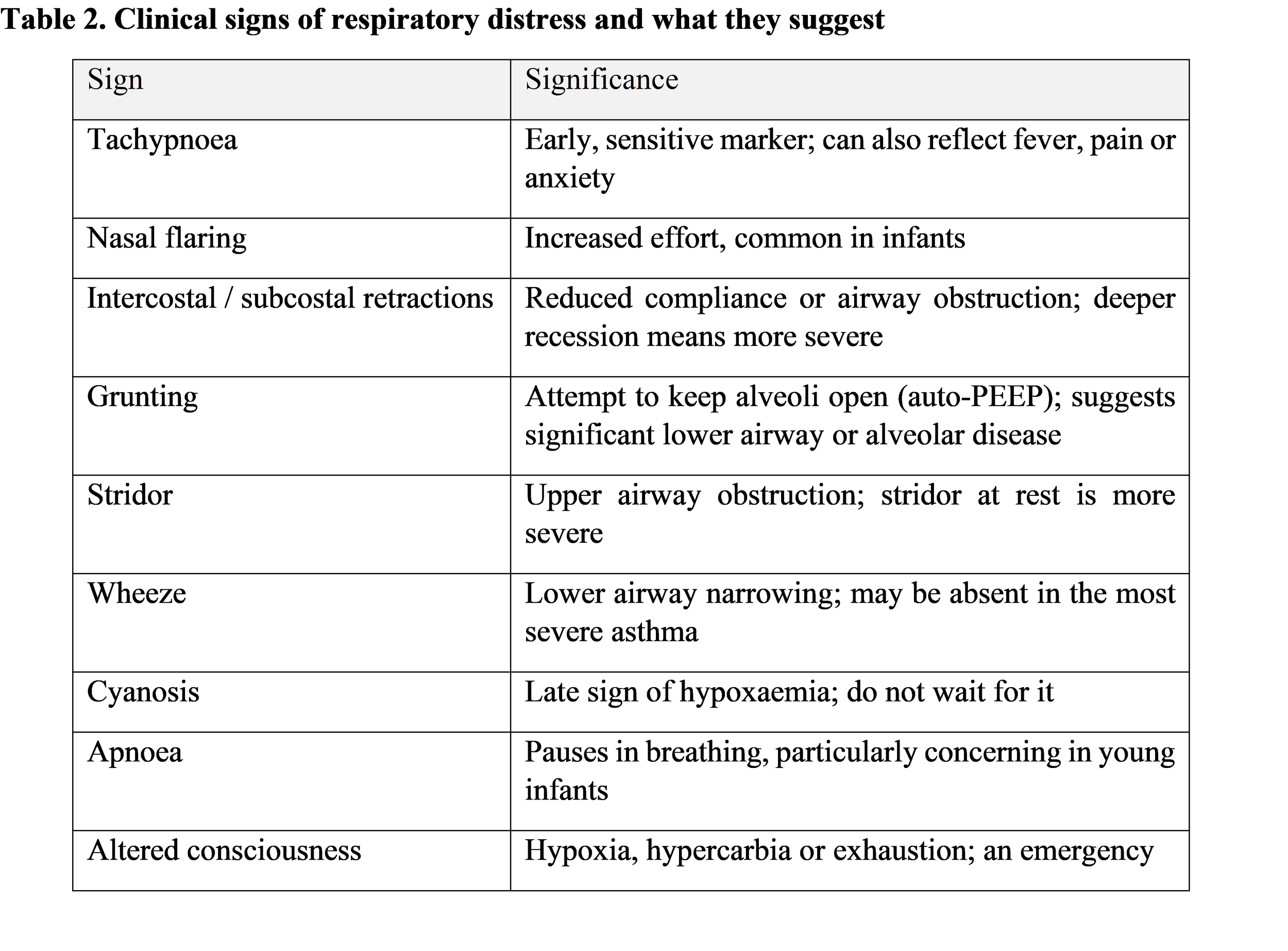
Tachypnoea and nasal flaring are usually the first things to appear, followed by retractions. Grunting, heard as a short expiratory noise at the end of each breath, indicates a child who is trying to stop the lungs collapsing and should rarely be ignored. Stridor in a child at rest suggests significant upper airway narrowing.
5.2 The late signs
Cyanosis is a late and unreliable sign. It depends on the amount of deoxygenated haemoglobin, so anaemic children may be severely hypoxic without looking blue, and skin tone can make it harder to see. Apnoea and irregular breathing, a falling respiratory rate in a child who is getting worse, drowsiness, floppiness and not recognising parents all point to impending failure.
5.3 Impending respiratory failure
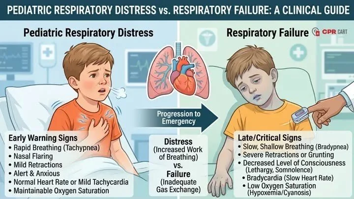
The typical sequence is escalating effort, then tiring, then slowing. A respiratory rate that drops while the child looks worse is not improvement. Bradycardia is a pre-arrest sign in children, and a child who becomes unexpectedly calm after a period of agitation may be becoming hypoxic and exhausted.
6. Diagnostic and Monitoring Approaches
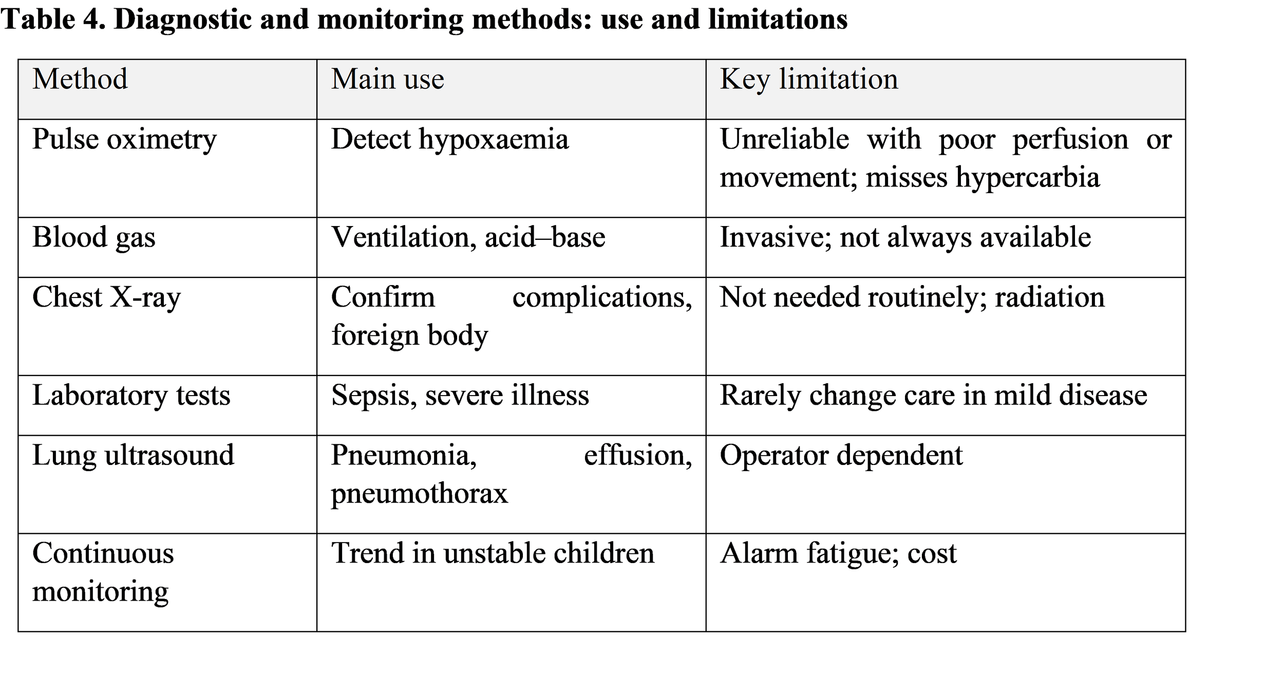
6.1 Pulse oximetry
Pulse oximetry is the most important monitoring tool for hypoxaemia. Clinical signs alone miss a substantial number of hypoxaemic children, and hypoxaemia substantially increases the risk of death in pneumonia [5,10]. WHO recommends oxygen for children with saturations below 90%, and many high-income guidelines use 92% as a threshold [3,7]. Oximetry has limits: probes may misread with poor perfusion, movement, nail polish or darker skin pigmentation, and a normal saturation on oxygen can mask worsening ventilation.
6.2 Blood gas analysis
Arterial or capillary gases tell us about ventilation and acid-base status, which oximetry does not. A rising carbon dioxide level in a tiring child is an important late finding. In most mild to moderate illness gases are unnecessary, and they should not delay treatment.
6.3 Chest radiography
Radiography is not required for every child with a respiratory infection. Randomised data in ambulatory children showed no benefit in routine chest radiographs for outcome [11]. It is most useful when the diagnosis is uncertain, when there is a suspected complication such as effusion or pneumothorax, when a foreign body is suspected, or in severe or non-responding illness.
6.4 Laboratory tests
Blood counts and inflammatory markers rarely change management in uncomplicated respiratory illness, but may help in sepsis, suspected bacterial pneumonia needing admission, or when planning for severe disease. Viral testing is mainly useful for infection control and epidemiology rather than individual treatment.
6.5 Lung ultrasonography
Point-of-care lung ultrasound has become a valuable bedside tool. A meta-analysis found good sensitivity and specificity for paediatric pneumonia, without radiation [12]. It does depend on operator skill and equipment, and its role in routine low-resource practice is still being defined.
6.6 Continuous monitoring
Continuous saturation, heart rate and respiratory rate monitoring help in high-dependency areas, but alarms generate fatigue. Monitors are an aid to nursing observation rather than a replacement for it.
7. Assessing Severity and Using Early Warning Systems
7.1 Grading severity
A practical way to think about severity is in three steps. In mild distress the child is alert, with mild tachypnoea and minimal recession, and is feeding. In moderate distress there is visible recession, nasal flaring, reduced feeding and perhaps saturations in the low 90s. In severe distress there is marked recession, grunting, saturations below 90%, exhaustion or altered consciousness. Severity should be reassessed repeatedly, because children move between categories in both directions.
7.2 Recognising hypoxaemia
Restlessness, irritability, poor feeding and head nodding are common clues to hypoxaemia in the absence of an oximeter, and WHO also highlights inability to drink, lower chest indrawing and central cyanosis as danger signs [3]. Without oximetry, a clinician should lower the threshold to give oxygen in any child with these signs.
7.3 Paediatric Early Warning Score (PEWS)
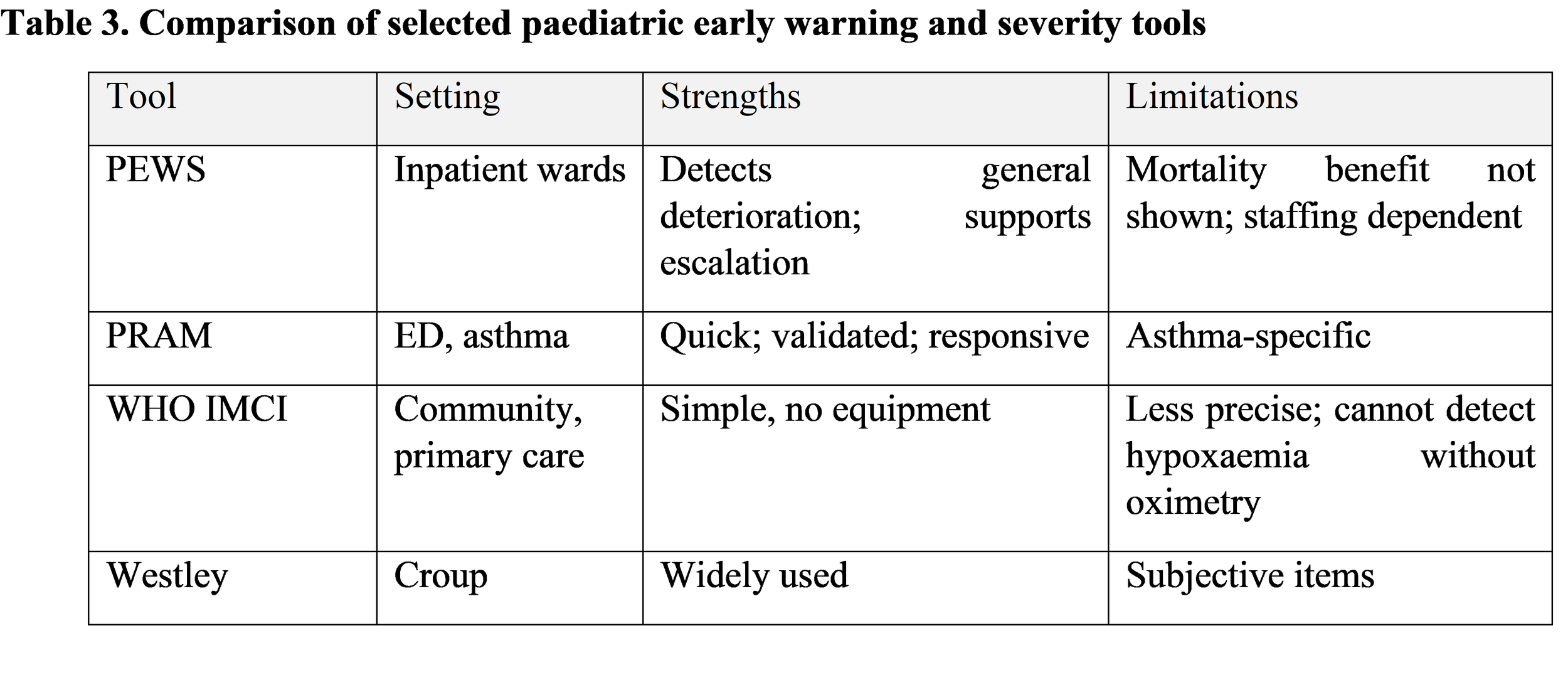
PEWS combines behaviour, cardiovascular and respiratory scores into a single number that triggers review. The Bedside PEWS showed good discrimination for critical deterioration in hospitalised children [13]. However, a systematic review found limited high-quality evidence, and the large cluster randomised EPOCH trial found that implementing a PEWS-based system did not reduce all-cause hospital mortality, though it was associated with lower rates of clinical deterioration events in some analyses [14,15].
7.4 Paediatric Respiratory Assessment Measure (PRAM)
PRAM scores suprasternal retraction, scalene muscle contraction, air entry, wheezing and oxygen saturation, and was validated for acute asthma from toddlers to teenagers [9,16]. It is quick, relies on observation, and is responsive to change, which makes it practical in emergency departments. It is asthma-specific, so it should not be used for croup or bronchiolitis without validation.
7.5 Other tools
Other clinical prediction rules include WHO IMCI classification for community settings, the Westley score for croup, and bronchiolitis severity scores. Each is designed for a particular population, and applying a tool outside its intended group tends to reduce its accuracy.
7.6 Strengths and weaknesses of scores
Scores standardise language, prompt escalation and help junior staff. But they can create false reassurance when the number is low yet the clinician is worried, and they should never override clinical concern. They work best as a prompt for conversation, not a substitute for judgement.
8. Early Management
8.1 Start with ABCDE
Initial stabilisation follows an Airway, Breathing, Circulation, Disability, Exposure approach. Treat life-threatening problems as you find them, call for help early, and keep reassessing. Where possible, keep the child with a parent, since calm children breathe better.
8.2 Airway and positioning
Let a child with respiratory distress choose their own position of comfort. In infants a neutral head position, sometimes with a small shoulder roll, helps keep the airway open. Clear nasal secretions gently, since infants are obligate nose breathers. Avoid distressing interventions in suspected epiglottitis or severe croup, where agitation may worsen obstruction.
8.3 Oxygen
Oxygen is the single most important intervention for hypoxaemic children. Evidence from improved oxygen systems in hospitals showed lower pneumonia mortality [17], and WHO has produced practical guidance for delivery by nasal prongs, catheters or masks [4,18]. Targets should be explicit, and the response should be monitored.
8.4 Bronchodilators and other drugs
Inhaled short-acting beta-agonists, delivered by spacer where possible, remain first-line for wheezy asthma [9]. Early oral or inhaled corticosteroids help in moderate to severe asthma. In bronchiolitis, routine bronchodilators and systemic steroids are not recommended [6,7]. In croup, a single dose of dexamethasone is recommended, with nebulised adrenaline for severe cases [8].
8.5 Non-invasive support
High-flow nasal cannula oxygen is widely used, and a randomised trial in infants with bronchiolitis found it reduced treatment failure on the ward compared with standard oxygen, though it did not shorten length of stay [19]. CPAP and non-invasive ventilation may help children with more severe disease, in settings with experienced staff and monitoring.
8.6 When to escalate
Consider advanced respiratory support in a child with worsening work of breathing despite treatment, rising carbon dioxide, persistent hypoxaemia, exhaustion, apnoea or reduced consciousness. The real art is not the technical procedure but the decision to call early, and to transfer to a higher level of care before the child has crashed.
8.7 Referral
Clear criteria for referral, a structured handover (for example SBAR) and safe transport with oxygen and an experienced escort all reduce risk. In smaller facilities, a call to a senior colleague earlier rather than later changes outcomes.
9. Challenges in Resource-Limited Settings
9.1 Equipment and oxygen
Many hospitals in low-income settings lack pulse oximeters, reliable oxygen supply or even functioning power. Studies suggest that a substantial proportion of children with severe pneumonia are hypoxaemic, and without oximetry they are often missed [5,10].
9.2 People and training
Shortages of trained nurses and doctors mean one clinician may be responsible for many unwell children. Training in structured assessment, such as WHO's emergency triage assessment and treatment approach, has been shown to be feasible and effective when it is supported over time [3].
9.3 Delay in seeking care
Families may delay because of cost, distance, previous poor experience, traditional beliefs or simply not recognising danger signs. Community education on danger signs, such as fast breathing, chest indrawing and inability to feed, is a low-cost but powerful intervention.
9.4 Financial and geographical barriers
Travel costs, lost income and informal fees prevent timely access. Delays are compounded where referral roads are poor or ambulances are unavailable.
9.5 Practical strategies
Strategies with evidence or strong rationale include: low-cost oximeters at primary care level; oxygen concentrators with solar power backup; clear, laminated danger-sign charts; mentoring and supportive supervision; community health worker training; and affordable referral transport. These work best when combined rather than introduced one at a time.
10. Discussion
10.1 What the evidence tells us
Taken together, the evidence points in a consistent direction. The simplest elements of assessment, which are respiratory rate, signs of increased work of breathing and the child's overall behaviour, carry most of the weight. They cost nothing, can be taught to anyone, and are present in nearly every well-validated tool. The more sophisticated the intervention, the more it tends to depend on context, staffing and the quality of the underlying observation.
10.2 Comparing clinical assessment methods
Bedside examination is fast and universal but varies between observers. Auscultation and percussion in particular show only moderate agreement, and respiratory rate is easily mis-measured. Structured tools reduce this variation by telling clinicians what to look for, and WHO danger signs have been shown to work well in settings where nothing else is available. The trade-off is that simpler tools sacrifice precision, while complex ones demand time and training that busy units may not have.
10.3 How effective is monitoring?
Pulse oximetry has the strongest case. It finds hypoxaemia that examination misses, and when linked to a reliable oxygen supply it is associated with lower mortality [10,17]. Its value falls sharply when oximeters are available but oxygen is not, or when staff have not been trained to act on the reading. Lung ultrasound is promising but depends on the operator. Continuous monitoring is useful in selected high-risk children but is no cure for under-staffing.
10.4 How reliable are early warning systems?
This is where honesty matters. PEWS tools predict deterioration reasonably well in the settings where they were developed, yet the largest randomised trial did not show a mortality benefit [14,15]. Possible explanations include limited baseline mortality, variation in how teams responded to a high score, and the fact that a score is only as good as the escalation culture behind it. In other words, the score is rarely the weak link; the response is.
10.5 What this means for practice
For clinicians, a few messages stand out:
• Count the respiratory rate properly, every time, and record it.
• Look at the child before the chart, and listen to parents.
• Use pulse oximetry wherever available, and give oxygen on clinical grounds where it is not.
• Treat a falling respiratory rate in a worsening child as an emergency.
• Reassess often, and escalate early without apologising for it.
10.6 Limitations and research gaps
This is a narrative rather than a systematic review, so selection of studies may reflect the authors' judgement and some relevant work may have been missed. The evidence base is uneven: most validation of scoring systems has come from high-income tertiary hospitals, whereas most deaths occur elsewhere. Important gaps include accuracy of oximetry across skin tones, validated tools for low-resource wards and primary care, the effect of early warning systems on outcomes outside hospitals, and economic evaluation of oxygen programmes.
11. Future Perspectives
11.1 Artificial intelligence
Machine learning models that use vital signs, electronic records or even audio and video of breathing are under development, and early studies suggest they may detect deterioration earlier than conventional scores. Prospective validation in diverse populations is still needed before routine use, and attention must be paid to bias and to clinician trust.
11.2 Digital health and portable devices
Smartphone-based oximeters, automated respiratory rate counters, handheld ultrasound and solar-powered oxygen concentrators could bring monitoring to places where it has never existed. Mobile decision-support apps for community health workers may help standardise IMCI practice. Their promise is real, but so is the danger of introducing devices without maintenance, training or supply chains.
11.3 Better prediction and standard protocols
Future prediction models should be built on data from the populations in which they will be used, and should be tested against outcomes that matter, such as death, intensive care admission and parental experience. International agreement on a minimum standard for assessing the breathless child, adaptable to local resources, would make research easier to compare and training easier to deliver.
12. Conclusion
Recognising respiratory distress early is, at heart, a matter of attention. The tools discussed here, from a clinician's eyes and hands to oximeters and early warning scores, all serve one purpose: to notice a struggling child soon enough for simple treatment to work. The evidence supports careful observation, routine use of pulse oximetry where available, context-appropriate scoring systems and a culture that responds quickly to concern. In low-resource settings, the priorities are oxygen, training and community awareness. Technology will help, but only if it strengthens, rather than replaces, the basics.
Recommendations for practice
Teach every clinician a standard approach to the breathless child, built around rate, work of breathing and mental state.
Make pulse oximetry and a reliable oxygen supply a minimum standard at every level of care.
Use early warning and severity tools as prompts, not substitutes for clinical judgement.
Educate caregivers about danger signs, and make it easy to seek care.
Invest in research that includes the children most at risk, in the places they live.
References
Perin J, Mulick A, Yeung D, Villavicencio F, Lopez G, Strong KL, et al. Global, regional, and national causes of under-5 mortality in 2000–19: an updated systematic analysis with implications for the Sustainable Development Goals. Lancet Child Adolesc Health. 2022;6(2):106–15.
Nair H, Simões EA, Rudan I, Gessner BD, Azziz-Baumgartner E, Zhang JSF, et al. Global and regional burden of hospital admissions for severe acute lower respiratory infections in young children in 2010: a systematic analysis. Lancet. 2013;381(9875):1380–90.
World Health Organization. Pocket book of hospital care for children: guidelines for the management of common childhood illnesses. 2nd ed. Geneva: WHO; 2013.
Fleming S, Thompson M, Stevens R, Heneghan C, Plüddemann A, Maconochie I, et al. Normal ranges of heart rate and respiratory rate in children from birth to 18 years of age: a systematic review of observational studies. Lancet. 2011;377(9770):1011–8.
Subhi R, Adamson M, Campbell H, Weber M, Smith K, Duke T. The prevalence of hypoxaemia among ill children in developing countries: a systematic review. Lancet Infect Dis. 2009;9(4):219–27.
Ralston SL, Lieberthal AS, Meissner HC, Alverson BK, Baley JE, Gadomski AM, et al. Clinical practice guideline: the diagnosis, management, and prevention of bronchiolitis. Pediatrics. 2014;134(5):e1474–502.
Fernandes RM, Bialy LM, Vandermeer B, Tjosvold L, Plint AC, Patel H, et al. Glucocorticoids for acute viral bronchiolitis in infants and young children. Cochrane Database Syst Rev. 2013;(6):CD004878.
Gates A, Gates M, Vandermeer B, Johnson C, Hartling L, Johnson DW, et al. Glucocorticoids for croup in children. Cochrane Database Syst Rev. 2018;(8):CD001955.
Global Initiative for Asthma. Global strategy for asthma management and prevention. Updated annually. Available from: https://ginasthma.org
Duke T, Subhi R, Peel D, Frey B. Pulse oximetry: technology to reduce child mortality in developing countries. Ann Trop Paediatr. 2009;29(3):165–75.
Swingler GH, Hussey GD, Zwarenstein M. Randomised controlled trial of clinical outcome after chest radiograph in ambulatory acute lower-respiratory infection in children. Lancet. 1998;351(9100):404–8.
Pereda MA, Chavez MA, Hooper-Miele CC, Gilman RH, Steinhoff MC, Ellington LE, et al. Lung ultrasound for the diagnosis of pneumonia in children: a meta-analysis. Pediatrics. 2015;135(4):714–22.
Parshuram CS, Hutchison J, Middaugh K. Development and initial validation of the Bedside Paediatric Early Warning System score. Crit Care. 2009;13(4):R135.
Chapman SM, Wray J, Oulton K, Peters MJ. Systematic review of paediatric track and trigger systems for hospitalised children. Resuscitation. 2016;109:87–109.
Parshuram CS, Dryden-Palmer K, Farrell C, Gottesman R, Gray M, Hutchison JS, et al. Effect of a pediatric early warning system on all-cause mortality in hospitalized pediatric patients: the EPOCH randomized clinical trial. JAMA. 2018;319(10):1002–12.
Ducharme FM, Chalut D, Plotnick L, Savdie C, Kudirka D, Zhang X, et al. The Pediatric Respiratory Assessment Measure: a valid clinical score for assessing acute asthma severity from toddlers to teenagers. J Pediatr. 2008;152(4):476–80.
Duke T, Wandi F, Jonathan M, Matai S, Kaupa M, Saavu M, et al. Improved oxygen systems for childhood pneumonia: a multihospital effectiveness study in Papua New Guinea. Lancet. 2008;372(9646):1328–33.
World Health Organization. Oxygen therapy for children: a manual for health workers. Geneva: WHO; 2016.
Franklin D, Babl FE, Schlapbach LJ, Oakley E, Craig S, Neutze J, et al. A randomized trial of high-flow oxygen therapy in infants with bronchiolitis. N Engl J Med. 2018;378(12):1121–31.