
Anatomical Variations of the Brachial Plexus: Clinical and Surgical Implications: Morphological Diversity, Prevalence Patterns, and Impact on Modern Peripheral Nerve Surgery
1. Tanabaeva Aiperi
2. Juyal Sheikh
3. Ilias Ahmed
4. Syed Abdullah Hussaini
(1. Professor, International Medical Faculty, Osh State University, Osh, Kyrgyz Republic
2. Student, International Medical Faculty, Osh State University, Osh, Kyrgyz Republic
3. Student, International Medical Faculty, Osh State University, Osh, Kyrgyz Republic
4. Student, International Medical Faculty, Osh State University, Osh, Kyrgyz Republic)
Abstract
The brachial plexus represents the most complex neural network in the peripheral nervous system, serving as the critical conduit for motor and sensory innervation of the entire upper limb. Despite its fundamental importance, the anatomical configuration of the brachial plexus demonstrates remarkable variability that challenges the classical textbook description of trunks, divisions, and cords. This comprehensive review examines the anatomical variations of the brachial plexus through the lens of contemporary surgical anatomy, synthesizing cadaveric dissection studies, intraoperative observations, and advanced imaging data to elucidate the prevalence and clinical significance of variant configurations. The article presents detailed analysis of variations occurring at every level of the plexus, including prefixed and postfixed formations, trunk anomalies, cord variations, and terminal branch anomalies, with particular attention to the prevalence rates reported in recent large-scale anatomical studies. Recent epidemiological data indicate that prefixed plexus configurations occur in approximately 20 to 30 percent of individuals, while postfixed configurations occur in 10 to 15 percent, with significant ethnic and population-specific variations documented across global anatomical studies. The review further explores the surgical implications of these variations, including their impact on brachial plexus block anesthesia, surgical approaches to the axilla and arm, nerve transfer procedures, and the interpretation of electrodiagnostic studies. Variant anatomy of the musculocutaneous nerve, including its absence, fusion with the median nerve, and variable relationship to the coracobrachialis muscle, occurs in 10 to 30 percent of individuals and represents a critical consideration in nerve transfer planning. By integrating classical anatomical knowledge with contemporary imaging studies and surgical series, this article provides orthopedic surgeons, plastic surgeons, anesthesiologists, and anatomists with a robust framework for understanding the brachial plexus not as a static idealized structure, but as a dynamic neural network whose individual configuration dictates surgical strategy, anesthetic approach, and patient outcomes in upper limb surgery.
Introduction
The brachial plexus stands as a testament to the intricate elegance of human neuroanatomy, a complex interweaving of neural elements that transforms the ventral rami of five spinal nerves into the sophisticated peripheral nerves governing the movement and sensation of the upper extremity. First described in systematic detail by the ancient anatomist Galen and later refined through the centuries by Vesalius, Eustachi, and the meticulous dissections of the nineteenth century, this neural network has captivated anatomists and challenged surgeons who seek to repair its injuries or navigate its complexities during surgical approaches to the shoulder and arm. The classical description of the brachial plexus, with its elegant progression from roots to trunks to divisions to cords to branches, represents an idealized pattern that, while pedagogically valuable, fails to capture the remarkable morphological diversity encountered in clinical practice and anatomical dissection.
The clinical importance of understanding brachial plexus variations has intensified in the modern era of peripheral nerve surgery, where nerve transfer procedures and sophisticated reconstructive techniques demand precise knowledge of normal and variant anatomy. The surgeon planning an Oberlin transfer to restore elbow flexion must know not only the typical location of the biceps branch of the musculocutaneous nerve but also anticipate variations in its origin and relationship to surrounding structures. Similarly, the anesthesiologist performing ultrasound-guided brachial plexus blockade must recognize that the arrangement of neural elements within the interscalene groove or axillary sheath may deviate significantly from textbook descriptions, with implications for block efficacy and risk of complications including vascular puncture or nerve injury.
The study of brachial plexus variations has evolved from anecdotal case reports in early anatomical literature to systematic large-scale dissection studies utilizing standardized classification systems. Recent investigations incorporating three-dimensional imaging, intraoperative nerve monitoring, and electrophysiological mapping have refined our understanding of these variations, revealing that certain configurations previously considered rare occur with sufficient frequency to mandate constant vigilance during surgical dissection. The prevalence of variations differs significantly across populations, with studies from Asia, Africa, Europe, and the Americas demonstrating distinct patterns that may reflect genetic, developmental, or environmental influences on plexus formation.
The embryological basis for brachial plexus variations lies in the complex process of limb bud development and nerve growth cone navigation during weeks four to eight of gestation. The plexus forms through the intermingling of ventral rami from C5 through T1, with contributions from C4 in prefixed configurations and T2 in postfixed configurations. The segmental origin of each terminal branch demonstrates variability, with the axillary nerve typically arising from C5 and C6 but occasionally receiving contributions from C4, and the radial nerve typically arising from C5 through T1 but showing variable contributions from each root. This developmental plasticity explains the frequent occurrence of variations in trunk formation, cord organization, and branching patterns that characterize the human brachial plexus.
This review aims to provide a comprehensive examination of the anatomical variations of the brachial plexus, integrating classical cadaveric studies with contemporary imaging research and surgical observations. The article will explore variations at each anatomical level of the plexus, examine the prevalence and distribution of these variants across populations, and discuss the profound clinical implications for nerve block anesthesia, surgical approaches, nerve transfer procedures, and the management of brachial plexus injuries. By synthesizing data from anatomical dissections, intraoperative findings, and radiological studies, this work seeks to provide a reference framework that acknowledges the brachial plexus as a structure of remarkable variability, where knowledge of variant configurations is not merely academic but essential for safe and effective surgical practice.
Materials and Methods
This review article was constructed through systematic analysis of the contemporary anatomical and surgical literature, with emphasis on publications from the past two decades regarding brachial plexus anatomy, variations, and clinical applications. The methodological approach involved comprehensive examination of peer-reviewed journals in anatomy, orthopedic surgery, plastic surgery, neurosurgery, and anesthesiology, including Clinical Anatomy, Journal of Anatomy, Plastic and Reconstructive Surgery, Journal of Hand Surgery, and Regional Anesthesia and Pain Medicine. Particular attention was directed toward studies utilizing cadaveric dissection with standardized protocols, intraoperative observations during brachial plexus surgery, and advanced imaging including magnetic resonance neurography and high-resolution ultrasound for in vivo assessment of plexus anatomy.
Inclusion criteria for anatomical data encompassed studies reporting quantitative measurements of variation prevalence in cohorts exceeding thirty specimens or subjects, with preference given to studies utilizing standardized classification systems such as the Todd or Kerr classifications for describing plexus variations. Clinical correlation data were derived from surgical series reporting anatomical findings during brachial plexus exploration, nerve transfer procedures, and tumor resections, with specific attention to studies documenting variant anatomy encountered intraoperatively and its impact on surgical strategy. When discrepancies existed between classical anatomical descriptions and contemporary findings, preference was given to recent studies incorporating modern imaging techniques and large sample sizes.
The synthesis of this literature was organized according to the anatomical levels of the brachial plexus, progressing from spinal nerve roots through trunks, divisions, cords, and terminal branches. All anatomical descriptions are presented according to standard surgical orientation, with prevalence rates reported as percentages. The reference list comprises foundational anatomical works, contemporary dissection studies, and clinical series that collectively represent the current state of knowledge regarding brachial plexus variations and their surgical implications.
Results
Variations at the Level of the Roots and Trunks
The formation of the brachial plexus begins with the ventral rami of C5 through T1, which emerge from the intervertebral foramina and course through the interscalene triangle between the anterior and middle scalene muscles. Variations at this proximal level fundamentally alter the subsequent architecture of the entire plexus. The prefixed brachial plexus, characterized by a significant contribution from C4 to the plexus with correspondingly reduced contribution from T1, occurs in approximately 20 to 30 percent of individuals according to recent large-scale dissection studies. In this configuration, the C4 ventral ramus joins the C5 root either directly or through a communicating branch, creating a plexus that appears shifted superiorly with respect to the vertebral column. The clinical implications of this variation are profound, as injuries to the upper trunk in a prefixed plexus may affect C4 function, leading to phrenic nerve involvement and diaphragmatic paralysis, or conversely, lower trunk injuries may spare the hand intrinsic muscles that receive their innervation from T1 in typical configurations.
The postfixed brachial plexus, conversely, receives significant contribution from T2 with reduced or absent contribution from C5, occurring in approximately 10 to 15 percent of individuals. This configuration creates a plexus that extends more caudally, with the lower trunk forming at a more inferior level and the T2 contribution joining the C6 or C7 roots. Postfixed plexus anatomy has significant implications for thoracic outlet syndrome, as the lower trunk may be compressed at the level of the first rib or cervical rib with greater frequency due to its more caudal position. Additionally, the presence of a cervical rib, occurring in 0.5 to 1 percent of the population, interacts with plexus configuration to produce variable patterns of neurovascular compression, with postfixed plexus anatomy potentially exacerbating symptoms due to the altered relationship between neural elements and the bony thoracic outlet.
Variations in trunk formation represent another significant category of proximal plexus anomalies. The classical description of three trunks, superior from C5 and C6, middle from C7, and inferior from C8 and T1, is observed in approximately 70 to 80 percent of dissections. However, variations including the absence of a distinct middle trunk, with C7 joining the superior or inferior trunk directly, occur in 5 to 10 percent of cases. More rarely, four trunks may be formed when C7 contributes to both superior and inferior trunks through early branching. These variations in trunk organization influence the pattern of nerve fiber distribution to the divisions and cords, potentially affecting the somatotopic organization of the plexus and the pattern of neurological deficit following injury.
Communicating branches between adjacent roots or trunks occur with surprising frequency, with studies documenting anastomoses between C5 and C6 roots in 10 to 20 percent of specimens, and between C8 and T1 in 5 to 10 percent. These communications, while typically asymptomatic, create potential pathways for aberrant reinnervation following nerve injury and may complicate electrodiagnostic interpretation by providing alternative routes for nerve fiber conduction.
Variations at the Level of Divisions and Cords
The divisions of the brachial plexus, formed by the bifurcation of each trunk into anterior and posterior divisions, represent a critical crossroads where nerve fibers are sorted into anterior and posterior compartments destined for flexor and extensor muscle groups respectively. Variations at this level, while less common than root or trunk variations, significantly impact the organization of the cords and the distribution of terminal branches. The absence of bifurcation of a trunk, with C7 continuing as a single structure to join the lateral or medial cord, occurs in 2 to 5 percent of specimens and creates anomalous pathways for nerve fibers that may result in unexpected patterns of muscle innervation.
The cords of the brachial plexus, named for their relationship to the axillary artery, demonstrate remarkable variability in their formation and relationship to the second part of the axillary artery. The classical description of lateral, posterior, and medial cords surrounding the axillary artery is observed in approximately 75 percent of dissections. However, variations including the absence of the posterior cord, with posterior divisions joining the lateral and medial cords directly, occur in 3 to 7 percent of cases. More commonly, the relationship of the cords to the axillary artery varies, with the medial cord passing posterior to the artery rather than medial to it in 5 to 10 percent of specimens, or the lateral cord crossing anterior to the artery to join the medial cord in rare cases.
The musculocutaneous nerve, a terminal branch of the lateral cord, demonstrates the most frequent and clinically significant variations of any terminal branch. The classical description of the musculocutaneous nerve arising from the lateral cord, piercing the coracobrachialis muscle, and innervating the biceps, brachialis, and coracobrachialis before continuing as the lateral antebrachial cutaneous nerve, is observed in only 60 to 70 percent of specimens. Variations include the absence of the musculocutaneous nerve as a distinct entity, with its muscular branches arising directly from the median nerve, occurring in 10 to 20 percent of cases. In this configuration, the median nerve provides branches to the coracobrachialis, biceps, and brachialis, and the lateral antebrachial cutaneous nerve arises from the median nerve rather than as a continuation of the musculocutaneous nerve. This variation has profound implications for nerve transfer surgery, as the Oberlin transfer utilizing ulnar nerve fascicles to the biceps branch requires identification of a distinct biceps branch that may not exist in this variant configuration.
Fusion of the musculocutaneous nerve with the median nerve, with the two nerves running together for a variable distance before separating, occurs in 10 to 15 percent of specimens. In some cases, the fusion extends throughout the arm, with the musculocutaneous nerve not separating from the median nerve until the level of the elbow. This variation complicates surgical approaches to the arm and may lead to inadvertent injury during procedures requiring exposure of the median nerve, such as anterior interosseous nerve syndrome release or vascular repair.
The relationship of the musculocutaneous nerve to the coracobrachialis muscle demonstrates significant variability, with the nerve piercing the muscle in 70 to 80 percent of cases, passing medial to the muscle without piercing in 10 to 15 percent, and passing lateral to the muscle in 5 to 10 percent. In some specimens, the nerve divides into multiple branches before entering the muscle, creating a plexiform arrangement that complicates surgical dissection. The clinical significance of this variation lies in the risk of nerve compression within the coracobrachialis muscle, particularly in athletes or individuals with hypertrophy of this muscle, producing a musculocutaneous nerve entrapment syndrome that may require surgical release.
Variations at the Level of Terminal Branches
The terminal branches of the brachial plexus, including the axillary, radial, median, and ulnar nerves, demonstrate variations in their origin, course, and branching patterns that influence surgical approaches and nerve transfer options. The axillary nerve, typically arising from the posterior cord with contributions from C5 and C6, may receive contributions from C4 in prefixed plexus configurations or from C7 in postfixed configurations. The relationship of the axillary nerve to the posterior humeral circumflex vessels varies, with the nerve passing anterior to the vessels in 80 percent of cases and posterior in 20 percent, influencing the risk of vascular injury during surgical exposure of the quadrangular space.
The radial nerve, the largest branch of the brachial plexus, demonstrates variations in its origin from the posterior cord, with high origin occurring in 5 to 10 percent of cases where the nerve arises at the level of the axilla rather than the posterior cord proper. The branching pattern of the radial nerve in the arm varies significantly, with the posterior cutaneous nerve of the arm arising from the radial nerve proper in 60 percent of cases, from the axillary nerve in 20 percent, and from the posterior cord directly in 20 percent. These variations influence the pattern of sensory loss following radial nerve injury and complicate the interpretation of electrodiagnostic studies.
The median nerve, formed by the union of lateral and medial roots from the lateral and medial cords respectively, demonstrates variations in its formation that occur in 5 to 10 percent of specimens. These variations include the absence of a distinct lateral root, with the median nerve arising entirely from the medial cord, or the presence of accessory roots contributing to the median nerve from the lateral or posterior cords. The most clinically significant variation involves the presence of a median nerve branch to the pronator teres arising proximal to the elbow joint, occurring in 10 to 15 percent of cases, which may be compressed by the lacertus fibrosus or pronator teres muscle producing median nerve entrapment.
The ulnar nerve, typically arising from the medial cord with contributions from C8 and T1, may receive contributions from C7 in 10 to 20 percent of cases, creating a nerve with a more proximal origin that may be vulnerable to injury at the level of the thoracic outlet. The branching pattern of the ulnar nerve in the forearm varies, with the nerve dividing into its terminal branches at the level of the wrist in 70 percent of cases and within the Guyon canal in 30 percent. This variation influences the pattern of motor and sensory loss following ulnar nerve injury at the wrist and complicates surgical planning for nerve repair or transfer.
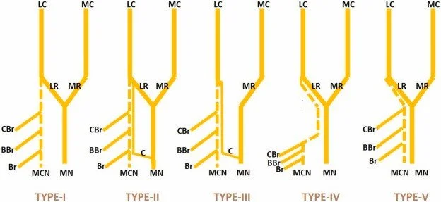
Fig.1. Diagrammatic Presentation Of The Anatomical Variations Of Median & Musculocutaneous Nerves According To Le Minor’s Classification.
Type I- No communication between MCN and MN. The MCN penetrates the CBr muscle and divides into three branches to innervate the CBr, BBr and Br Muscles.
Type II- This pattern is similar to the normal but an extra ramus connects the MCN and MN while it gives a branch to Brachialis muscle
Type III- The lateral root fibres follow the MCN distally and leave it to unite with the median nerve only after MCN gives rami to CBr, BBr and Br Muscles.
Type IV- MCN fibres join the lateral root of the MN and after some distance the MCN arises from the MN as a proper and independent nerve.
Type V- The MCN is absent and the entire fibres of the MCN first pass through the lateral root and then run into the MN along its course All the brachial flexors are innervated by median nerve rami.
LC- Lateral Cord; MC- Medial Cord; LR- Lateral Root of the Median Nerve; MR- Medial Root of the Median Nerve; MCN- Musculocutaneous Nerve; MN- Median Nerve; CBrM- Coracobrachialis Muscle; BBrM- Biceps Brachii Muscle; BrM- Brachialis Muscle; C: Communicating Ramus
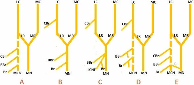
Fig.2. Diagrammatic Presentation Of The Findings Of The Present Study.
A- Normal Pattern. B- Absent MCN; CBr-By Direct Branches From LC; BB & Br-By Direct Branches From MN (Partially Type-V Le Minor). C- Absent MCN; CBr-By Direct Branches From LC; BB-By Direct Branches of MN; Br-By LCNF (Partially Type-V Le Minor). D- CBr- By Direct Branches From LC; BB & Br-By Direct Branches From MCN (Present Study). E− CBr, BB & Br-By Direct Branches From MCN; Communication between MCN &MN at the level of Brachialis (Type-II Venieratos & Anagnostopoulou and Type-II Le Minor). LC- Lateral Cord; MC- Medial Cord; LR- Lateral Root of the Median Nerve; MR- Medial Root of the Median Nerve; MCN- Musculocutaneous Nerve; MN- Median Nerve; CBrM: Coracobrachialis Muscle; BBrM- Biceps Brachii Muscle; BrM- Brachialis Muscle; C- Communicating Ramus.
Clinical and Surgical Implications
The anatomical variations of the brachial plexus carry profound implications for clinical practice across multiple specialties, from anesthesiology to orthopedic surgery to plastic surgery. In regional anesthesia, the performance of brachial plexus blockade requires recognition of variant anatomy to achieve reliable blockade and avoid complications. The interscalene approach to brachial plexus blockade targets the nerve roots at the level of C5 through C7, but in prefixed plexus configurations, the C4 contribution may be blocked as well, increasing the risk of phrenic nerve paralysis and respiratory compromise. Conversely, in postfixed plexus configurations, the T2 contribution may be missed by the interscalene approach, resulting in incomplete blockade of the medial arm and forearm.
The axillary approach to brachial plexus blockade, targeting the terminal branches surrounding the axillary artery, requires recognition of variations in cord and terminal branch anatomy. The presence of a median nerve branch to the pronator teres arising proximal to the block site may result in incomplete motor blockade despite adequate sensory anesthesia. Similarly, variations in the relationship of the musculocutaneous nerve to the coracobrachialis muscle may result in failure to block this nerve using the axillary approach, as the nerve may be separated from the axillary artery by the muscle belly.
In brachial plexus surgery, whether for trauma reconstruction or tumor resection, recognition of variant anatomy is essential to avoid iatrogenic injury and to optimize functional outcomes. During exploration for brachial plexus injury, the surgeon must identify not only the typical structures but also anticipate variations such as a prefixed or postfixed configuration that may alter the relationship of the plexus to surrounding landmarks. The presence of communicating branches between roots or trunks may provide alternative pathways for nerve fiber conduction that can be exploited in nerve transfer procedures, or conversely, may complicate the interpretation of intraoperative nerve stimulation studies.
Nerve transfer procedures, which have revolutionized the treatment of brachial plexus injuries, rely upon precise knowledge of variant anatomy for donor and recipient nerve identification. The Oberlin transfer, utilizing ulnar nerve fascicles to the biceps branch of the musculocutaneous nerve, requires identification of a distinct biceps branch that may not exist in cases of musculocutaneous nerve absence or fusion with the median nerve. In such cases, alternative donor nerves or direct muscle transfer may be required. Similarly, the double fascicular transfer, utilizing median and ulnar nerve fascicles to biceps and brachialis branches, requires recognition of variations in the branching pattern of the musculocutaneous nerve to ensure appropriate matching of donor and recipient fascicles.
In surgical approaches to the shoulder and arm, recognition of brachial plexus variations is essential to avoid nerve injury. The deltopectoral approach to the shoulder exposes the cephalic vein and deltoid muscle, but in cases of variant axillary nerve anatomy, the nerve may be more anteriorly placed and vulnerable to injury during retraction. The posterior approach to the shoulder, utilized for posterior capsulorrhaphy or tumor resection, requires careful identification of the axillary nerve as it passes through the quadrangular space, with recognition that the nerve may pass posterior to the posterior humeral circumflex vessels in 20 percent of cases.
Discussion
The anatomical variations of the brachial plexus, ranging from prefixed and postfixed configurations to variant branching patterns of terminal nerves, represent a fundamental aspect of human anatomical diversity that challenges the classical textbook description of this complex neural network. The prevalence of these variations, with prefixed plexus occurring in 20 to 30 percent and postfixed in 10 to 15 percent of individuals, mandates that surgeons and anesthesiologists approach each case with anticipation of variant anatomy rather than expectation of the standard configuration. The integration of advanced imaging techniques including magnetic resonance neurography and high-resolution ultrasound has enhanced our ability to identify these variations preoperatively, permitting surgical planning that accounts for individual anatomy.
The clinical implications of brachial plexus variations extend across multiple specialties, from the anesthesiologist performing regional blockade to the surgeon performing nerve transfer procedures for brachial plexus injury reconstruction. The recognition that the musculocutaneous nerve may be absent, fused with the median nerve, or demonstrate variable relationship to the coracobrachialis muscle in up to 30 percent of individuals has profound implications for nerve transfer surgery, requiring flexibility in surgical strategy and willingness to adapt to unexpected anatomical findings. Similarly, the recognition that the axillary nerve may demonstrate variable relationship to the posterior humeral circumflex vessels influences surgical exposure of the quadrangular space and the risk of vascular injury during nerve exploration.
The study of brachial plexus variations also informs our understanding of the developmental biology of the peripheral nervous system, providing insights into the mechanisms of nerve growth cone navigation and target recognition during embryogenesis. The frequent occurrence of communicating branches between adjacent roots and trunks suggests a developmental plasticity that permits alternative pathways for nerve fiber routing, potentially explaining the variable patterns of reinnervation observed following nerve injury. The ethnic and population-specific variations in plexus configuration documented in recent studies suggest genetic influences on plexus development that warrant further investigation.
Future directions in the study of brachial plexus variations likely involve the integration of advanced imaging techniques with intraoperative nerve monitoring to create individualized maps of plexus anatomy for each patient. The development of artificial intelligence algorithms for image recognition may assist in identifying variant anatomy on preoperative imaging, while augmented reality systems may overlay anatomical information onto the surgical field to guide dissection. However, these technological advances will remain adjuncts to, rather than replacements for, the fundamental anatomical knowledge and surgical judgment that have defined safe brachial plexus surgery since the era of Seddon and Sunderland.
Conclusion
The brachial plexus demonstrates remarkable anatomical variability that challenges classical descriptions and mandates constant vigilance in clinical practice. Prefixed and postfixed configurations, variations in trunk and cord formation, and anomalies of terminal branching occur with sufficient frequency to influence every aspect of brachial plexus surgery and regional anesthesia. The surgeon who understands these variations, who anticipates the unexpected, and who adapts surgical strategy to individual anatomy will achieve the best outcomes for patients undergoing brachial plexus exploration, nerve transfer, or regional blockade. As the field of peripheral nerve surgery continues to evolve, the importance of anatomical mastery remains undiminished, ensuring that future generations of surgeons can navigate the complexities of this remarkable neural network with confidence and precision.
References
Kerr AT. The brachial plexus of nerves in man, the variations in its formation and branches. Am J Anat. 1918;23(2):285-395.
Todd TW. The descriptive anatomy of the brachial plexus. Anat Rec. 1920;19(2):89-102.
Fazan VP, Amadeu AD, Caleffi AL, et al. Brachial plexus variations in its formation and main branches. Acta Cir Bras. 2003;18(5):441-447.
Uysal II, Seker M, Karabulut AK, et al. Brachial plexus variations in human fetuses. Neurosurgery. 2003;53(3):676-681.
Lee HY, Chung IH, Sir WS, et al. Variations of the ventral rami of the brachial plexus. J Korean Med Sci. 1992;7(1):19-24.
Nakatani T, Tanaka S, Mizukami S. Bilateral four-trunked brachial plexus. Anat Sci Int. 1997;72(4):299-302.
Bergman RA, Thompson SA, Afifi AK, et al. Compendium of Human Anatomic Variation: Text, Atlas, and World Literature. Baltimore: Urban and Schwarzenberg; 1988.
Ballesteros LE, Forero PL, Buitrago ER. Communication between the musculocutaneous and median nerves in the arm: anatomical study and clinical implications. Rev Bras Anestesiol. 2013;63(3):239-243.
Venieratos D, Anagnostopoulou S. Classification of communications between the musculocutaneous and median nerves. Clin Anat. 1998;11(5):327-331.
Guerri-Guttenberg RA, Ingolotti M. Classifying musculocutaneous nerve variations. Clin Anat. 2009;22(6):671-683.
Choi D, Rodriguez-Niedenführ M, Vazquez T, et al. Patterns of connection between the musculocutaneous and median nerves in the axilla and arm. Clin Anat. 2002;15(1):11-17.
Aslan A, Yalcin B, Elhan A, et al. A variation of the brachial plexus: the relationship between the musculocutaneous nerve and the coracobrachialis muscle. Neurosurgery. 2005;56(2 Suppl):E409.
Loukas M, Aqueelah H. Musculocutaneous and median nerve connections within, proximal and distal to the coracobrachialis muscle. Folia Morphol. 2005;64(2):101-108.
Tubbs RS, Salter EG, Wellons JC, et al. Anomalous intramuscular innervation of the musculocutaneous nerve to the biceps brachii. Clin Anat. 2003;16(4):308-311.
Oberlin C, Beal D, Leechavengvongs S, et al. Nerve transfer to biceps muscle using a part of ulnar nerve for C5-C6 avulsion of the brachial plexus: anatomical study and report of four cases. J Hand Surg Am. 1994;19(2):232-237.
Leechavengvongs S, Witoonchart K, Uerpairojkit C, et al. Nerve transfer to deltoid muscle using the nerve to the long head of triceps, part I: an anatomic feasibility study. J Hand Surg Am. 2003;28(4):628-632.
Mackinnon SE, Novak CB, Myckatyn TM, et al. Results of reinnervation of the biceps and brachialis muscles with a double fascicular transfer for elbow flexion. J Hand Surg Am. 2005;30(5):978-985.
Moayeri N, Groen GJ. Difference in quantity and distribution of extradural and intradural nerve roots of the brachial plexus. Clin Anat. 2008;21(1):55-62.
Piskač P, Vraždil J, Hanušová J, et al. High-resolution ultrasound of the brachial plexus: normal appearance and practical applications. Biomed Pap Med Fac Univ Palacky Olomouc Czech Repub. 2012;156(2):107-117.
Standring S, editor. Gray's Anatomy: The Anatomical Basis of Clinical Practice. 42nd ed. London: Elsevier; 2020. p. 920-935.